Digital Posters
Digestive, Diabetes & Pancreas
ISMRM & SMRT Annual Meeting • 15-20 May 2021

| Concurrent 5 | 13:00 - 14:00 |
1884.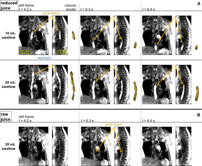 |
Accelerated Dynamic MRI for the Assessment of Esophageal Peristalsis During Swallowing
Ethan M I Johnson1, Sourav Halder2, Peter J Kahrilas1, John E Pandolfino1, Neelesh A Patankar2, and Michael Markl1
1Northwestern University, Chicago, IL, United States, 2Northwestern University, Evanston, IL, United States
Pure pineapple juice was reduced by heating to a range of concentrations, and the T1 values were characterized for each concentration. A log-linear relationship between concentration factor and T1 reduction factor was shown. In vivo dynamic MR imaging of esophageal peristalsis was demonstrated using a modified MR angiography sequence and a 50% volume-reduced concentration of pineapple juice as swallowable contrast agent.
|
|||
1885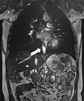 |
Reaal-time MRI for assessment of patients with gastroesophageal reflux disease:a feasibility research Video Permission Withheld
Chao Wu1, Xinyu Wang1, Chen Zhang2, Cheng Cheng3, Wei Zhao4, and Haoran Sun1
1Medical Imaging Department, Tianjin Medical University General Hospital, Tianjin, China, 2MR Scientific Marketing, Siemens Healthcare, Beijing, China, 3Clinical Application, Siemens Healthcare, Tianjin, China, 4Department of Gastroenterology, Tianjin Medical University General Hospital, Tianjin, China
This study was aim to observe swallowing processes in volunteers and patients with gastroesophageal reflux through real-time magnetic resonance imaging (MRI), and to assess the transport and reflux of pineapple juice through the gastroesophageal junction during Valsalva. Gastroesophageal reflux was detected in all patients included in this study.
|
|||
1886.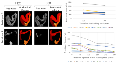 |
MRI Characterisation of the Reformation of Colonic Content after Bowel Purgation
Hannah Grace Williams1,2, Caroline Hoad1,3, Neele Dellschaft1,3, Christabella Ng3,4, Alan Smyth3,4, Giles Major2,3, and Penny Gowland1,3
1Sir Peter Mansfield Imaging Centre, University of Nottingham, Nottingham, United Kingdom, 2Nottingham Digestive Diseases Centre, University of Nottingham, Nottingham, United Kingdom, 3National Institute for Health Research (NIHR) Nottingham Biomedical Research Centre, Nottingham University Hospitals NHS Trust and the University of Nottingham, Nottingham, United Kingdom, 4Division of Child Health, Obstetrics and Gynaecology, University of Nottingham, Nottingham, United Kingdom
A key colonic function is the formation of faeces and the growth and proliferation of the microbiota. This process is little described as it is difficult to study in vivo. We aimed to investigate it using MRI which can non-invasively monitor changes in the colonic content. In 8 healthy volunteers we observed the initial process of faecal formation following purgation. The results suggest that MRI can provide new insights into the important physiological process of faeces formation and establishment of the microbiome in the colon.
|
|||
1887.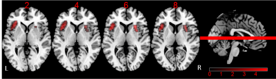 |
Disrupted resting-state salience network in type 2 diabetes with and without mild cognitive impairment
Yumeng Lei1, Dongsheng Zhang1, Fei Qi2, Man Wang2, Jie Gao1, Min Tang1, Yu Su2, Zhirong Shao2, Kai Ai3, and Xiaoling Zhang1
1Department of MRI, Shaanxi Provincial People’s Hospital, Xi'an, China, 2Xi'an Medical University, Xi’an, China, 3Philips Healthcare, Xi'an, China
To investigate the underlying mechanisms of cognitive decline in patients with diabetes, data-driven independent component analysis (ICA) was applied to resting-state fMRI images from 34 type 2 diabetes mellitus (T2DM) patients with normal cognition (DMCN) and 31 T2DM patients with cognitive impairment (DMCI), and 31 healthy controls (HC) to identify the difference in salience network (SN). The resting-state functional connectivity (rs-FC) alteration are different and complicated among HC, DMCN and DMCI groups, and also are correlated to neuropsychological scores, indicated that altered SN rs-FC in T2DM patients are closely related to cognitive impairment.
|
|||
1888.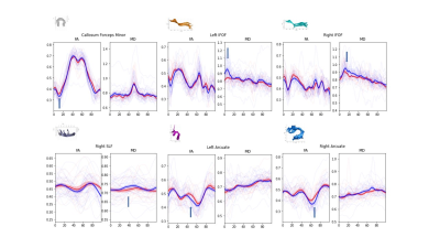 |
Study on the white matter integrity in T2DM using automated fiber quantification tractography
Jun Wang1, Wenjuan Bai2, Pengfei ZHANG1, Wenjing HUANG1, Wanjun HU1, Guangyao LIU1, and Jing ZHANG1
1Department of Magnetic Resonance, Lanzhou University Second Hospital, Lan Zhou, China, 2Second School of Clinical Medicine, Lanzhou University, Lan Zhou, China
The objective of this study was to explore the effect of glucose fluctuation on T2DM patients using a new method named automated fiber quantification (AFQ). To explore the value of different DTI parameters in the classification of T2DM patients and normal patients using SVM model.
|
|||
1889.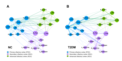 |
Modeling Central Olfactory Network Alteration in Type 2 Diabetes Mellitus: From Primary to Advanced Cortex
Wen Zhang1, Jiaming Lu1, Jilei Zhang2, and Bing Zhang1
1Drum Tower Hospital, The Affiliated Hospital of Nanjing University Medical School, Nanjing, China, 2Philips Healthcare, Shanghai, China
Type 2 diabetes mellitus (T2DM) is associated with olfactory dysfunction and cognitive decline. We proposed one functional connectivity (FC) model that links the primary olfactory cortex (POC) to advanced cognition related cognitive brain area. The whole olfactory network was divided into three sub-networks: POC consists of the bilateral anterior olfactory nucleus, piriform cortex, entorhinal cortex, and amygdala; secondary olfactory cortex (SOC) include the bilateral hippocampus, parahippocampus, thalamus, and hypothalamus; advanced olfactory cortex (AOC) consists of bilateral orbitofrontal cortex, insula and dorsolateral prefrontal cortex(DLPFC). Our results suggested that FC was heterogeneously disrupted in the POC-SOC-AOC olfactory pathway in T2DM.
|
|||
| 1890. | Amide Proton Transfer weighted imaging analysis of cerebral metabolism changes in T2DM patients
weiwei wang1, Yanwei Miao1, JianLin Wu2, and JianLin Wu2
1The First affiliated hospital of Dalian Medical University, Dalian, China, 2Affiliated Zhongshan Hospital of Dalian University, Dalian, China
APTw can reflect the abnormal changes of brain metabolism in T2DM patients from the molecular level, providing new imaging evidence for understanding the neuropathological mechanism of diabetic brain injury
|
|||
1891.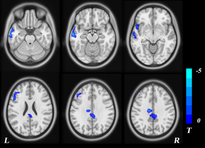 |
Study on cerebral perfusion characteristic network of type 2 diabetes mellitus using 3D arterial spin labeling imaging
hang qu1, wenjuan ba1, weiqiang Dou2, and wei wang1
1The Affiliated Hospital of Yangzhou University, yangzhou, China, 2GE Healthcare, MR Research China, Beijing, Beijing, China
This study aimed to investigate the cerebral microcirculatory blood flow perfusion and perfusion patterns in patients with type 2 diabetes by using 3D arterial spin labeling (ASL) imaging, and to analyze the correlation between perfusion changes and biochemical indexes. We obtained 3D-ASL derived cerebral blood flow (CBF) and perfusion feature network using principal component analysis (PCA) on each subject accordingly. These diabetic patients showed low regional CBF, and PCA-based perfusion characteristics could identify patients with diabetes. Changed perfusion patterns reflected the remodeling of cerebral blood flow perfusion, revealing clinical value and significance for early diagnosis and intervention of diabetic microangiopathy.
|
|||
1892.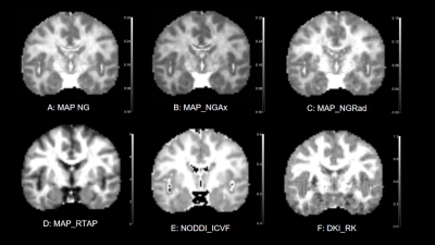 |
Early Detection of Type 2 Diabetes Mellitus with Mild Cognitive Impairment based on Multiple Advanced Diffusion Models: DKI, MAP-MRI and NODDI
Wen-jiao Lyu1, Yunzhu Wu2, Xiao-meng Ma1, Yue Feng1, Yu-na Chen1, Shi-jun Qiu1, Xu Yan2, Min-xiong Zhou3, and Guang Yang4
1Department of Radiology, The First Affiliated Hospital of Guangzhou University of Chinese Medicine, Guangzhou, China, 2MR Scientific Marketing, SIEMENS Healthcare, Shanghai, China, 3Shanghai University of Medicine & Health Sciences, Shanghai, China, 4Shanghai Key Laboratory of Magnetic Resonance, East China Normal University, Shanghai, China
Compare to pathological examination and cognitive assessment scale, diffusion imaging can detect microscopic diffusion properties of biological tissues noninvasively and repeatably. This study aimed to evaluate the ability of multiple advanced diffusion models reconstructed by diffusion sequence: MAP-MRI, NODDI, and DKI in the early detection of Type 2 diabetes mellitus (T2DM) patients with mild cognitive impairment (MCI).
|
|||
1893.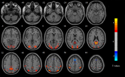 |
Inter-hemispheric Functional Connections are more Vulnerable to Attack than Structural Connection in Irritable Bowel Syndrome Patients
Guangyao Liu1, Shan Li2, Hong Liu1, Laiyan Ma1, Zhijun Yao2, Jing Zhang1, Shaoyu Wang3, and Dekui Zhang4
1Department of Magnetic Resonance, Lanzhou University Second Hospital, Lanzhou, China., Lanzhou, China, 2Gansu Provincial Key Laboratory of Wearable Computing, School of Information Science and Engineering, Lanzhou University, Lanzhou, China., Lanzhou, China, 3MR Scientific Marketing, Siemens Healthineers, Shanghai, China., xian, China, 4Department of Gastroenterology, Lanzhou University Second Hospital, Lanzhou, China., Lanzhou, China
In this study, five types of MRI features, including voxel-mirrored homotopic connectivity (VMHC) from functional MRI (fMRI), asymmetry index (AI) from structural MRI (sMRI), FA, fiber length and fiber number (FN) from diffusion tensor imaging (DTI) were used to evaluate the asymmetry and inter-hemispheric connectivity of IBS patients. Through taking comprehensive analysis of the bilateral brain in Irritable bowel syndrome (IBS) patients, we speculated that inter-hemispheric functional connectivity is more vulnerable to IBS than anatomical connectivity, while the structural morphology of brain is the most stable. Meanwhile, the affected areas were concentrated in default mode network (DMN) and sensorimotor network. The results of our study are only preliminary, but it may provide theoretical basis for future research on the regulation of gut-brain axis (GBA) and pathophysiology in functional intestinal diseases.
|
|||
1894.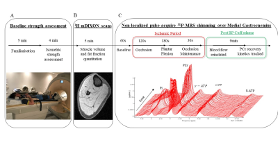 |
31P MRS and MRI phenotyping of muscle metabolic quality in Inflammatory Bowel Disease fatigue
Jordan J McGing1,2,3, Rosemary Nicholas2, Sébastien Serres4, Paul L Greenhaff5,6,7, Gordon W Moran1,7, and Susan T Francis2
1Nottingham Digestive Diseases Centre, Queens Medical Centre, Nottingham, United Kingdom, 2Sir Peter Mansfield Imaging Centre, Nottingham, United Kingdom, 3School of Medicine, University of Nottingham, Nottingham, United Kingdom, 4School of Life Sciences, University of Nottingham, Nottingham, United Kingdom, 5MRC Versus Arthritis Centre for Musculoskeletal Ageing Research, Nottingham, United Kingdom, 6Centre for Sport, Exercise and Osteoarthritis Research Versus Arthritis, Nottingham, United Kingdom, 7National Institute of Health Research (NIHR) Nottingham Biomedical Research Centre, Nottingham, United Kingdom
Fatigue is a prevalent and debilitating symptom in Inflammatory Bowel Disease (IBD) with an unclear aetiology. Sarcopenia and muscle deconditioning are common in IBD, implicating peripheral mechanisms in IBD fatigue. We carried out functional, 31P MRS and MRI phenotyping of quiescent IBD patients with fatigue complaints and a healthy control group, to characterise peripheral contributions to fatigue aetiology. Collectively, the reduced rate of PCr resynthesis and concomitant maintenance of muscle mass and strength in IBD patients suggests that IBD fatigue may be attributable to peripheral muscle deconditioning, which could potentially be restored by exercise training intervention.
|
|||
1895.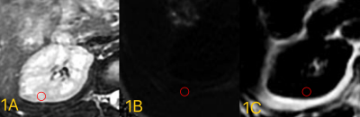 |
Quantifieation research of the severity of diabetic nephropathy by IDEAL-IQ sequence
Xinmiao Bu1, Ailian Liu1, Ye Ju1, Wenjun Hu1, Changyu Du2, Haoyang Jiang2, and Lingli Qi2
1The First Affiliated Hospital of Dalian Medical University, Dalian, China, 2Dalian Medical University, Dalian, China
IDEAL-IQ technology is a new scanning sequence which corrects many mixed factors that interfere with fat quantification to realize the quantitative measurement of fat fraction. We retrospectively analyzed 42 patients with diabetic nephropathy in our hospital from March 2017 to December 2019. According to the presence of proteinuria, they were divided into severe group and mild group.The R2* and FF value of the severe group was greater than that of the mild group, the difference was statistically significant (P <0.05).The AUC value of severe value and mild value for identifying severe with mild groups are 0.792 and 0.823。
|
|||
1896.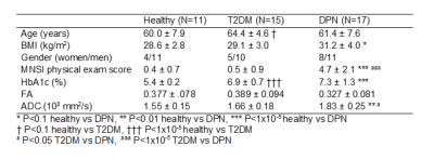 |
Diffusion tensor imaging in the tibial nerve in patients with diabetic peripheral neuropathy
Nathan Davis1, Steven Baete2,3, Smita Rao4, Jill Slade5, Prodromos Parasoglou2,3, and Ryan Brown2,3
1New York Institute of Technology, Old Westbury, NY, United States, 2Department of Radiology, Bernard and Irene Schwartz Center for Biomedical Imaging, New York University Grossman School of Medicine, New York, NY, United States, 3Department of Radiology, Center for Advanced Imaging Innovation and Research, New York University Grossman School of Medicine, New York, NY, United States, 4Department of Physical Therapy, New York University, New York, NY, United States, 5Department of Radiology, Michigan State University, East Langsing, MI, United States
Prolonged type 2 diabetes mellitus (T2DM) can result in diabetic peripheral neuropathy (DPN), a disease in which ischemic conditions degrade peripheral nerves. Diffusion tensor imaging (DTI) studies have demonstrated significant microstructural changes in upper extremity pathology, yet limited research has focused on the tibial nerve. We found cross-sectional differences in apparent diffusion coefficient between controls and T2DM and DPN. In a separate longitudinal study, insignificant DTI changes were found in DPN patients that were scanned before and after a 10-week exercise intervention.
|
|||
1897.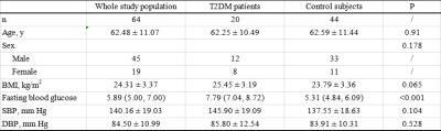 |
Quantitative assessment of the pancreas in T2DM patients using DWI and T2 mapping
Zihao Xu1, Qinhe Zhang1, Chao Liang1, Shuangyi Li1, Yaru You1, Jiazheng Wang2, Liangjie Lin2, Ailian Liu1, and Qingwei Song1
1Department of Radiology, the First Affiliated Hospital of Dalian Medical University, Dalian Liaoning, China, 2Philips Healthcare, Beijing, China
This study measured the ADC and T2 values of the pancreas in T2DM patients and control subjects. Distribution of T2 values were uneven in different locations of pancreas in both T2DM patients and control subjects , and the T2 values in head and neck were higher than those of body and tail. While distribution of ADC values was even in both of the two groups (P>0.05). ADC values of pancreas in T2DM patients were lower than those of control subjects, and it is inversed for T2 values . It may indicate that pancreatic cells are more closely arranged with reduced cell gaps.
|
|||
1898. |
Diffusion-Weighted Imaging of the Abdomen using Echo Planar Imaging with Compressed SENSE (EPICS)
Yoshifumi Noda1, Takayuki Mori1, Nobuyuki Kawai1, Kimihiro Kajita2, Yuta Akamine3, Masami Yoneyama3, Fuminori Hyodo4, and Masayuki Matsuo1
1Department of Radiology, Gifu University, Gifu, Japan, 2Department of Radiology Services, Gifu University Hospital, Gifu, Japan, 3Philips Japan, Tokyo, Japan, 4Department of Frontier Science for Imaging, Gifu University, Gifu, Japan
Diffusion-weighted imaging (DWI) is one of the important sequences in abdominal magnetic resonance (MR) imaging, especially in unenhanced examination. Recently, echo planar imaging with Compressed SENSE (EPICS) has been introduced and it can reduce noise-like artifacts and improve image quality. In this study, we compared qualitative and quantitative imaging parameters between conventional echo planar DWI (C-EPI-DWI) and EPICS. Our results showed that EPICS significantly reduce noise-like artifacts and improve the accuracy of ADC values compared with C-EPI-DWI.
|
|||
1899.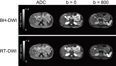 |
A single breath-hold abdominal diffusion-weighted imaging with simultaneous multislice echo-planar imaging
Naoki Ohno1, Kotaro Yoshida1, Yu Ueda2, Tosiaki Miyati1, Yuki Koshino1, Toshifumi Gabata1, and Satoshi Kobayashi1
1Kanazawa University, Kanazawa, Japan, 2Philips Japan, Tokyo, Japan
Although respiratory-triggered (RT)-diffusion-weighted imaging (DWI) has often been used to minimize respiratory motion in abdomen, unstable respiration leads to prolonged scan time without resolving the problem of respiratory motion. Therefore, we proposed a single breath-hold (BH)-DWI with simultaneous multislice (SMS) echo-planar imaging to allow faster acquisition and reliable quantification of apparent diffusion coefficient (ADC) in abdominal DWI. The BH-DWI with SMS technique reduced the scan time by more than fourfold and demonstrated comparable accuracy and repeatability in ADC quantification with conventional RT-DWI. The proposed method enables to acquire abdominal diffusion-weighted images in a single breath-holding.
|
|||
1900.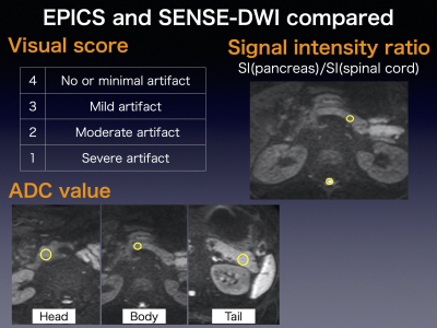 |
Reduction of susceptibility artifact using echo-planar imaging with compressed SENSE (EPICS) in the upper abdomen
Hazuki Takishima1, Hajime Yokota2, Takayuki Sakai3, Masami Yoneyama4, and Takashi Uno2
1Department of Radiology, Chiba University Hospital, Chiba, Japan, 2Department of Diagnostic Radiology and Radiation Oncology, Graduate School of Medicine, Chiba University, Chiba, Japan, 3Eastern Chiba Medical Center, Togane, Japan, 4Philips Japan, Tokyo Japan, Tokyo, Japan
Intestinal gas in upper abdomen interferes with diffusion-weighted image. Echo-planar imaging with compressed SENSE (EPICS) can reduce the susceptibility artifacts by shortening the data sampling time. In this study, EPICS and conventional SENSE-DWI were compared visually and quantitatively. The visual score of EPICS were significantly higher than that of SENSE in the pancreatic head and body. The signal ratio between the pancreas and spinal cord was significantly higher in the pancreatic tail in SENSE. The ADC value of EPICS was significantly higher in the pancreatic head. EPICS is feasible for reducing gas-producing artifact in the upper abdomen.
|
|||
1901.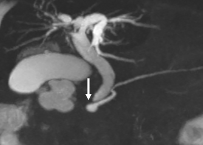 |
Value of MRCP in Santorinicele and Wirsungocele
Xinzhu Zhao1, Xing Wan1, Min Luo1, Mu Du1, Zhongxian Yang1, Qiuxia Xie1, Qian Zou1, Aiwen Guo1, Yingjie Mei2, and Yubao Liu1
1Medical Imaging Center, Shenzhen Hospital of Southern Medical University, Southern Medical University, Shenzhen, China, 2Philips Healthcare, Guangzhou, China
The study investigated the potential association among pancreatitis, pancreas divisum (PD), and santorinicele or wirsungocele (two types of pancreatic ductal variations) based on three-dimensional magnetic resonance cholangiopancreatography (3D-MRCP). The clinical and imaging samples of 75 patients were collected and sorted into santorinicele and wirsungocele groups. Potential factors related to wirsungocele and santorinicele were investigated. MRCP showed good performance in visualizing ductal anomalies. Patients with santorinicele had higher risks of developing pancreatitis compared with those with wirsungocele. Additionally, santorinicele itself might be more closely associated with pancreatitis than with PD.
|
|||
1902.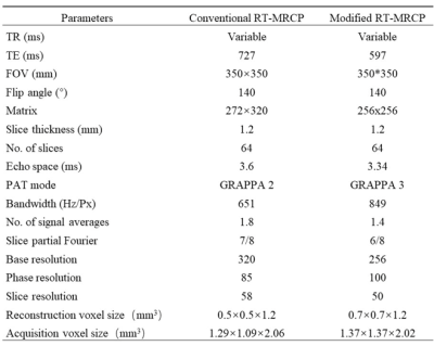 |
Comparation of the image quality between a modified 3D RT-SPACE-MRCP and routine 3D RT-SPACE-MRCP sequence in bile duct disease
Yue Qin1, Xin Li1, Yinhu Zhu1, Dayong Jin1, Liyao Liu1, Yanqiang Qiao1, Juan Tian1, Yifan Qian1, and Shaoyu Wang2
1XIAN DAXING HOSPITAL, Xi'an, China, 2MR Scientific Marketing, Siemens Healthineers, Xi'an, China
MR cholangiopancreatography (MRCP) is an method allowing noninvasive investigation of biliary and pancreatic disorders. However, the respiratory-triggered 3D MRCP technique requires long acquisition time depending on the breathing rate of patients, which leads to image blurring and motion artifacts. The purpose of this study was to compare the modified RT-SPACE-MRCP protocol and the conventional RT-SPACE-MRCP protocol with respect to image quality as well as the acquisition time.
|
|||
1903.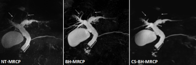 |
Comparing compressed sensing Breath-hold 3D MR cholangiopancreatography with two parallel imaging MRCP strategies
Zhiyong Chen1, Bin Sun1, Qing Duan1, Yunjing Xue1, ZhongShuai Zhang2, and guijin li3
1Radiology, Union Hospital, School of Medical Technology and Engineering, Fujian Medical University, Fuzhou, China, 2Diagnostic imaging, Siemens Healthcare, Shanghai, China., Shanghai, China, 3MR application, Siemens Healthineers Ltd,Guangzhou,China, guangzhou, China
The overall imaging quality of BH CS-MRCP, BH-MRCP and NT MRCP protocols were not significant different. Both the two breath-hold approaches were considering the time-saving advantages without deterioration of image quality. Compared with BH CS-MRCP, BH-MRCP yielded significantly better visualization of the segment 2 and 3 branch of the intrahepatic duct, and performed better consistency in main pancreatic duct and common bile duct morphology. Therefore, BH-MRCP in terms of superior visualization and morphological consistency can be recommended to improve workflow and diagnostic efficacy, and be preferred for patients with irregular breathing pattern.
|
The International Society for Magnetic Resonance in Medicine is accredited by the Accreditation Council for Continuing Medical Education to provide continuing medical education for physicians.