Digital Posters
Parallel Imaging Reconstruction
ISMRM & SMRT Annual Meeting • 15-20 May 2021

Digital Posters
Parallel Imaging Reconstruction
| Concurrent 1 | 15:00 - 16:00 |
 |
1153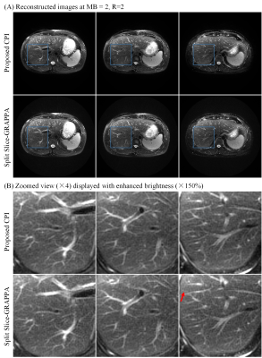 |
Calibrationless Parallel Imaging Reconstruction for Simultaneous Multi-slice PROPELLER of Upper Abdomen Video Permission Withheld
Yilong Liu1,2, Kun Zhou3, Dehe Weng3, Hua Guo4, and Ed X. Wu1,2
1Laboratory of Biomedical Imaging and Signal Processing, The University of Hong Kong, Hong Kong, China, 2Department of Electrical and Electronic Engineering, The University of Hong Kong, Hong Kong, China, 3Siemens Shenzhen Magnetic Resonance Ltd, Shenzhen, China, 4Center for Biomedical Imaging Research, Department of Biomedical Engineering, Tsinghua University, Beijing, China
This study presents a calibrationless parallel imaging (CPI) reconstruction for simultaneous multi-slice (SMS) PROPELLER MRI of the upper abdomen. With simultaneously excited slices having different blipped-CAIPI shifts, the inherent incoherency of SMS PROPELLER data enables CPI reconstruction via low rank matrix approximation. The proposed method was evaluated with both simulated phantom and acquired abdominal MR data. Compared to conventional split slice-GRAPPA, the proposed method jointly reconstructs all blades, providing significantly improved SNR and reduced artifact level.
|
||
1154.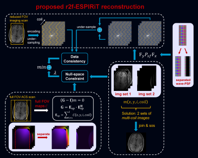 |
Reduced Field of View Parallel Imaging with Wave Encoded k-Space Trajectory
Zhilang Qiu1,2, Sen Jia1, Haifeng Wang1, Lei Zhang1, Xin Liu1, Hairong Zheng1, and Dong Liang1
1Shenzhen Institutes of Advanced Technology, Chinese Academy of Sciences, Shenzhen, China, 2University of Chinese Academy of Sciences, Beijing, China
Parallel imaging with wave encoded k-space trajectory can use most of the existing reconstruction methods such as SENSE, ESPIRiT and SPIRiT. However, these reconstruction methods are not suitable to reduced field-of-view (FOV) imaging scenario, where the imaging FOV is smaller than the object size, and severe aliasing artifacts remain in the reconstructed images. This work proposes to extend the existing reconstruction methods to treat the reduced FOV case, by accurately modeling and resolving the aliased components. Without increasing scan time, the artifact-free and full FOV images can be reconstructed by the proposed methods.
|
|||
1155.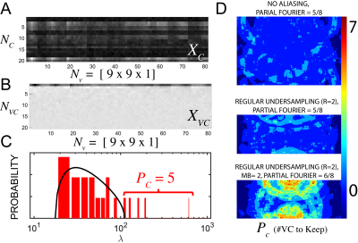 |
Marchenko-Pastur Virtual Coil Compression (MP-VCC)
Gregory Lemberskiy1, Jelle Veraart1, Benjamin Ades-aron1, Els Fieremans1, and Dmitry S Novikov1
1Radiology, NYU School of Medicine, New York, NY, United States
We propose a method of virtual coil compression using random matrix theory, MP-VCC, in which the Marchenko-Pastur distribution defines how many virtual coils may be discarded without loss beyond the PCA precision. MP-VCC is evaluated for partial Fourier, regular undersampling, and multiband acceleration.
|
|||
1156.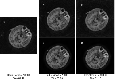 |
non-Cartesian Parallel imaging and compressed sensing adapted for accelerating hybrid trajectory PETRA
Fang Dong1, Dehe Weng1, and Nan Xiao1
1Siemens Shenzhen Magnetic Resonance Ltd., Shen Zhen, China
PETRA sequence, with ultra-short TE and ultra-low acoustic noise, has wide applications such as dental imaging, lung imaging, bone imaging, pediatric imaging as well as the routine anatomical imaging. However, the long scanning time and the vulnerability to motion hampered its usage. In this study, the combining non-Cartesian parallel imaging and Compressed sensing technique is adapted for the hybrid trajectory PETRA sequence. The image quality is clinically adequate for 2-5 folds acceleration.
|
|||
|
1157.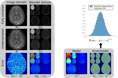 |
Near-optimal tuning-free multicoil compressed sensing MRI with Parallel Variable Density Approximate Message Passing
Charles Millard1,2, Aaron T Hess2, Jared Tanner1, and Boris Mailhe3
1Mathematical Institute, University of Oxford, Oxford, United Kingdom, 2Oxford Centre for Clinical Magnetic Resonance Research, University of Oxford, Oxford, United Kingdom, 3Digital Technology and Innovation, Siemens Healthineers, Princeton, NJ, United States
We present the Parallel Variable Density Approximate Message Passing (P-VDAMP) algorithm for compressed sensing MRI, which extends the recently proposed single-coil VDAMP algorithm to multiple coils. We evaluate the performance of P-VDAMP on eight datasets at a number of undersampling factors and find that it converges to a mean-squared error similar to the Fast Iterative Shrinkage Thresholding Algorithm (FISTA) with an optimally tuned sparse weighting, but in around 5x fewer iterations and without the need to tune model parameters.
|
||
1158.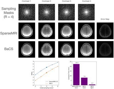 |
Accelerating Bayesian Compressed Sensing for Fast Multi-Contrast Reconstruction
Alexander Lin1, Demba Ba1, and Berkin Bilgic2,3
1Harvard University, Cambridge, MA, United States, 2Department of Radiology, Massachusetts General Hospital, Martinos Center for Biomedical Imaging, Boston, MA, United States, 3Harvard Medical School, Boston, MA, United States We propose Bayesian accelerated Compressed Sensing (BaCS) to improve the computational speed of Bayesian CS by two orders of magnitude. We achieve this by circumventing a costly matrix inversion problem using conjugate gradients and Monte Carlo sampling, which lend themselves well to parallel processing using GPUs. Exploiting parallelism renders BaCS even faster than sparseMRI, while having the ability to exploit similarities between multi-contrast images to improve reconstruction performance. Further, we extend BaCS to multi-channel reconstruction by synergistically combining it with SENSE to enable yet higher acceleration rates. |
|||
1159.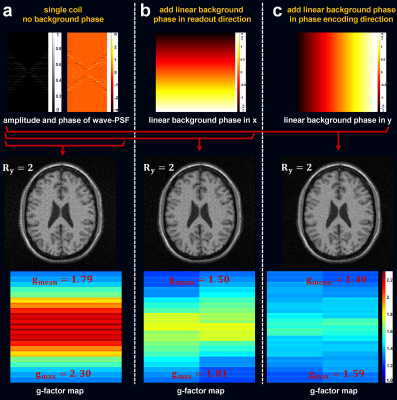 |
VCC-Wave for Improved Parallel MRI of High Resolution and High Bandwidth
Zhilang Qiu1,2, Sen Jia1, Shi Su1, Yanjie Zhu1, Xin Liu1, Hairong Zheng1, Haifeng Wang1, and Dong Liang1
1Shenzhen Institutes of Advanced Technology, Chinese Academy of Sciences, Shenzhen, China, 2University of Chinese Academy of Sciences, Beijing, China
Wave encoding is less efficient in situations of high resolution and high bandwidth. In this work, a novel model (named as VCC-Wave) which combines virtual conjugate coil (VCC) and wave encoding (Wave) was proposed. It can not only combine both advantages of VCC and Wave, but also exploit more priors of Wave under the VCC framework. Further significant improvement is achieved, and the limitation of Wave in situations of high resolution and high bandwidth is alleviated.
|
|||
1160.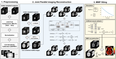 |
Accelerated 3D Myelin Water Imaging using Joint Parallel Imaging and Variable Splitting Network
Jae-Hun Lee1, Jaeuk Yi1, Kanghyun Ryu2, Soozy Jung1, and Dong-Hyun Kim1
1Department of Electrical & Electronic Engineering, Yonsei Univ., Seoul, Korea, Republic of, 2Department of Radiology, Stanford Univ., Stanford, CA, United States
Myelin water imaging using multi-cho GRE (mGRE) requires long acquisition times. In this study, we explored a multi-step reconstruction method using both advanced parallel imaging and deep learning, which can utilize joint information between the multi-echo images to further accelerate the acquisition. The proposed method shows acceptable image quality with improved quantitative values compared to conventional methods. The proposed method can achieve high acceleration factors for mGRE based 3D myelin water imaging.
|
|||
1161.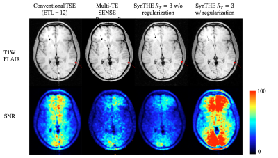 |
Simultaneous FLAIR T1W and T2W imaging using Temporal Harmonic Encoding
Tzu-Cheng Chao1 and James G. Pipe1
1Department of Radiology, Mayo Clinic, Rochester, MN, United States
A temporal harmonic encoding scheme is proposed to improve T1W FLAIR imaging with TSE acquisition. The imaging method reduces blurring in T1W FLAIR and provides an extra T2W FLAIR contrast at the same time with no need of additional scan time.
|
|||
1162.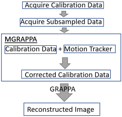 |
MGRAPPA: Motion Corrected GRAPPA for MRI
Michael Rawson1, Xiaoke Wang2, Ze Wang2, Radu Balan1,3, and Thomas Ernst2
1Department of Mathematics, University of Maryland at College Park, College Park, MD, United States, 2Department of Diagnostic Radiology and Nuclear Medicine, University of Maryland School of Medicine, Baltimore, MD, United States, 3Center for Scientific Computation and Mathematical Modeling, University of Maryland at College Park, College Park, MD, United States
We introduce an approximation and resulting method called MGRAPPA to allow high speed MRI scans robust to subject motion using prospective motion correction and GRAPPA [1,2]. In experiments on both simulated data and in vivo data, we observe high accuracy and robustness to subject movement in L2 (Frobenius) norm error including a 41% improvement in the in vivo experiment.
|
|||
1163.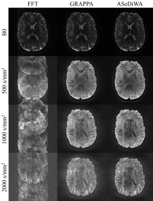 |
Autocalibrating Segmented Diffusion Weighted Acquisitions (ASeDiWA)
Michael Herbst1
1Bruker BioSpin MRI GmbH, Ettlingen, Germany
Segmented EPI enables high resolution DWI. However, phase differences between segments can lead to severe artifacts. This work investigates an algorithm to enable reconstruction of interleaved segmented acquisitions without the need of additional calibration or navigator measurements. Given a limited number of interleaves, the initial phase estimates can be calculated by a traditional parallel imaging reconstruction, using the unweighted scan of the DWI measurement as a reference. The ASeDiWA jointly reconstructs all segments of one DWI frame maintaining their phase information. Therefore, the algorithm allows for an iterative improvement of the phase estimates included in the joint reconstruction.
|
|||
1164.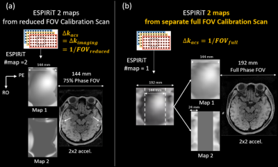 |
Resolving fold-over artefacts for Reduced Field-of-View Parallel Imaging with Cartesian Sampling
Sen Jia1, Zhilang Qiu1,2, Lei Zhang1, Haifeng Wang1, Xin Liu1, Hairong Zheng1, and Dong Liang1
1Paul C. Lauterbur Research Center for Biomedical Imaging, Shenzhen Institutes of Advanced Technology, Shenzhen, China, 2University of Chinese Academy of Sciences, Beijing, China
Equipping the existing parallel imaging methods such as ESPIRiT and SPIRiT with a full field-of-view (FOV) calibration could resolve the fold-over artefacts induced by reducing the imaging FOV to be smaller than the object size. Full-FOV images could be reconstructed by accurately resolving the aliased components in image space, or by reconstructing the kspace at a finer sampling interval corresponding to full-FOV. Both approaches requires a separate full-FOV calibration data which could be acquired efficiently. Reduced FOV Parallel imaging methods with full-FOV calibration may provide an alternative approach to treat the common FOV aliasing problem in practice.
|
|||
1165.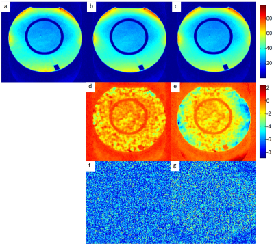 |
Accurate Quantitative G-factor Calculation in Dual-kernel Slice-GRAPPA Reconstruction
Wei Liu1, Simon Bauer2, and Stephan Kannengiesser2
1Siemens Shenzhen Magnetic Resonance Ltd, Shenzhen, China, 2Siemens Healthcare GmbH, Erlangen, Germany
We describe a more accurate analytical method to calculate the g-factor in dual kernel Slice-GRAPPA (SG-DK) reconstruction for blipped-CAIPI simultaneous multi-slice EPI data. To account for the effect of EPI phase correction, the Slice-GRAPPA kernels are phase corrected before the combination with the in-plane GRAPPA kernel. The experimental results based on a phantom study highlight that there is excellent agreement between SNR maps calculated with the standard pseudo multiple-replica method and the proposed method.
|
|||
1166.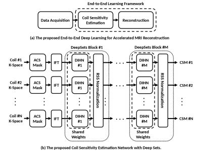 |
Coil Sensitivity Estimation with Deep Sets Towards End-to-End Accelerated MRI Reconstruction
Mahmoud Mostapha1, Boris Mailhe1, Simon Arberet1, Dominik Nickel2, and Mariappan S. Nadar 1
1Digital Technology and Innovation, Siemens Healthineers, Princeton, NJ, United States, 2Magnetic Resonance, Siemens Healthineers, Erlangen, Germany Parallel Imaging (PI) is a crucial technique for accelerating data acquisition in Magnetic Resonance Imaging (MRI), which is exceedingly time-consuming. With current SENSE-based MRI reconstruction formulated as a trainable unrolled optimization framework with several cascades of regularization networks and varying data consistency layers, coils sensitivity maps (CSMs) are needed at each cascade. Therefore, we propose a deep sets CSM estimation network (DS-CSME in short), enabling an end-to-end deep learning solution that allows for further MRI acceleration while preserving the overall reconstructed image quality. |
|||
1167. |
Two-Dimensional Coil-signature-based Phase Cycled Reconstruction for Inherent Correction of Echo-Planar Imaging Nyquist Ghost Artifacts
Silu Han1, Chidi Patrick Ugonna1, Mahesh Bharath Keerthivasan2,3, and Nan-kuei Chen1,3
1Biomedical Engineering Department, The University of Arizona, Tucson, AZ, United States, 2Siemens Medical Solutions USA, New York, NY, United States, 3Medical Imaging Department, The University of Arizona, Tucson, AZ, United States
A novel two-dimensional (2D) coil-signature-based phase cycled correction method has been developed for Nyquist artifact removal in echo planar imaging (EPI). Our method uses already available coil sensitivity information, without requiring extra reference scans, to correct 2D phase errors and can be applied equally well to single-shot and multi-shot EPI. Our results show that the developed method can effectively reduce Nyquist artifacts in EPI data acquired using a variety of acceleration schemes, such as through-plane Multi-band Imaging (MB) and in-plane parallel SENSitivity Encoding Imaging (SENSE).
|
|||
1168.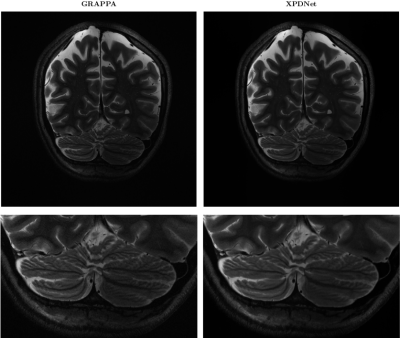 |
Is good old GRAPPA dead?
Zaccharie Ramzi1,2,3, Philippe Ciuciu1,2, Jean-Luc Starck3, and Alexandre Vignaud1
1Neurospin, Gif-Sur-Yvette, France, 2Parietal team, Inria Saclay, Gif-Sur-Yvette, France, 3Cosmostat team, CEA, Gif-Sur-Yvette, France
We perform a qualitative analysis of performance of XPDNet, a state-of-the-art deep learning approach for MRI reconstruction, compared to GRAPPA, a classical approach. We do this in multiple settings, in particular testing the robustness of the XPDNet to unseen settings, and show that the XPDNet can to some degree generalize well.
|
|||
1169.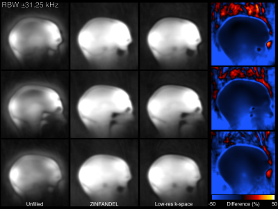 |
ZTE Infilling From Auto-calibration Neighbourhood Elements
Tobias C Wood1, Emil Ljungberg1, and Mark Chiew2
1Neuroimaging, King's College London, London, United Kingdom, 2Wellcome Centre for Integrative Neuroimaging, FMRIB, Nuffield Department of Clinical Neurosciences, University of Oxford, Oxford, United Kingdom
We present a method for filling the dead-time gap in ZTE imaging using a parallel imaging technique. We present results in phantoms and a human brain demonstrating high fidelity reconstructions comparable to existing methods that require collection of additional data.
|
|||
1170.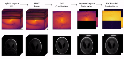 |
Hybrid K-space EPI (HyKE) Reconstruction for Accelerated Imaging
Tyler E Cork1,2, Matthew J Middione1, Michael Loecher1, Kévin J Moulin1, John M Pauly3, and Daniel B Ennis1,4
1Radiology, Stanford University, Stanford, CA, United States, 2Bioengineering, Stanford University, Stanford, CA, United States, 3Electrical Engineering, Stanford University, Stanford, CA, United States, 4Radiology, Veterans Affairs Health Care System, Palo Alto, CA, United States
Echo planar imaging (EPI) is amongst the fastest ways to reduce scan times for long imaging techniques, but it is also susceptible to geometric distortion caused by phase accrual while traversing k-space over long echo times (TE). The objective was to determine the feasibility of a new EPI data acquisition technique that is able to: (1) maintain the SNR efficiency compared to traditional EPI methods, and (2) create a break in the trajectory linearity to allow for geometric distortion correction from one acquisition. Herein, we demonstrate a feasible reconstruction pipeline.
|
|||
1171.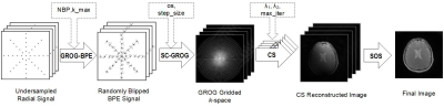 |
Highly undersampled GROG-BPE radial data reconstruction using Compressed Sensing
Yumna Bilal1,2, Ibtisam Aslam1,3, Muhammad Faisal Siddiqui1, and Hammad Omer1
1Medical Image Processing Research Group (MIPRG), Department of Electrical & Computer Engineering, COMSATS University Islamabad, Islamabad, Pakistan, 2Department of Electrical Engineering, University of Gujrat, Gujrat, Pakistan, 3Service of Radiology, Geneva University Hospitals and Faculty of Medicine, University of Geneva, Geneva, Switzerland
This work aims at reconstructing the undersampled non-Cartesian k-space signals by generating a cloud of randomly located additional points through GRAPPA Operator Gridding (GROG) facilitated Bunch Phase Encoding (BPE) collectively termed as GROG-BPE scheme. Gridding of this data is performed onto a Cartesian grid. Inherent randomness in the gridded BPE data is exploited using Compressed Sensing (CS) to obtain the solution image at higher acceleration factors and the results are compared with conventional CG-INNG method. Every step in the proposed method (right from BPE generation to reconstruction) is self-calibrating and does not require additional calibration signals.
|
The International Society for Magnetic Resonance in Medicine is accredited by the Accreditation Council for Continuing Medical Education to provide continuing medical education for physicians.