Digital Posters
Pulse Sequence II
ISMRM & SMRT Annual Meeting • 15-20 May 2021

| Concurrent 1 | 19:00 - 20:00 |
4165.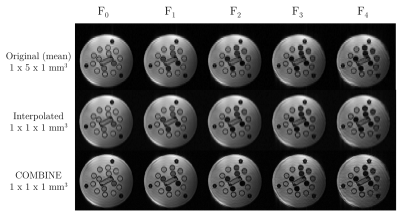 |
High-resolution, low-SAR 3D T2 relaxometry with COMBINE
Peter J Lally1, Matthew Grech-Sollars2,3, Joely Smith3,4, Ben Statton5, Paul M Matthews1,6, Karla L Miller7, and Neal K Bangerter4
1Department of Brain Sciences, Imperial College London, London, United Kingdom, 2Department of Surgery and Cancer, Imperial College London, London, United Kingdom, 3Department of Imaging, Imperial College Healthcare NHS Trust, London, United Kingdom, 4Department of Bioengineering, Imperial College London, London, United Kingdom, 5MRC London Institute of Medical Sciences, London, United Kingdom, 6UK Dementia Research Institute Centre at Imperial College, London, United Kingdom, 7Wellcome Centre for Integrative Neuroimaging, Nuffield Department of Clinical Neurosciences, University of Oxford, Oxford, United Kingdom
Here we describe a super-resolution 3D T2 relaxometry approach using an unbalanced SSFP acquisition with very low flip angle RF pulses (α ≤ 1°). We then apply this to obtain 1mm isotropic T2 maps in a reference phantom, and compare this to both the reference values and a 2D multi-echo spin echo approach. The proposed approach provides new options for high-resolution, low-SAR T2 relaxometry experiments in a range of tissues.
|
|||
4166.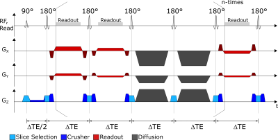 |
Radial Fast Spin Echo MRI with Compressed Sensing for Simultaneous ADC and T2 Mapping
Lars Bielak1,2, Thomas Lottner1, and Michael Bock1,2
1Dept.of Radiology, Medical Physics, Medical Center University of Freiburg, Faculty of Medicine, University of Freiburg, Freiburg, Germany, 2German Cancer Consortium (DKTK), Partner Site Freiburg, Freiburg, Germany
A novel sequence design based on radial fast spin echo with interleaved diffusion sensitization for simultaneous ADC and T2 mapping is presented. Additionally, a model restriction to a conventional compressed sensing reconstruction is implemented to support higher undersampling during acquisition. Simulations and phantom measurements show accurate measurement of diffusion ADC and T2 with as few as 11 spokes per TE, and 45 different TEs.
|
|||
4167.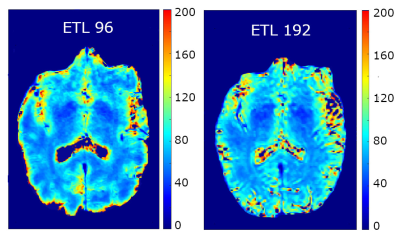 |
T2 Quantification in Brain Using Three Dimensional Fast Spin Echo Imaging with Long Echo Trains
Jeff Snyder1 and Alan H Wilman1
1Biomedical Engineering, University of Alberta, Edmonton, AB, Canada
A 3D FSE method using variable flip angle trains is proposed for quantification of T2 in brain. Two images are acquired at different echo times to allow for decay curve fitting using Bloch equation and Echo Phase Graph (EPG) simulations. Echo train lengths of 96 and 192 were investigated (total scan times of 8:12 and 2:38) in phantom and healthy subjects at 3 T, with isotropic resolutions of 0.9 and 1.3 mm3, respectively. RF was optimized to reduce blurring, sustain signal and allow T2 resolution. Comparison with previous methods was excellent, with good resolution and contrast in the 96 case.
|
|||
4168.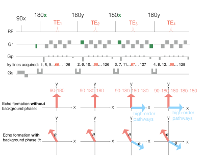 |
Improved echo-split GRASE imaging: a single-shot parametric T2 mapping protocol with removal of contamination from multiple echo pathways
Mei-Lan Chu1, Tzu-Cheng Chao2, Nan-kuei Chen3, and Hsiao-Wen Chung1
1Graduate Institute of Biomedical Electronics and Bioinformatics, National Taiwan University, Taipei, Taiwan, 2Mayo Clinic, Rochester, MN, United States, 3University of Arizona, Tucson, AZ, United States
Our novel single-shot T2 mapping framework, integrating ES-GRASE acquisition and parametric mapping, has the following advantages. First, signal contamination of high-order echoes is removed in the propose framework. Second, T2 relaxation times can be accurately measured by incorporating parallel imaging and multi-echo-pathway signal modeling into the reconstruction procedure.
|
|||
4169.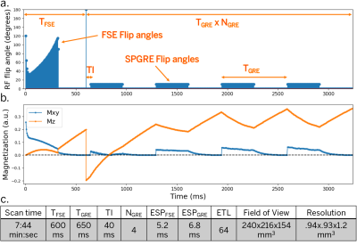 |
T2-Shuff-LL: Multi-contrast 3D Shuffling Combining Fast Spin-Echo and Look-Locker Gradient Echo
Jonathan Tamir1,2,3, Ken-Pin Hwang4, Naoyuki Takei5, and Suchandrima Banerjee6
1Electrical and Computer Engineering, The University of Texas at Austin, Austin, TX, United States, 2Diagnostic Medicine, Dell Medical School, The University of Texas at Austin, Austin, TX, United States, 3Oden Institute for Computational Engineering and Sciences, Austin, TX, United States, 4Imaging Physics, MD Anderson Cancer Center, Houston, TX, United States, 5MR Applications and Workflow, GE Healthcare, Hino, Tokyo, Japan, 6MR Applications and Workflow, GE Healthcare, Menlo Park, CA, United States
Fast, volumetric multi-contrast and quantitative imaging has a broad range of applications, but applying them in 3D remains challenging. Most volumetric approaches rely on gradient-echo-based acquisitions to achieve high scan-efficiency, even though spin-echo imaging provides higher SNR. Here we explore a hybrid acquisition that combines a fast-spin-echo acquisition block with multiple spoiled gradient-echo blocks to acquire spin- and gradient-echo images. We use a shuffled acquisition ordering together with a global subspace constraint and local low rank regularization to accelerate the scan. Following reconstruction, we fit the time series of images to quantitative parameters directly in the subspace using dictionary matching.
|
|||
4170.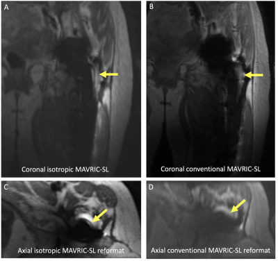 |
Clinical Evaluation of Isotropic MAVRIC-SL at 3T
Zoe Doyle1, Daehyun Yoon 1, Philip Lee1, Brian Hargreaves1, and Kathryn Stevens1
1Stanford University, Stanford, CA, United States
For the past decade, the development of 3D multispectral imaging (MSI) approach has facilitated the early detection of complications around metallic implants. However, the long acquisition time and need for multi-planar acquisitions make it difficult for symptomatic, elderly patients to tolerate. In this preliminary study, we evaluate the use of a fast isotropic 3D MSI technique at 3T MRI, which can potentially replace multiple sequences of orthogonal imaging planes with a single acquisition. Our results show promise for the improved visualization of various hardware complications using image reformats on arbitrary planes with diagnostically acceptable image quality.
|
|||
4171.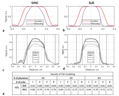 |
Can we achieve a better performance in metamaterial-assisted MRI combined to an SLR-based fast spin-echo sequence?
Ekaterina A. Brui1, Stanislas Rapacchi2, David Bendahan2, and Anna Andreychenko1,3
1Department of Physics and Engineering, ITMO University, Saint Petersburg, Russian Federation, 2Aix-Marseille Universite, CNRS, Centre de Résonance Magnétique Biologique et Médicale, Marseille, France, 3Center of Diagnostics and Telemedicine Technologies, Department of Health Care of Moscow, Research and Practical Clinical, Moscow, Russian Federation
There has been previously proved that a metamaterial-based wireless coil (WLC) provided a 48-fold lower SAR in 1.5T wrist MRI in comparison to conventional RF setup. This can allow to extend the acceptable threshold of energy deposition for SAR-demanding pulse sequences. This study demonstrates that SLR-based FSE together with the WLC allowed to increase the slice selectivity while still being within the safe SAR limits. The actual energy deposition was decreased as compared to a conventional RF setup. The combination of this coil and SLR-based FSE offers an interesting alternative for investigations which require scanning in a “Low SAR” regim.
|
|||
4172.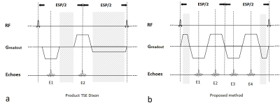 |
Combined Echo Two-Point Dixon method for high efficiency water/fat separation
Shi Cheng1, Kun Zhou1, Wei Liu1, and Dehe Weng1
1Siemens Shenzhen Magnetic Resonance Ltd., Shenzhen, China Spin-echo-based Dixon sequences may suffer from reduced SNR efficiency, due to dead time necessary for the readout shifts. In this abstract, an imaging method utilizing two pairs of fast-switching bipolar readout gradients and partially-opposed-phase and in-phase Dixon (CETD) is proposed to further reduce dead time. More attractively, the novel steps in joint reconstruction of the two pairs ensure the consistency of water-fat separation. |
|||
4173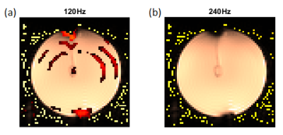 |
Neuronal current imaging on clinical whole body scanners: capabilities and limitations Video Permission Withheld
Milena Capiglioni1, Claus Kiefer2, and Roland Wiest1
1Institute for Diagnostic and Interventional Neuroradiology, Support Center for Advanced Neuroimaging (SCAN), University of Bern, Bern, Switzerland, 2Institute for Diagnostic and Interventional Neuroradiology, Support Center for Advanced Neuroimaging (SCAN), Inselspital, Bern, Bern, Switzerland
We evaluated the performance of a new contrast based on the rotary saturation technique to observe oscillating fields induced by neuronal currents. We used Bloch simulations and phantom experiments to study and observe the double resonance effect and the influences of external factors such as field inhomogeneities and relaxation phenomena. We detected oscillating fields in the nT range using the proposed Spin lock on/off contrast. We conclude that this technique can be used to observe oscillating neuronal fields and we propose different methods to reduce the effect of external influences on the signal.
|
|||
4174.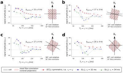 |
Quantifying lactate using double quantum filtered 1H MRS with adiabatic refocusing pulses at 7 T
Fabian Niess1, Albrecht Ingo Schmid1, Graham Kemp2, Ewald Moser1, Maxim Zaitsev1, and Martin Meyerspeer1
1High Field MR Center, Center for Medical Physics and Biomedical Engineering, Medical University Vienna, Vienna, Austria, 2Department of Musculoskeletal & Ageing Science, Institute of Life Course and Medical Sciences, University of Liverpool, Liverpool, United Kingdom
The purpose of this study was to develop a novel single-shot acquisition scheme to quantify the lactate doublet at 1.3 ppm in skeletal muscle at ultrahigh field of 7 T. A double quantum filtered 1H MRS sequence was implemented using a Shinnar-Le-Roux optimised 90° pulse and adiabatic full-passage 180° pulses for slice-selective excitation and refocusing, respectively. The lactate resonance was successfully quantified in phantom (free lactate in solution) and ex vivo measurements (lactate injected into a meat specimen). Muscle fibre orientation and corresponding effects on the observed lactate resonance were in good agreement with the literature.
|
|||
4175.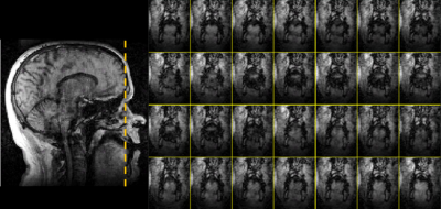 |
Increasing three-dimensional coverage of dynamic speech magnetic resonance imaging
Riwei Jin1, Zhi-pei Liang1, and Bradley P. Sutton1
1University of Illinois Urbana-Champaign, Champaign, IL, United States
We managed to increase the 3D coverage of dynamic speech magnetic resonance imaging to 32 slices with 96 mm full thickness, spatial resolution to 1.875×1.875×3mm and temporal resolution of 35 fps by applying sparsely sampling the temporal navigators with four k-space lines and and apply to a low-rank constraint Partial-Separability (PS) model for reconstruction. This enables visualization approaching isotropic resolution with the full vocal tract covered from side to side, enabling dynamic visualizations of complex motions that do not stay within a single imaging plane.
|
|||
4176.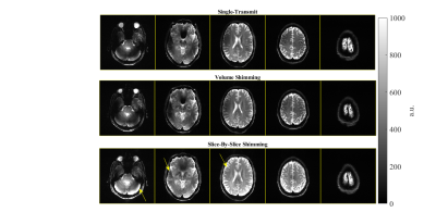 |
Multi-Slice 2D pTx Readout-Segmented Diffusion-Weighted Imaging Using Slice-by-Slice B1+ Shimming
Sydney Nicole Williams1, Iulius Dragonu2, Patrick Liebig3, and David A. Porter1
1Imaging Centre of Excellence, University of Glasgow, Glasgow, United Kingdom, 2Siemens Healthcare Ltd., Frimley, United Kingdom, 3Siemens Healthineers, Erlangen, Germany
For ultra-high field, parallel transmit (pTx) plays a key role in the mitigation of B1+ field inhomogeneity. Slice-selective 2D imaging is particularly challenging, where common solutions such as spokes haven’t been robustly implemented in standard multi-slice acquisitions. In this abstract we use slice-by-slice shimming to combat B1+ homogeneity in a multi-slice readout-segmented diffusion-weighted sequence with preliminary results shown in a human subject. Slice-by-slice shimming is compared to single-transmit and volumetric-shimming, with promising improvements in image quality.
|
|||
4177.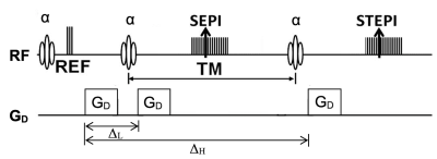 |
Simultaneous Measurements of Low-b Spin-Echo and High-b Stimulated-Echo DWI for Ultrahigh-b DWI
Kyle Jeong1, Noel Carlson2, John Rose3, and Eun-Kee Jeong4
1Utah Center for Advanced Imaging Research, University of Utah, Salt Lake City, UT, United States, 2NeuroImunology and NeuroVirology, University of Utah, Salt Lake City, UT, United States, 3Neurology, University of Utah, Salt Lake City, UT, United States, 4Radiology and Imaging Sciences, University of Utah, Salt Lake City, UT, United States
The signal-b curve of the Ultrahigh-b DWI (UHb-DWI) with bmax = ~ 10,000 s/mm2 is sensitive to the water exchange at the myelin sheath in white-matter. Ultrahigh-b DWI is obtained using DW-stimulated-echo (DW-STE) sequence, however, a half the prepared magnetization is discarded in conventional STE imaging. Therefore, the main objective of this work has been to measure low-b DW-spin-echo and DW-stimulated-echo in a single sequence, and to correct/combine DWSE and DWSTE for ultrahigh-b DWI (UHb-DWI) of cervical spinal cord (CSC).
|
|||
4178.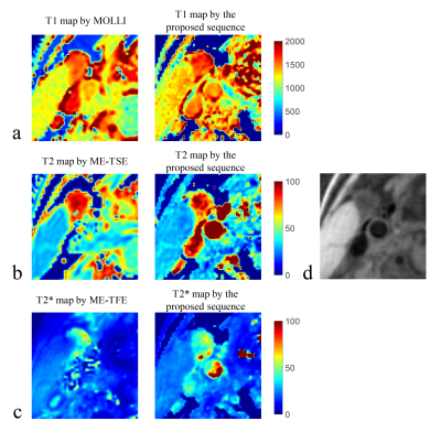 |
Simultaneous T1, T2 and T2* Mapping of the Carotid Plaque Using Combined Single- and Multi-echo 3D Golden Angle Radial Acquisition
Yajie Wang1, Yishi Wang2, Haikun Qi3, Rui Guo4, Huiyu Qiao1, Dongyue Si1, and Huijun Chen1
1Center for Biomedical Imaging Research, Department of Biomedical Engineering, School of Medicine, Tsinghua University, Beijing, China, 2Philips Healthcare, Beijing, China, 3School of Biomedical Engineering and Imaging Sciences, King's College London, London, United Kingdom, 4Department of Medicine, Beth Israel Deaconess Medical Center and Harvard Medical School, Boston, MA, United States
Previous quantitative techniques for carotid plaque T1 mapping or T1, T2 mapping have been achieved using only one scan. Besides T1 and T2 quantification, T2* mapping of the carotid plaque is also important for the detection of iron deposition, which plays an important role in plaque progression. In this study, a new quantitative technique using combined single- and multi-echo 3D golden angle radial acquisition has been proposed for simultaneous T1, T2 and T2* mapping of the carotid plaque. The quantitative accuracy and the in-vivo feasibility of the proposed sequence have been demonstrated in phantom and volunteer studies.
|
|||
4179.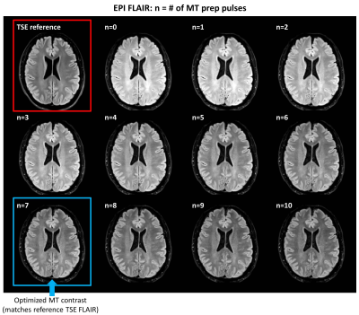 |
Optimization of Magnetization Transfer Contrast for EPI FLAIR Brain Imaging
Serdest Demir1, Bryan Clifford2, Thorsten Feiweier3, Tom Hilbert4, Zahra Hosseini5, Augusto Lio Goncalves Filho1, Azadeh Tabari1, Wei-Ching Lo2, Maria Gabriela Figueiro Longo1, Michael Lev1, Pamela Schaefer1, Otto Rapalino1, Kawin Setsompop6,
Berkin Bilgic6, Stephen Cauley6, Susie Huang1, and John Conklin1
1Radiology, Massachusetts General Hospital, Boston, MA, United States, 2Siemens Medical Solutions, Boston, MA, United States, 3Siemens Healthcare GmbH, Erlangen, Germany, 4Siemens Healthcare AG, Lausanne, Switzerland, 5Siemens Medical Solutions, Atlanta, GA, United States, 6Radiology, A. A. Martinos Center for Biomedical Imaging, Massachusetts General Hospital, Boston, MA, United States
EPI FLAIR images have lower tissue contrast than conventional TSE images due to the absence of significant magnetization transfer (MT) effects, which limits their acceptability to radiologists and clinicians. In this study, we developed an MT-prepared EPI FLAIR acquisition, and determined the optimal parameters of the MT-preparation module to match the tissue contrast of clinical reference TSE FLAIR images. This approach may facilitate clinical adoption of EPI FLAIR images for a variety of applications, including those where ultrafast imaging is desired (e.g., acute stroke, motion prone patients, pediatrics).
|
|||
4180.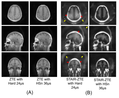 |
A Short TR Adiabatic Inversion Recovery Zero Echo Time (STAIR-ZTE) Sequence with Interleaved Encoding and a Modulated RF Pulse for Myelin Imaging
Hyungseok Jang1, Yajun Ma1, Michael Carl2, Saeed Jerban1, Roland Lee1, Eric Y Chang1,3, Jody Corey-Bloom1, and Jiang Du1
1University of California, San Diego, San Diego, CA, United States, 2GE Healthcare, San Diego, CA, United States, 3Veterans Affairs San Diego Healthcare System, San Diego, CA, United States
It is challenging to directly image myelin due to its extremely short T2* (<300µs at 3T). Adiabatic Inversion Recovery prepared Ultrashort Echo Time (IR-UTE) imaging has been proposed for direct myelin imaging in human brain. More recently, Short Repetition Time Adiabatic Inversion Recovery (STAIR) has been proposed as a novel contrast mechanism for myelin imaging with improved suppression of long T2 signal. In this study, we explored feasibility of STAIR based Zero Echo Time (STAIR-ZTE) combined with an amplitude- and phase-modulated RF pulse and interleaved Water- and Fat-Suppressed Proton Projection MRI (WASPI) for myelin imaging in human brain.
|
|||
4181.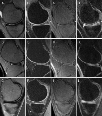 |
Knee osteochondral junction imaging using a 3D dual adiabatic inversion recovery ultrashort echo time cones (3D DIR-UTE-cones) sequence at 3T
Alecio F. Lombardi1,2, Zhao Wei1, Hyungseok Jang1, Saeed Jerban1, Lillian Gong1, Jiang Du1, Eric Y. Chang1,2, and Ya-Jun Ma1
1Radiology, University of California, San Diego, CA, United States, 2Radiology Service, Veterans Affairs, San Diego, CA, United States
To overcome the limitations of imaging short T2/T2* tissues with conventional MRI and increase contrast between the osteochondral junction and adjacent tissues, we developed a 3D dual adiabatic inversion recovery prepared ultrashort echo times cones (3D DIR-UTE-Cones) sequence for volumetric imaging at a 3T scanner. We expected the proposed DIR-UTE-Cones sequence to generate higher OCJ contrast than the IR-FS-UTE Cones, especially in between OCJ and fat.
|
|||
4182.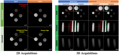 |
Phantom validation of a novel user-configurable ultrashort echo time (UTE) sequence
Lumeng Cui1, Emily J. McWalter1,2, Gerald R. Moran3, and Niranjan Venugopal4
1Division of Biomedical Engineering, University of Saskatchewan, Saskatoon, SK, Canada, 2Department of Mechanical Engineering, University of Saskatchewan, Saskatoon, SK, Canada, 3Research Collaboration Manager, Siemens Healthcare Limited, Oakville, ON, Canada, 4Department of Radiology, University of Manitoba, Winnipeg, MB, Canada
Ultrashort echo time (UTE) pulse sequences can capture the signal from short T2 tissues. Many UTE techniques have been developed for achieving an ultrashort TE (less than 0.1 ms) or enhancing the short T2 contrast. However, these approaches have not been extensively compared head-to-head due to the lack of a flexible UTE sequence that integrates the different approaches. Therefore, in this work, we developed a versatile UTE sequence that can directly compare UTE approaches and provide options for researchers to choose the appropriate UTE that fit the particular application.
|
|||
4183.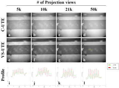 |
Volume-Selective 3D Ultrashort Echo-time Imaging
Jinil Park1 and Jang-Yeon Park2,3
1Biomedical Institute for Convergence at SKKU, Sungkyunkwan University, Suwon, Korea, Republic of, 2Department of Biomedical engineering, Sungkyunkwan University, Suwon, Korea, Republic of, 3Department of Intelligent Precision Healthcare Convergence, Sungkyunkwan University, Suwon, Korea, Republic of
Non-Cartesian radial sampling can preserve the spatial resolution without aliasing when under-sampling, but makes streak artifacts and internal structure identification difficult.Because C-UTE uses non-selective rectangular pulses to acquire projection data containing information about the entire object, setting the ROI smaller than the object further increases the under-sampling problem. the recently developed VS-UTE uses selective SINC pulses, and it's a method to minimize under-sampling problems. In this study, we demonstrate the ability of VS-UTE to maintain image quality when the number of projection views is under-sampled or when the imaging volume is chosen smaller than the imaging target.
|
The International Society for Magnetic Resonance in Medicine is accredited by the Accreditation Council for Continuing Medical Education to provide continuing medical education for physicians.