Digital Posters
Data Acquisition Sampling Trajectories
ISMRM & SMRT Annual Meeting • 15-20 May 2021

Digital Posters
Data Acquisition Sampling Trajectories
| Concurrent 1 | 13:00 - 14:00 |
0879.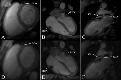 |
Fast 3D whole Heart Imaging using low-discrepancy Sampling Strategies
Tobias Speidel1, Dagmar Bertsche1, Patrick Metze1, Kilian Stumpf1, Wolfgang Rottbauer1, and Volker Rasche1
1Internal Medicine II, Ulm University Hospital, Ulm, Germany Three-dimensional MRI is a valuable tool for the diagnosis of heart diseases, which furthermore offers advantages such as intrinsically higher signal-to-noise ratios, less scan planning efforts and the possibility to reconstruct arbitrary anatomical planes from one dataset. Despite these advantages, inherently long acquisition durations often limit clinical applications. This abstract highlights the possibility of acquiring qualitative 3D images at an isotropic resolution of 1.09 mm within 250 heartbeats (ECG-gating), using specifically generated k-space interleaves with low-coherent and low-discrepancy sampling properties, based on a generalised form of the previously introduced Seiffert-Spirals. Cardiac image segmentations are furthermore presented to emphasise image qualities. |
|||
0880.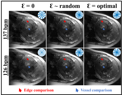 |
Reducing clustering of readout trajectories in non-Cartesian CINE MRI
Datta Singh Goolaub1,2 and Christopher Macgowan1,2
1Medical Biophysics, University of Toronto, Toronto, ON, Canada, 2Translational Medicine, The Hospital for Sick Children, Toronto, ON, Canada
In this study, we demonstrate a new non-Cartesian trajectory scheme to reduce clustering of trajectory arms during CINE reconstruction, while still allowing for real-time reconstructions. In this scheme, trajectory arms are incremented within temporal blocks and additional angles are played between blocks. These added angles are optimized to perturb trajectory coherence during CINE reconstruction, as demonstrated through simulations showing reduced clustering and improved CINE image quality.
|
|||
0881.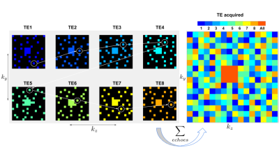 |
Fast $$$T_{2}^{*}$$$ Mapping Using Complementary Poisson Disk Sampling and ADMM Reconstruction
Charles Iglehart1, Ali Bilgin1, Evan Levine2, and Manojkumar Saranathan3
1Electrical and Computer Engineering, University of Arizona, Tucson, AZ, United States, 2Microsoft Research, Redmond, WA, United States, 3Department of Medical Imaging, University of Arizona, Tucson, AZ, United States
Using Complementary Poisson Disk (CPD) sampling, we accelerate multi-echo gradient recalled echo acquisitions (ME GRE) by efficiently undersampling k-space in such a way that lends itself to practically realizable sampling trajectories. We reconstruct $$$T_{2}^{*}$$$ maps from these undersampled acquisitions and validate against fully sampled data.
|
|||
0882.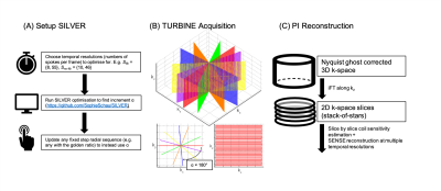 |
Optimizing the fixed angular increment between k-space spokes can lead to improved SNR in radial imaging
S Sophie Schauman1, Thomas W Okell1, and Mark Chiew1
1Nuffield Department of Clinical Neurosciences, University of Oxford, Oxford, United Kingdom
Many dynamic MRI methods use golden ratio radial sampling. Here, we show that golden ratio sampling is not always ideal from an SNR point of view, and that optimizing the fixed angular increment between spokes can lead to more uniform coverage of k-space, which in turn can lead to lower g-factors in a non-Cartesian parallel imaging reconstruction. The method we present, the Set Increment with Limited Views Encoding Ratio method, SILVER, maintains the flexibility of reconstructing a single dataset at multiple temporal resolutions but improves SNR compared with the golden ratio method.
|
|||
0883.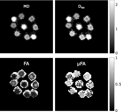 |
Boosting the SNR-efficiency of Free Gradient Waveform Diffusion MRI using Spiral Readouts and Ultra-Strong Gradients
Lars Mueller1, Maryam Afzali1, Malwina molendowska1, Chantal M.W. Tax1,2, Fabrizio Fasano3,4, Suryanarayana Umesh Rudrapatna1, and Derek K. Jones1,5
1CUBRIC, Cardiff University, Cardiff, United Kingdom, 2Image Sciences Institute, University Medical Center Utrecht, Utrecht, Netherlands, 3Siemens Healthcare Ltd, Camberly, United Kingdom, 4Siemens Healthcare GmbH, Erlangen, Germany, 5Mary McKillop Institute for Health Research, Faculty of Health Sciences, Australian Catholic University, Melbourne, Melbourne, Australia
Free gradient waveforms for diffusion MRI offer new insights in the underlying microstructure compared the classical Stejskal-Tanner linear encoding. A drawback of this approach is the prolonged echo time and thus decreased SNR. Here we present an approach to shorten the echo time by employing spiral readouts instead of echo-planar imaging and using the ultra-strong gradients of the Connectom scanner. The feasibility of this approach is demonstrated in a biomimetic phantom.
|
|||
0884. |
Variable Density Phase Encoding for High Resolution Single-Shot EPI
Mark Chiew1
1Wellcome Centre for Integrative Neuroimaging, FMRIB, Nuffield Department of Clinical Neurosciences, University of Oxford, Oxford, United Kingdom
A new variable density phase-encoding (vdPE) scheme for single-shot EPI is presented, in which phase encoding blips scale with a decaying exponential. This serves to reshape the signal decay envelope in the PE direction, resulting in shorter achievable TEs and reduced T2* blurring, without the need for partial Fourier sampling. We demonstrate this approach in high-resolution single-shot 2D EPI data, using a virtual-coil sensitivity encoded reconstruction, and show improved image fidelity and fewer artefacts compared to conventional EPI with ¾ partial Fourier. This approach could have utility for high-resolution, high-field brain imaging for applications such as layer-specific fMRI.
|
|||
0885.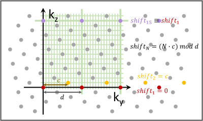 |
Mono-planar T-Hex EPI
Maria Engel1, Lars Kasper1, and Klaas Prüssmann1
1Institute for Biomedical Engineering, ETH Zurich, Zurich, Switzerland
In this work, we show high-resolution stacks of EPIs on a tilted hexagonal grid. The scheme provides flexibility in balancing readout and scan time, thereby allowing for high-quality images in a temporal resolution regime suitable for fMRI. 0.7 mm whole-brain coverage is achieved in below 5s.
|
|||
0886. |
Whole-brain fMRI at 5 frames per second using T-Hex spiral acquisition
Maria Engel1, Lars Kasper1, Franz Patzig1, Samuel Bianchi1, and Klaas Prüssmann1
1Institute for Biomedical Engineering, ETH Zurich, Zurich, Switzerland
In this work we show high-temporal resolution fMRI using T-Hex spiral-in trajectories. 3mm-resolved whole-brain volumes are acquired at a frame rate of 5Hz.
|
|||
0887.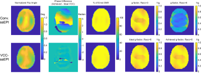 |
High Resolution EPI with Multi-spoke Parallel Transmission and Virtual Coil Reconstruction
Zhipeng Cao1
1Vanderbilt University, Nashville, TN, United States
Novel encoding method based on multi-spoke parallel transmission is presented for distortion-minimized high resolution single and multi- shot GRE-EPI. By jointly designing flip angle patterns and excitation phase dictated by a 32-channel receive array using an 8-channel transmit array, the acceleration ratio of parallel imaging is drastically increased, with x6 for ssEPI and x8 for msEPI, potentially enabling sub-mm resolution and motion insensitive fMRI and diffusion MRI at UHF.
|
|||
0888.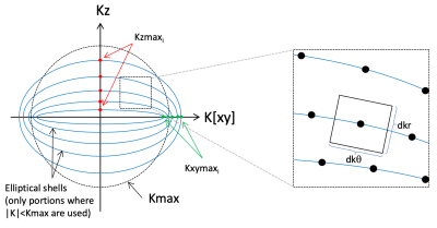 |
Elliptical shells: a fast single-shot 3D readout for Arterial Spin Labeling perfusion imaging
Joseph G. Woods1, Divya Bolar1, and Eric C. Wong1
1Department of Radiology, University of California San Diego, San Diego, CA, United States
Single shot 3D acquisitions are ideal for ASL because they provide insensitivity to motion and allow for effective background suppression. Current state of the art implementations use stack-of-spiral FSE based imaging for time efficiency and insensitivity to resonance offsets. We introduce here a further improvement in time efficiency by using spirals that are designed to approximate a spherically symmetrical variable density function. Spirals are constrained to elliptical shells, and are numerically optimized for time efficiency. An example trajectory is twice as fast as a cylindrical stack-of-spirals with similar density parameters (excluding refocusing time), and results in high quality ASL images.
|
|||
0889.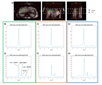 |
3D choline metabolite imaging in the liver by MRSI with selective excitation using spectral-spatial RF pulses at 7T
Lieke van den Wildenberg1, Arjan Hendriks1, Wybe van der Kemp1, Dennis Klomp1, and Jeanine Prompers1
1Radiology Department, UMC Utrecht, Utrecht, Netherlands
MR measurement of increased concentrations of choline containing compounds (tCho) in the liver could be used as a non-invasive tumor tissue biomarker. However, 3D 1H-MRSI with conventional water suppression and volume selection does not yield the sensitivity gain expected at higher fields like 7T. As a result, it cannot be performed in clinically feasible scan times with full coverage of the liver and at sufficient spatial resolution to detect tumor metastases. With spectral-spatial pulses, it is possible to spatially map tCho much faster and at higher SNR than with conventional MRSI, with excellent suppression of both water and lipid signals.
|
|||
0890. |
Towards Accelerating 3D 1H-MRSI Using Randomly Undersampled Spatial and Spectral Spirals with Low-rank Subspaces
Yamin Arefeen1, Borjan Gagoski2,3, and Elfar Adalsteinsson1,4,5
1Massachusetts Institute of Technology, Cambridge, MA, United States, 2Fetal-Neonatal Neuroimaging and Developmental Science Center, Boston Children’s Hospital, Boston, MA, United States, 3Department of Radiology, Harvard Medical School, Boston, MA, United States, 4Harvard-MIT Health Sciences and Technology, Cambridge, MA, United States, 5Institute for Medical Engineering and Science, Cambridge, MA, United States
1H Magnetic Resonance Spectroscopic Imaging (MRSI) suffers from low signal-to-noise ratio (SNR) and long acquisition times. Time-varying gradient trajectories reduce acquisition time by more efficiently sampling the k-space data. Meanwhile, a subspace approach to high-resolution MRSI (SPICE) elegantly enables high resolution MRSI despite low SNR. To accelerate 3D MRSI, we propose a randomly undersampled, time-varying, 3D-trajectory that synergizes with the SPICE-subspace framework by inducing incoherent aliasing across all four imaging dimensions. This enables ~3x acceleration in analytic experiments and ~2x acceleration in retrospectively undersampled phantom experiments with minimal degradation in image quality in comparison to fully sampled 3D-spiral MRSI.
|
|||
0891. |
Lipid and water separation through an SMS-like approach in 7T to reduce SAR in brain EPI
Amir Seginer1, Edna Furman-Haran2,3, Ilan Goldberg4, and Rita Schmidt3,5
1Siemens Healthcare, Rosh Ha'ayin, Israel, 2Life Sciences Core Facilities, Weizmann Institute of Science, Rehovot, Israel, 3The Azrieli National Institute for Human Brain Imaging and Research, Weizmann Institute of Science, Rehovot, Israel, 4Deparment of Neurology, Wolfson medical center, Holon, Israel, 5Neurobiology Department, Weizmann Institute of Science, Rehovot, Israel
We examine the potential to significantly reduce SAR in 7T fMRI (GRE-EPI) by circumventing the fat-suppression pulse. The resulting (shifted) lipid artifact is resolved by a parallel-imaging based reconstruction which separates the lipid and water images. Simulations, phantom experiments, and fMRI experiments were performed with the suggested method. SAR was shown to be reduced to less than half, allowing to shorten the repetition time and/or increase the volume coverage in fMRI studies.
|
|||
0892.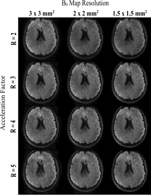 |
On the impact of B0 map resolution and undersampling on reconstructions using expanded encoding models
Paul Dubovan1,2, Lars Kasper3, Kamil Uludag3,4, and Corey Baron1,2
1Medical Biophysics, Western University, London, ON, Canada, 2Center for Functional and Metabolic Mapping, Robarts Research Institute, London, ON, Canada, 3Techna Institute, University Health Network, Toronto, ON, Canada, 4Medical Biophysics, University of Toronto, Toronto, ON, Canada
Spiral diffusion images are known to suffer from image distortions due to the enhanced sensitivity to magnetic field inhomogeneities. Application of parallel imaging to accelerate readout times, and inclusion of robust B0 field maps are two techniques that generally contribute to spiral image improvements. In this work, the impact of field map resolution and acceleration factors are investigated to identify acquisition parameters that optimize image quality. A moderate acceleration factor (3 or 4) coupled with a field map on the order of the imaging resolution (1.5 mm in-plane) provided the best trade-off between geometric accuracy, blurring reduction, and noise amplification.
|
|||
0893. |
Pushing the acceleration performance of WAVE-CAIPI using a single-axis gradient insert – a simulation study
Alejandro Monreal Madrigal1, Edwin Versteeg1, and Jeroen Siero1,2
1Radiology, University Medical Center Utrecht, Utrecht, Netherlands, 2Spinoza Centre for Neuroimaging, Amsterdam, Netherlands
Wave-CAIPI achieves high acceleration factors by using wave-gradients to spread out aliasing in 3D. In this work, we present a simulation study investigating the gains in acceleration performance when using a single-axis gradient insert (Gmax = 200 mT/m and SRmax = 1300 T/m/s) for Wave-CAIPI. The simulations were performed by retrospectively under-sampling data with different combinations of wave-gradients while assessing the image quality after reconstruction. Here, a 16-fold acceleration was simulated without a noticeable decrease in image quality. Furthermore, the high gradient performance allowed for a 5-fold shorter readout time, which might make a combination of EPI and Wave-CAIPI feasible.
|
|||
0894. |
The Design of an Acentric Cartesian Spiral Sampling with Partial Fourier for Highly Accelerated Single-breath-hold Isotropic 3-D Cardiac Cine
Yu Ding1, Qi Liu1, Jingyuan Lyu1, Yuan Zheng1, and Jian Xu1
1UIH America, Inc, Houston, TX, United States
We design a novel acentric spiral pattern and combine with partial Fourier to form a new sampling strategy in 3-D Cartesian acquisition. This new strategy was implemented and tested in isotropic 3-D single breath hold cardiac cine imaging with isotropic spatial resolution and whole heart coverage. Volunteer study shows that this new sampling strategy greatly reduced the number of samples and have acceptable image quality.
|
|||
0895. |
Optimization of scan parameters to reduce acquisition time for resolve-based DKI in NPC
Yaoyao He1, Hao Chen1, Huiting Zhang2, Robert Grimm2, Cecheng Zhao3, Xiaofang Guo1, Yulin Liu1, and Zilong Yuan1
1Hubei Cancer Hospital, Wuhan, China, 2Siemens Healthcare, Erlangen, Germany, 3Huazhong Agricultural University, Wuhan, China
This study aimed to shorten acquisition time of RESOLVE-based diffusion kurtosis imaging (DKI) in nasopharyngeal carcinoma (NPC), and we explored the influence of b value combinations on the scan time and parameters from DKI. The results showed that the group with b-value (200, 400, 800, 2000) had excellent agreement with the actual acquisition with b-value (200, 400, 800, 1500, 2000) and the scan time saved 28% (3min46s vs 5min13s), and was recommended in the clinical DKI research in NPC.
|
|||
| 0896. | Strategically Acquired Gradient Echo imaging with Compressed Sensing: Comparison of quantitative images with different acceleration factors
bingbing gao1, Yuhan Jiang1, Nan Zhang1, Yanwei Miao1, Qingwei Song1, Ailian Liu1, and Peng Sun2
1the First Affiliated hospital of Dalian Medical university, Dalian, China, 2Philips Healthcare, BeiJing, China
Strategically Acquired Gradient Echo (STAGE) imaging is a novel and useful MR sequence which can provide seven different key modalities at the same time, T1w, PDw, SWI, T1 mapping, PD mapping, T2* mapping, and SWIM. However, the acquisition time is relatively long (about 8 minutes). The compressed sensing (CS) technique is now wildly applied for accelerating MR acquisition. This study aimed to evaluate the feasibility of CS accelerated STAGE technique, and find an optimal acceleration factor (AF). According to our preliminary results, the AF of 4 was recommended.
|
|||
0897.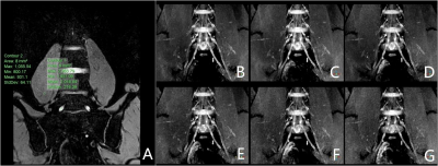 |
Optimization of Compressed Sensing Acceleration Factors for Lumbosacral Plexus 3D MRI
Renwang PU1, Qingwei SONG2, Ailian LIU1, Hao nan ZHANG1, Nan ZHANG1, and Jiazheng WANG3
1the First Affiliated Hospital of Dalian Medical University, Dalian, China, 2the First Affiliated Hospital of Dalian Medical University, DALIAN, China, 3Philips Healthcare, BEIJING, China
MRI, particularly the 3D-T2*-FFE sequence is widely used imaging tool for evaluating lumbosacral plexus because of its multiplanar capabilities and excellent soft-tissue contrast. However, conventional 3D-T2*-FFE scan time is long, despite the SENSE acceleration, which may cause spontaneous or involuntary movement due to the patient's discomfort and which may ultimately lead to imaging artifacts. Compressed Sensing (CS) is able to further reduce the imaging time by pseudo-random under-sample through the acquisition , but the image quality may be degraded when the acceleration factor (AF) is over-stretched. In this study we explored the optimal CS acceleration factor for lumbosacral plexus 3D-T2*-FFE imaging.
|
|||
0898.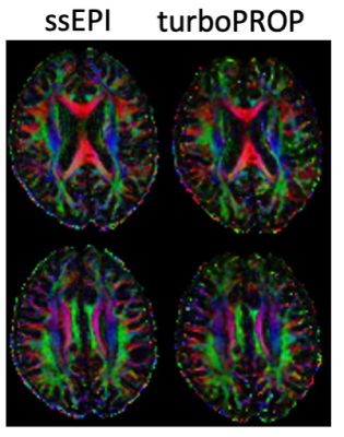 |
Rapid and robust DTI using a turboPROP technique with blade sharing and whole-blade acquisition
Zhiqiang Li1 and John P Karis1
1Neuroradiology, Barrow Neurological Institute, Phoenix, AZ, United States
DTI is a vital tool in many neurological applications. ssEPI is the method of choice in clinical DTI but suffers from strong geometric distortions. FSE and PROPELLER-based techniques are free from geometric distortions but have low scan efficiency. Recently, a split-blade turboPROP technique has been enhanced for brain DWI with improved image quality. In this work we develop a whole-blade acquisition mode and a blade sharing strategy with turboPROP for DTI at a speed comparable to an ssEPI-based clinical protocol. In vivo results demonstrate good image quality.
|
The International Society for Magnetic Resonance in Medicine is accredited by the Accreditation Council for Continuing Medical Education to provide continuing medical education for physicians.