Digital Posters
Motion: Methods
ISMRM & SMRT Annual Meeting • 15-20 May 2021

| Concurrent 1 | 17:00 - 18:00 |
1352.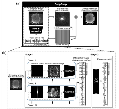 |
DeepResp: Deep Neural Network for respiration-induced artifact correction in 2D multi-slice GRE
Hongjun An1, Hyeong-Geol Shin1, Sooyeon Ji1, Woojin Jung1, Sehong Oh2, Dongmyung Shin1, Juhyung Park1, and Jongho Lee1
1Department of Electrical and computer Engineering, Seoul National University, Seoul, Korea, Republic of, 2Division of Biomedical Engineering, Hankuk University of Foreign Studies, Gyeonggi-do, Korea, Republic of
Respiration-induced B0 fluctuation can generate artifacts by inducing phase errors. In this study, a new deep-learning method, DeepResp, is proposed to correct for the artifacts in multi-slice GRE images without any modification in sequence or hardware. DeepResp is designed to extract the phase errors from a corrupted image using deep neural networks. This information was applied to k-space data, generating an artifact-corrected image. When tested, DeepResp successfully reduced the artifacts of in-vivo images, showing improvements in normalized-root-mean-square error (deep breathing: from 13.9 ± 4.6% to 5.8 ± 1.4%; natural breathing: from 5.2 ± 3.3% to 4.0 ± 2.5%).
|
|||
1353.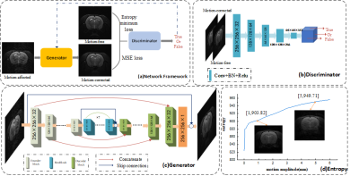 |
Retrospective motion correction for Fast Spin Echo based on conditional GAN with entropy loss
Qingjia Bao1, Yalei Chen2, Pingan Li2, Kewen Liu2, Zhao Li3, Xiaojun Li2, Fang Chen3, and Chaoyang Liu3
1Department of Chemical and Biological Physics, Weizmann Institute of Science, Rehovot, Israel, 2School of Information Engineering, Wuhan University of Technology, Wuhan, China, 3State Key Laboratory of Magnetic Resonance and Atomic and Molecular Physics, Wuhan Center for Magnetic Resonance, Wuhan Institute of Physics and Mathematics, Innovation Academy for Precision Measurement Science and Technology, Chinese Academy of Sciences., Wuhan, China
We proposed a new end-to-end motion correction method based on conditional generative adversarial network (GAN) and minimum entropy of MRI images for Fast Spin Echo(FSE) sequence. The network contains an encoder-decoder generator to generate the motion-corrected images and a PatchGAN discriminator to classify an image as either real (motion-free) or fake(motion-corrected). Moreover, the image's entropy is set as one loss item in the cGAN's loss as the entropy increases monotonically with the motion amplitude, indicating that entropy is a good criterion for motion. The results show that the proposed method can effectively reduce the artifacts and obtain high-quality motion-corrected images from the motion-affected images in both pre-clinical and clinical datasets.
|
|||
1354.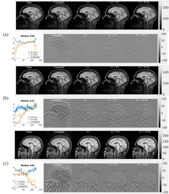 |
Motion correction in MRI with large movements using deep learning and a novel hybrid loss function
Lei Zhang1, Xiaoke Wang1, Michael Rawson2, Radu Balan3, Edward H. Herskovits1, Linda Chang1, Ze Wang1, and Thomas Ernst1
1Department of Diagnostic Radiology & Nuclear Medicine, University of Maryland School of Medicine, Baltimore, MD, United States, 2Department of Mathematics, University of Maryland, College Park, MD, United States, 3Department of Mathematics and Center for Scientific Computation and Mathematical Modeling, University of Maryland, College Park, MD, United States Patient motion continues to be a major problem in MRI. We propose and validate a novel deep learning approach for the correction of large movements in brain MRI. Training pairs were generated using in-house MRI data of high quality, and simulated images with artifacts based on real head movements. The images predicted by the proposed DL method from motion-corrupted data have improved image quality compared with the original corrupted images in terms of a quantitative metric and visual assessment by experienced readers. |
|||
1355.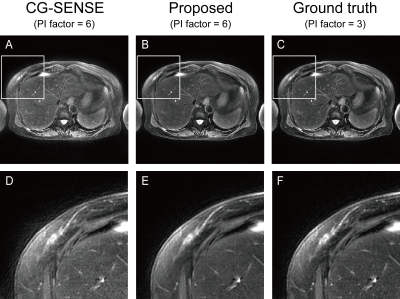 |
Fast abdominal T2 weighted PROPELLER using deep learning-based acceleration of parallel imaging
Motohide Kawamura1, Daiki Tamada1, Masahiro Hamasaki2, Kazuyuki Sato2, Tetsuya Wakayama3, Satoshi Funayama1, Hiroyuki Morisaka1, and Hiroshi Onishi1
1Department of Radiology, University of Yamanashi, Chuo, Japan, 2Division of Radiology, University of Yamanashi Hospital, Chuo, Japan, 3MR Collaboration and Development, GE Healthcare, Hino, Japan
Respiratory motion is a big problem in abdominal T2W imaging. PROPELLER sequence is an excellent solution for motion artifact. However, it requires longer acquisition than FSE, limiting its wider adoption in clinical situations. Here, we propose to use a deep learning-based parallel imaging reconstruction for accelerating PROPELLER. Our approach applies deep learning to the reconstruction of blade images. Thus, training is robust to respiratory motion because blade data can be obtained with single shot. Preliminary results showed that the proposed method significantly outperformed SENSE reconstruction.
|
|||
1356.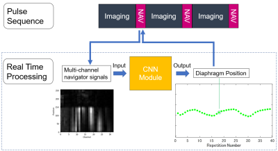 |
Deep Learning-Based Respiratory Navigator Echo (DLnav) for Robust Free-Breathing Abdominal MRI
Yuji Iwadate1, Atsushi Nozaki1, Shigeo Okuda2, Tetsuya Wakayama1, and Masahiro Jinzaki2
1Global MR Applications and Workflow, GE Healthcare Japan, Hino, Japan, 2Department of Radiology, Keio University School of Medicine, Tokyo, Japan
We propose a deep learning-based respiratory navigator (DLnav) technique which uses a convolutional neuronal network (CNN) for respiratory motion detection. The pencil-beam navigator signals were transferred to the real time processing unit including a CNN module and a diaphragm position value was calculated there. DLnav was incorporated into prospectively navigator-gated 3D SPGR and its performance was evaluated in the volunteer scan. DLnav resulted in good synchronization with actual respiratory motion and reduced motion-induced blurring with two different tracker positions.
|
|||
1357.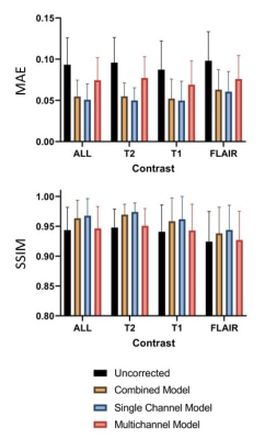 |
Deep Learning-Based Rigid-Body Motion Correction in MRI using Multichannel Data
Miriam Hewlett1,2, Ivailo E Petrov2, and Maria Drangova1,2
1Medical Biophysics, Western University, London, ON, Canada, 2Robarts Research Institute, London, ON, Canada
Motion artefacts remain a common problem in MRI. Deep learning presents a solution for motion correction requiring no modifications to image acquisition. This work investigates incorporating multichannel MRI data for motion correction using a conditional generative adversarial network (cGAN). Correcting for motion artefacts in the single-channel images prior to coil combination improved performance compared to motion correction on coil-combined images. The model trained for simultaneous motion correction of multichannel data produced the worst result, likely a result of its limited modelling capacity (reduced due to memory limitations).
|
|||
1358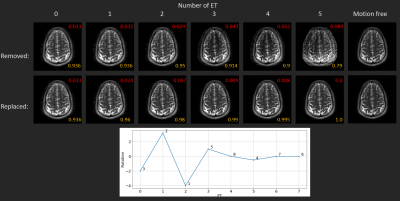 |
Prospective motion assessment within multi-shot imaging using coil mixing of the data consistency error and deep learning Video Permission Withheld
Julian Hossbach1,2,3, Daniel Nicolas Splitthoff3, Bryan Clifford4, Daniel Polak3, Stephan F. Cauley5, and Andreas Maier1
1Friedrich-Alexander-University Erlangen-Nuremberg, Erlangen, Germany, 2Erlangen Graduate School in Advanced Optical Technologies, Erlangen, Germany, 3Siemens Healthcare GmbH, Erlangen, Germany, 4Siemens Medical Solutions, Boston, MA, United States, 5Department of Radiology, Athinoula A. Martinos Center for Biomedical Imaging, Boston, MA, United States
In this work we investigate the effect of motion on the data consistency error coil-mixing matrix, obtained by singular value decomposition. More specifically, a Neural Network is trained to translate motion induced deviations of this coil-mixing matrix relative to a reference acquisition into a motion score. This score can be used for the prospective detection of the most corrupted echo trains for removal or triggering a replacement by reacquisition. We show that a selective removal/replacement using the prospective motion score increases the image quality.
|
|||
1359.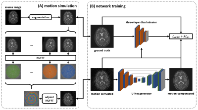 |
Retrospective motion compensation for spiral brain imaging with a deep convolutional neural network
Quan Dou1, Zhixing Wang1, Xue Feng1, John P. Mugler2, and Craig H. Meyer1
1Biomedical Engineering, University of Virginia, Charlottesville, VA, United States, 2Radiology & Medical Imaging, University of Virginia, Charlottesville, VA, United States
Head motion can severely degrade the quality of MR brain images. A deep convolutional neural network was implemented in this study to retrospectively compensate for motion in spiral imaging. The network was trained on images with simulated motion artifacts and tested on both simulated and in vivo data. The image quality was improved after the motion correction.
|
|||
1360. |
Rigid motion artifact correction in multi-echo GRE using navigator detection and Parallel imaging reconstruction with Deep Learning
Seul Lee1, Jae-Hun Lee1, Soozy Jung1, and Dong-Hyun Kim1
1Department of Electrical and Electronic Engineering, Yonsei University, Seoul, Korea, Republic of
Motion artifacts which are occurred in subject motion during MR data acquisition can cause significant image degradation. In this study, we propose a rigid motion artifact correction method, which eliminates the motion-corrupted phase encoding lines detected by navigator echoes and reconstructs motion-compensated images using parallel imaging with deep learning. According to evaluation of simulated motion data and real motion-corrupted data, the proposed method achieved competent compensation for motion artifacts.
|
|||
1361.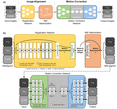 |
Motion Correction and Registration Networks for Multi-Contrast Brain MRI
Jongyeon Lee1, Byungjai Kim1, Wonil Lee1, and HyunWook Park1
1Korean Advanced Institute of Science and Technology, Daejeon, Korea, Republic of
Deep learning techniques have been applied to motion artifact correction without motion estimation or tracking. We previously studied the motion correction method for the multi-contrast brain MRI using NMI maximization and the multi-input neural network. However, as the previous work suffered from a prolonged alignment time and a training inconvenience, we adopt the registration network to reduce alignment time and the multi-output neural network to be trained only once. Our proposed method successfully reduces motion artifacts in the multi contrast images.
|
|||
1362.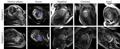 |
Learning-based automatic field-of-view positioning for fetal-brain MRI
Malte Hoffmann1,2, Daniel C Moyer3, Lawrence Zhang3, Polina Golland3, Borjan Gagoski1,4, P Ellen Grant1,4, and André JW van der Kouwe1,2
1Department of Radiology, Harvard Medical School, Boston, MA, United States, 2Department of Radiology, Massachusetts General Hospital, Boston, MA, United States, 3Computer Science and Artificial Intelligence Laboratory, MIT, Cambridge, MA, United States, 4Fetal-Neonatal Neuroimaging and Developmental Science Center, Boston Children's Hospital, Boston, MA, United States
Unique challenges of fetal-brain MRI include successful acquisition of standard sagittal, coronal and axial views of the brain, as motion precludes acquisition of coherent orthogonal slice stacks. Technologists repeat scans numerous times by manually rotating slice prescriptions but inaccuracies in slice placement and intervening motion limit success. We propose a system to automatically prescribe slices based on the fetal-head pose as estimated by a neural network from a fast scout. The target sequence receives the head pose and acquires slices accordingly. We demonstrate automatic acquisition of standard anatomical views in-vivo.
|
|||
1363.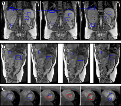 |
Computer vision object tracking for MRI motion estimation
Stefan Wampl1, Tito Körner1, Martin Meyerspeer1, Marcos Wolf1, Maxim Zaitsev1, and Albrecht Ingo Schmid1
1High Field MR Center, Center for Medical Physics and Biomedical Engineering, Medical University of Vienna, Vienna, Austria
Computer vision (CV) libraries such as OpenCV provide versatile algorithms for object tracking. We demonstrate the feasibility of out-of-the-box object trackers in different dynamic MR imaging scenarios. In contrast to specialized detection and registration algorithms, the generic implementation of object trackers enables targeting of different and challenging organs during respiratory motion, including the heart, liver and kidney. Apart from post-processing, fast algorithm and implementation allowed for application in our online and prospective motion compensation pipeline. By leveraging these open source libraries, MR applications can benefit from both the current powerful library and the continuous developments by the CV community.
|
|||
1364.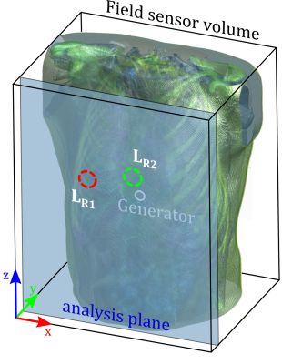 |
Listening in on the Pilot Tone: A Simulation Study
Mario Bacher1,2, Barbara Dornberger2, Jan Bollenbeck2, Matthias Stuber1, and Peter Speier2
1Department of Radiology, Lausanne University Hospital and University of Lausanne, Lausanne, Switzerland, 2Siemens Healthcare Magnetic Resonance, Erlangen, Germany
The Pilot Tone navigator is a novel electromagnetic motion sensing method capable of contactless respiratory and cardiac motion sensing. We demonstrate the physical processes underlying the Pilot Tone navigator using electromagnetic simulations on a realistic virtual human phantom and validate these simulation results in-vivo. These simulations can be used to investigate how the PT signal is shaped by motion of the underlying anatomy with the aim of using this information to aid in improving our processing pipeline.
|
|||
1365.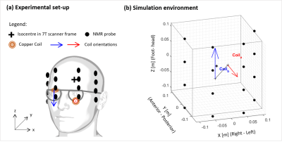 |
Simulating the use of active magnetic markers for motion correction using NMR field probes on a 7T scanner
Laura Bortolotti1 and Richard Bowtell1
1Sir Peter Mansfield Imaging Centre, University of Nottingham, Nottingham, United Kingdom
We use simulations to demonstrate the feasibility of a novel approach to motion tracking, which is based on using a 16-probe field camera to monitor the fields generated by briefly passing currents through small coils affixed to the head. Using previously recorded head motion data we show that changes in head position can be accurately estimated from the measured field changes. The benefits of this approach are that problematic line-of-sight access to markers is not required (cf. optical approaches) and that it could be implemented without modification of the MRI sequence (cf. navigators).
|
|||
1366.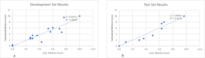 |
Perceptual motion scoring: An algorithm for automated detection and grading of MRI motion artifacts
Rafael Brada1, Michael Rotman1, Sangtae Ahn2, and Christopher J. Hardy2
1GE Reserach, Herzliya, Israel, 2GE Research, Niskayuna, NY, United States
We introduce a method for the automatic detection and scoring for motion artifacts in 2D FSE images. The method is based on analyzing the difference in k-space data between two coil array elements. The relative motion score is a parameter free calculation. To match human observer rankings, linear regression coefficients were calculated on a development set of seventeen T1 brain series. The normalized score was tested on nine T1-FLAIR FSE brain series achieving an R2 of 0.91. The ability to automatically detect and grade the severity of motion artifacts is important for better clinical workflows, and for research purposes.
|
|||
1367.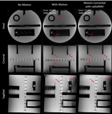 |
Radial Navigator (radNAV) for Rapid GRE (Turbo-FLASH) Sequence
Zhe Wu1, Lars Kasper1, and Kamil Uludag1,2,3
1Techna Institute, University Health Network, Toronto, ON, Canada, 2Koerner Scientist in MR Imaging, University Health Network, Toronto, ON, Canada, 3Center for Neuroscience Imaging Research, Institute for Basic Science and Department of Biomedical Engineering, Sungkyunkwan University, Suwon, Korea, Republic of
A radial navigator (radNAV) approach, which is easily implemented with minimal increase in TE/TR and without the need for non-Cartesian gradients or coil sensitivity extrapolation, is proposed. We demonstrate the feasibility of this approach to acquire motion information for image correction in turbo-FLASH sequences.
|
|||
1368.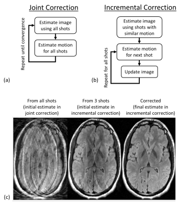 |
Incremental motion correction (iMoCo) for fast retrospective image reconstruction with reduced motion artifacts
Anuj Sharma1, Samir D Sharma1, and Andrew J Wheaton1
1Magnetic Resonance, Canon Medical Research USA, Inc., Mayfield Village, OH, United States
Retrospective rigid-body motion correction methods often alternate between updating the estimate of the motion parameters and updating the image. The alternating minimization reconstruction is time-consuming, which is problematic in the clinical setting. We propose a new method for retrospective rigid-body motion correction that makes use of the insight that many shots of the imaging data have similar motion values, and therefore a reference image can be created from the imaging data itself. By leveraging this insight, this method is able to quickly reconstruct motion-corrected images without requiring large amounts of ML training data and without requiring an additional scout scan.
|
|||
1369.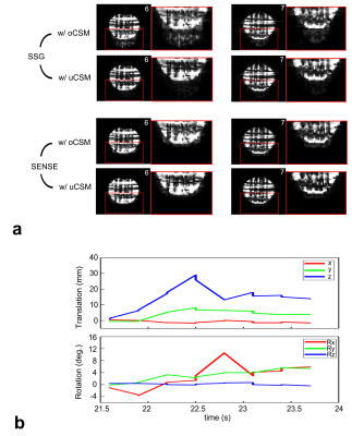 |
SMS-EPI real-time motion correction by receiver phase compensation and coil sensitivity interpolation
Bo Li1, Ningzhi Li2, Ze Wang1, and Thomas Ernst1
1Department of Diagnostic Radiology and Nuclear Medicine, University of Maryland, Baltimore, Baltimore, MD, United States, 2U.S. Food Drug Administration, Silver Spring, MD, United States
We investigated SMS-EPI real-time motion correction using phase compensation and SMS reconstruction with coil sensitivity interpolation for severe movements. A prospective motion correction system was used to provide motion information and update sequence parameters in real time. The receiver phase needs to match the cycled phase variation, and, when motion updates alter slice positions, an additional phase modulation is required. Coil sensitivity was initially calculated using single-slice reference images and then recalculated by spline interpolation for new slice position. Split slice-GRAPPA and SENSE with updated coil sensitivity maps show significantly improved results.
|
|||
1370.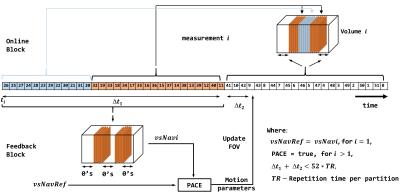 |
Self-navigating 3D-EPI Sequence for Prospective Motion Correction
Samuel Getaneh Bayih1, Ernesta Meintjes1, Marcin Jankiewicz1, and Andre van der Kouwe 2,3
1MRT/UCT Medical Imaging Research Unit, Department of Human Biology, University of Cape Town, Cape Town, South Africa, 2Athinoula A. Martinos Center for Biomedical Imaging, Charlestown, MA, United States, 3Department of Radiology, Harvard Medical School, Boston, MA, United States
Prospective motion correction typically requires additional pulses or hardware to track subject motion, incurring extra costs and increasing the complexity of an MRI experiment. We present a prospective motion tracking solution that instead constructs a volumetric self-navigator from a subset of the partitions acquired during 3D-EPI, thereby allowing motion during and between successive 3D-EPI measurements to be detected and corrected in real time. This work facilitates motion-robust 3D-EPI acquisition for functional MRI applications.
|
The International Society for Magnetic Resonance in Medicine is accredited by the Accreditation Council for Continuing Medical Education to provide continuing medical education for physicians.