Digital Posters
Novel Encoding Strategies
ISMRM & SMRT Annual Meeting • 15-20 May 2021

| Concurrent 1 | 13:00 - 14:00 |
0899.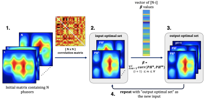 |
Nonlinear projection imaging with the Bloch-Siegert shift in an inhomogeneous B0 at low-field
Kartiga Selvaganesan1, Yonghyun Ha2, Basong Wu2, Kasey Hancock3, Charles Rogers III2, Sajad Hosseinnezhadian2, Gigi Galiana2, and Todd Constable2
1Biomedical Engineering, Yale University, New Haven, CT, United States, 2Radiology and Biomedical Imaging, Yale University, New Haven, CT, United States, 3Electrical Engineering, Yale University, New Haven, CT, United States
The ability to form an image without using spatial encoding gradients allows for cheaper MRI manufacturing costs, particularly in small low field devices suitable for point-of-care imaging. The Bloch-Siegert shift effect provides an RF alternative to spatial encoding with gradient coils. This work presents a new approach to evaluate and optimize nonlinear RF encoding schemes generated by transmit phased arrays. From thousands of possible encoding patterns, the algorithm finds the set that minimizes correlations between spatial patterns while maximizing the spatial encoding. Simulated images demonstrate that the nonlinear spatial encodings provided by Bloch-Siegert RF pulses can provide high image quality.
|
|||
0900.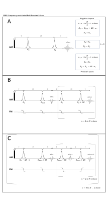 |
B1-gradient based MRI using Frequency-modulated Rabi Encoded Echoes (FREE)
Efraín Torres1,2, Taylor Froelich2, Lance DeLaBarre2, Michael Mullen2, Gregory Adriany2, Alberto Tannús3, Daniel Cosmo Pizetta3, Mateus Jose Martins3, and Michael Garwood2
1Biomedical Engineering, University of Minnesota, Minneapolis, MN, United States, 2Center for Magnetic Resonance Research, University of Minnesota, Minneapolis, MN, United States, 3Centro de Imagens e Espectroscopia por Ressonância Magnética - CIERMag - Sao Carlos Physics, São Carlos, Brazil
Gradient coils reduce available bore space, incur costs, and create acoustic noise, but their encoding ability requires them to be present in MRI. The encoding strategy introduced here (FREE) removes the reliance on these coils by using a B1-gradient to create a spatially-dependent nutation frequency. Double spin-echoes created with adiabatic full-passage pulses generate magnetization phase proportional to the difference in their time-bandwidth products (R). Modulating the R-value of one of these π pulses traverses k-space similar to standard MRI. Adiabatic pulses make this approach highly tolerant to B0 and B1 inhomogeneity. A family of new sequences using FREE is presented.
|
|||
0901.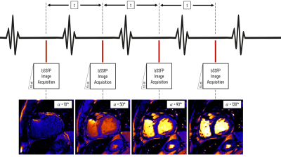 |
Rapid simultaneous T1 and T2 quantification (RAS-Q T1T2) of the myocardium using transient bSSFP with variable flip angles
Céline Marquet1,2,3, Jihye Jang1,2,4, Andrew J. Powell1,2, and Mehdi H. Moghari1,2
1Department of Pediatrics, Harvard Medical School, Boston, MA, United States, 2Department of Cardiology, Boston Children's Hospital, Boston, MA, United States, 3Department of Informatics, Technical University Munich, Munich, Germany, 4Philips Healthcare, Boston, MA, United States
We developed a novel MRI pulse sequence RAS-Q T1T2 for the simultaneous quantification of T1 and T2 of the myocardium using transient bSSFP imaging with a variable flip angle scheme. RAS-Q T1T2 was systematically analyzed based on a numerical simulation, as well as phantom and patient studies. We show that RAS-Q T1T2 yields accurate T2 estimates for the myocardium with a trade-off in precision compared to state-of-the-art methods, reducing scan time to less than 4s. The estimated T1 values reveal lower accuracy and precision than clinically established methods and need further
improvements.
|
|||
0902.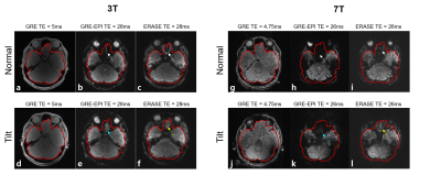 |
Susceptibility artifact-insensitive ultrafast 3D gradient-echo imaging by combination of head-tilting and ERASE acquisition
Jaeyong Yu1,2, Seulki Yoo1,2, Jae-Kyun Ryu3, Seung-Kyun Lee1,2,4, and Jang-Yeon Park1,2,3
1Department of Biomedical Engineering, Sungkyunkwan University, Suwon, Korea, Republic of, 2Department of Intelligent Precision Healthcare Convergence, Sungkyunkwan University, Suwon, Korea, Republic of, 3Biomedical Institute for Convergence at SKKU, Sungkyunkwan University, Suwon, Korea, Republic of, 4Center for Neuroscience Imaging Research, Institute for Basic Science (IBS), Suwon, Korea, Republic of
Equal-TE Rapid Acquisition with Sequential Excitation (ERASE) is a 3D ultrafast gradient-echo-based Spatiotemporal Encoding (SPEN) imaging technique with a constant TE and high tolerance to main magnetic field () inhomogeneity. However, misregistration of spin isochromat can occur due to large magnetic susceptibility-induced gradients in the prefrontal brain region. Here, we propose that shim improvement by chin-up head tilting can effectively mitigate misregistration in ERASE to realize fast prefrontal brain imaging with much higher image quality than conventional EPI.
|
|||
0903.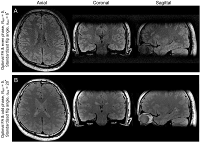 |
Optimization of Flip Angle and RF Pulse Phase in MP-SSFP for MRI in Inhomogeneous Magnetic Fields
Naoharu Kobayashi1 and Michael Garwood1
1CMRR, Radiology, University of Minnesota, Minneapolis, MN, United States
Flip angle and RF pulse phase in 3D MP-SSFP were optimized to maximize SNR. Optimal flip angle and pulse phase were estimated by numerically maximizing the steady-state magnetization in MP-SSFP with SAR penalty. The estimated optimal flip angle and phase were experimentally validated with an agar gel phantom at 3T. The optimal RF pulse setting improved SNR up to 41% under the fixed SAR conditions, depending on MP-SSFP sequence parameters. Finally, in vivo human brain imaging was conducted to demonstrate image contrasts in MP-SSFP images with the optimal flip angle and pulse phase.
|
|||
0904.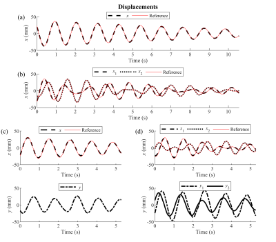 |
Spectro-Dynamic MRI: Characterizing Bio-Mechanical Systems on a Millisecond Scale
Max H. C. van Riel1,2, Niek R. F. Huttinga1, and Alessandro Sbrizzi1
1Department of Radiotherapy, Computational Imaging Group for MR diagnostics and therapy, UMC Utrecht, Utrecht, Netherlands, 2Department of Biomedical Engineering, Medical Image Analysis, Eindhoven University of Technology, Eindhoven, Netherlands
Measuring in vivo dynamics can yield valuable information about the cardiovascular or musculoskeletal system. MRI shows severe limitations when dealing with motion at high spatial and temporal resolutions. We propose spectro-dynamic MRI for the characterization of dynamics directly from k-space data. The sampling pattern is adjusted to achieve a temporal resolution of a few milliseconds. A measurement model, relating the measured data to the displacements, is combined with a dynamical model, introducing prior knowledge about the dynamics. Preliminary results show that spectro-dynamic MRI allows estimation of motion fields and dynamical system parameters from heavily undersampled data on a millisecond timescale.
|
|||
0905. |
2.5D MRI of the Vocal Fold Oscillation using Single Point Imaging with Rapid Encoding
Johannes Fischer1, Ali Caglar Özen1,2, Matthias Echternach3, Louisa Traser4, Bernhard Richter4, and Michael Bock1
1Dept.of Radiology, Medical Physics, Medical Center University of Freiburg, Faculty of Medicine, University of Freiburg, Freiburg, Germany, 2German Consortium for Translational Cancer Research Freiburg Site, German Cancer Research Center (DKFZ), Heidelberg, Germany, 3Division of Phoniatrics and Pediatric Audiology, Department of Otorhinolaryngology, Head and Neck Surgery, Ludwig-Maximilians-University, Munich, Germany, 4Institute of Musicians' Medicine, Freiburg University Medical Center, Germany Faculty of Medicine, University of Freiburg, Freiburg, Germany
Single point imaging with rapid encoding (SPIRE) can dynamically image the oscillations of the vocal folds in the coronal plane with sub-milisecond temporal resolution. To add spatial encoding in the third dimension, a slower frequency encoding gradient was applied in slice direction, where motion is minimal. Electroglottography and projection navigators are used to detect shifts in the larynx position, which are corrected during reconstruction. The velocity of the mucosal wave is estimated from the images.
|
|||
0906.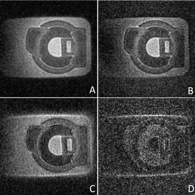 |
Zero Echo Time Imaging Using Low Resolution k-Space Interleaves
Hanna Frantz1, Thomas Huefken1, Patrick Metze1, Kilian Stumpf1, Tobias Speidel1, and Volker Rasche1
1Department of Internal Medicine II, Ulm University Medical Center, Ulm, Germany
Zero echo time (ZTE) imaging is an established approach for three-dimensional imaging of tissues and materials with ultrashort T2* relaxation times. An intrinsic disadvantage of this approach are missing data points within the center region of k-space due to hardware limitations. These missing points are often retrospectively interpolated or subsequently acquired using single-point imaging approaches or additional ZTE acquisitions with decreased resolutions. This abstract presents an approach that relies on the interleaved combination of ZTE read-outs with different resolutions combined with a Compressed Sensing reconstruction, to acquire more information around the k-space center, for high-quality ZTE without prolonged acquisition times.
|
|||
0907.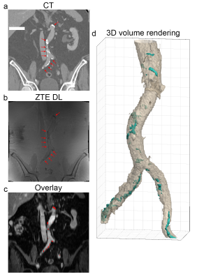 |
Arterial calcification imaging using ZTE on PET/MR
Edwin Eduard Gert Willem ter Voert1, Florian Wiesinger2, Graeme McKinnon3, Mathias Engström4, Jose Fernando de Arcos5, Marlena Hofbauer1, Ronny R Buechel1, and Philipp A Kaufmann1
1Department of Nuclear Medicine, University Hospital Zurich, Zurich, Switzerland, 2GE Healthcare, Munich, Germany, 3GE Healthcare, Milwaukee, WI, United States, 4GE Healthcare, Stockholm, Sweden, 5GE Healthcare, Little Chalfont, Amersham, United Kingdom Calcifications in atherosclerotic plaques are possibly associated with worsened clinical prognosis. In contrast to the often-used CT for diagnosis, calcifications cannot easily be detected with MRI. As PET becomes a major complementary modality for MRI in plaque imaging, and as integrated PET/MR scanners become increasingly available, PET/MR based calcification screening methods are desired. With zero TE (ZTE) MR imaging it is possible to image short T2 tissues like bone. In the current study we demonstrate that ZTE imaging on PET/MR, combined with Deep Learning reconstructed ZTE, can likely be applied to detect calcifications in peripheral vasculature. |
|||
0908.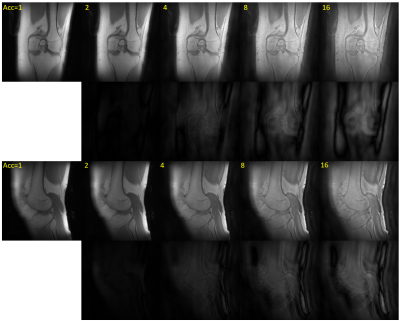 |
Compressed Sensing PETRA MRI
Serhat Ilbey1, Johannes Fischer1, Michael Bock1, and Ali Caglar Ozen1,2
1Dept. of Radiology, Medical Physics, Medical Center University of Freiburg, Faculty of Medicine, University of Freiburg, Freiburg, Germany, 2German Consortium for Translational Cancer Research Partner Site Freiburg, German Cancer Research Center (DKFZ), Heidelberg, Germany
PETRA is a combination of a radial zero-TE and single point imaging (SPI) acquisition, which is very silent and which can efficiently acquire MR signals with short T2*. In PETRA, the acquisition of SPI data constitutes a major part of the acquisition time especially for high-resolution protocols. To accelerate the SPI section while preserving the image quality, we combine SPI with compressed sensing (cs). csPETRA enables 3D imaging with isotropic sub-millimeter resolution within only a few minutes, e.g., for (0.5 mm)3 voxel-size with (20 cm)3 field-of-view, which is demonstrated with different acceleration factors for high-resolution imaging of the knee.
|
|||
0909.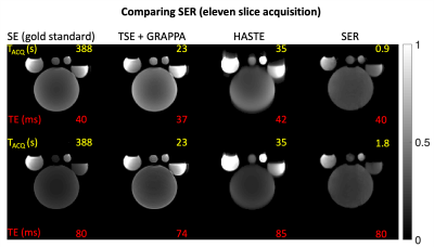 |
Single echo reconstruction for rapid and silent MRI
Sairam Geethanath1
1Columbia MR Center, Columbia University, New York, NY, United States
This work introduces a reconstruction-only approach, dubbed “single echo reconstruction” (SER) to demonstrate (i) the first, rapid 128 x 128 MRI without phase encoding using a 64-channel coil; (ii) significant reduction of RF power, PNS, and gradient noise; (iii) using only a commercially available coil with no external sensors (iv) comparison with gold-standard 2D spin-echo (SE) and accelerated acquisitions for $$$ T_2 $$$ weighted imaging as an application. For imaging 11 slices with TE = 80ms, the acquisition time was 1.8s with 10.8W total RF deposition, 12.09% peripheral nerve stimulation and no blurring artifacts.
|
|||
0910.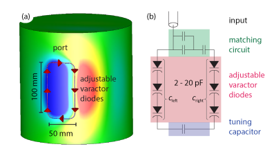 |
Investigations on accelerated imaging at 9.4T with electronically modulated time-varying receive sensitivities
Felix Glang1, Kai Buckenmaier1, Jonas Bause1, Alexander Loktyushin1, Nikolai Avdievich1, and Klaus Scheffler1,2
1High-field Magnetic Resonance Center, Max-Planck Institute for Biological Cybernetics, Tübingen, Germany, 2Department of Biomedical Magnetic Resonance, Eberhard Karls University Tübingen, Tübingen, Germany
In this work, it is assessed how electronically modulated time-varying receive sensitivities can improve parallel imaging reconstruction at 9.4T. The required sensitivity modulation is achieved by introducing variable capacitance diodes (varactors) in the receive loops that can be independently adjusted to modify B1- profiles. A prototype 4 channel receive array was built, and measured and simulated receive profiles were compared. Additionally, simulations were conducted regarding potential for g-factor improvement. It was found that potential improvements strongly depend on the B1- switching patterns during k-space acquisition, where strongest improvements are to be expected from fast B1- modulations.
|
|||
0911.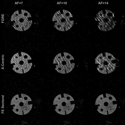 |
Feasibility of a Novel Sampling/Reconstruction Method Ensuring a SNR Benefit Over the Traditional Sampling Approach
Samuel Perron1, Matthew S. Fox1,2, Hacene Serrai1, and Alexei Ouriadov1,2,3
1Physics and Astronomy, The University of Western Ontario, London, ON, Canada, 2Lawson Health Research Institute, London, ON, Canada, 3School of Biomedical Engineering, The University of Western Ontario, London, ON, Canada
To produce high-SNR images, MRI systems usually employ many signal averages, expensive high field-strength coils, and/or expensive contrast agents. The proposed sampling and reconstruction method, requiring no hardware modifications, would produce images with higher SNR without increasing scan times. Nine under-sampled images are averaged for every combination of images, and the resulting SNR vs. averages function is fitted according to the Stretched-Exponential Model (SEM). The resulting reconstructed image yielded higher SNR than the original image for all three imaging schemes (FGRE, x-Centric, FE-Sectoral), demonstrating the feasibility of the proposed method for the first time.
|
|||
0912. |
A Deselecting Alias Approach to Volumetric Zoomed Imaging
Nicolas Arango1, Molin Zhang1, Jason Stockmann2,3, Jacob White1, and Elfar Adalsteinsson1,4
1Department of Electrical Engineering and Computer Science, Massachusetts Institute of Technology, Cambridge, MA, United States, 2A. A. Martinos Center for Biomedical Imaging, Massachusetts General Hospital, Charlestown, MA, United States, 3Harvard Medical School, Boston, MA, United States, 4Institute for Medical Engineering and Science, Massachusetts Institute of Technology, Cambridge, MA, United States
Reduced FOV imaging using ∆B0 control is limited by magnetostatics, but careful tracking of aliasing enables the vast reduction in the deselection volume required. A 2-sided spectral approach for the deselection enables the combination of strong linear gradients and adaptable multicoil shim array ∆B0 fields to perform selective excitation in concert. Simulations indicate potential for central volume rFOV where prior work in ∆B0-based rFOV imaging was limited to peripheral volumes.
|
|||
0913.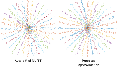 |
Efficient NUFFT Backpropagation for Stochastic Sampling Optimization in MRI
Guanhua Wang1, Douglas C. Noll1, and Jeffrey A. Fessler2
1Biomedical Engineering, University of Michigan, Ann Arbor, MI, United States, 2Electrical Engineering and Computer Science, University of Michigan, Ann Arbor, MI, United States
Optimization-based k-space sampling pattern design often involves the Jacobian matrix of non-uniform fast Fourier transform (NUFFT) operations. Previous works relying on auto-differentiation can be time-consuming and less accurate. This work proposes an approximation method using the relationship between exact non-uniform DFT (NDFT) and NUFFT, demonstrating improved results for the sampling pattern optimization problem.
|
|||
0914.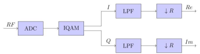 |
A digital MRI RF-receiver using an ordinary GPU
Annalena Erbrecht1, Enrico Pannicke1, and Christoph Dinh1
1Otto-von-Guericke University, Magdeburg, Germany
We want to present a CUDA based RF-receiver for a 1,5 T MRI consisting of an inverse quadrature modulation and the decimation step. We compared different decimation techniques based on a moving average, a CIC and an FIR filter. The filters were compared regarding their noise attenuation, their processing effort and their group delays. We conclude that the CIC filter is the optimal filter for our usage, because this filter provides the best compromise between noise attenuation and processing effort.
|
|||
0915.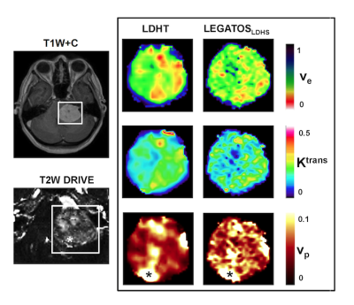 |
Feasibility of a single low dose dual temporal resolution DCE-MRI method for whole-brain, high-spatial resolution parametric mapping
Ka-Loh Li1, Daniel Lewis2, David J Coope3, Federico Roncaroli3, Omar N Pathmanaban2, Andrew T King2, Sha Zhao1, Erjon Agushi1, Alan Jackson1, Timothy Cootes1, and Xiaoping Zhu1
1Division of Informatics, Imaging and Data Sciences, The University of Manchester, Manchester, United Kingdom, 2Department of Neurosurgery, Salford Royal NHS Foundation Trust, Manchester, United Kingdom, 3Division of Neuroscience and Experimental Psychology, The University of Manchester, Manchester, United Kingdom
This study sought to evaluate the feasibility of using a low gadolinium-based contrast agent (GBCA) dose protocol with a newly developed analysis technique, the LEGATOS method, for deriving whole-brain, high-spatial resolution pharmacokinetic parameters from dual-temporal resolution (DTR) DCE-MRI. Through Monte Carlo simulations and an in vivo study incorporating histopathological data the accuracy of pharmacokinetic parameters derived using a low-dose interleaved protocol and LEGATOS was assessed. Our results demonstrate that this approach permitted the derivation of accurate, tissue-validated high-spatial resolution kinetic parameter estimates following a single low-dose injection, representing in some patients a greater than 80% reduction in GBCA dose.
|
|||
0916.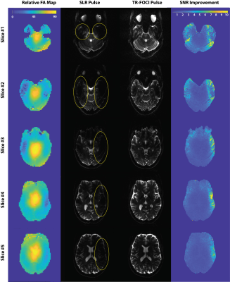 |
B1+ inhomogeneity mitigation using adiabatic refocusing RF pulses for diffusion weighted imaging at 7T
Shahrokh Abbasi-Rad1, Martijn Cloos1,2, Jin Jin3, Kieran O'Brien3, and Markus Barth1,2,4
1Centre for Advanced Imaging, University of Queensland, Brisbane, Australia, 2ARC Training Centre for Innovation in Biomedical Imaging Technology, The University of Queensland, Brisbane, Australia, 3Siemens Healthcare Pty Ltd, Brisbane, Australia, 4School of Electrical Engineering and Information Technology, The University of Queensland, Brisbane, Australia
Ultra-high field scanners offer a potential signal-to-noise ratio (SNR) improvement for Diffusion weighted Imaging (DWI). However, the application of DWI at 7T is hindered by several technical challenges. In particular, B1+ inhomogeneity can lead to signal dropouts that degrades image quality in the temporal lobes and base of the skull. In this work, we show that embedding time-resampled frequency offset corrected inversion pulses (TR-FOCI) in a twice-refocussed spin echo DWI sequence can recover the signal in these brain areas.
|
|||
0917.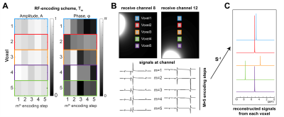 |
RF-encoding for improved multi-voxel separation in MR spectroscopy
Adam Berrington1, Penny Gowland1, and Richard Bowtell1
1Sir Peter Mansfield Imaging Centre, School of Physics and Astronomy, University of Nottingham, Nottingham, United Kingdom
The simultaneous acquisition and separation of signals from multiple voxels using receive sensitivities can accelerate MRS acquisition. However, separation is ill-conditioned when signals are acquired from voxels with similar receive-coil sensitivities. We propose a new approach which improves reconstruction using the transmitted RF to modulate the signals from different voxels. Simulations showed that for a small number of encoding steps, M, the g-factor of the reconstruction problem was reduced (~3-fold, M=2 for 5 regions). Reconstructed phantom data from two-voxels, acquired using phase-modulation, were of higher SNR than coil-encoding, indicating the potential of the proposed method for multi-voxel measurement in MRS.
|
|||
0918. |
Extended Accelerated Systematic Tracking using Experimental Radiology: an Encephalo-Graphic Generation
S. T. Claus1, Y. Eti2, and E. S. Terbuny3
1Dept. of Presents, North Pole Research Agency, Rovaniemi, Finland, 2Atopof Amountain, Himalaya, Bhutan, 3Dept. for Sweets, Greenfield Institute, Ostereistedt, Germany
Detecting Easter bunnies is a task that is difficult to impossible for humans - thus, the use of artificial intelligence is more than warranted. In this work we present a confounded nut-work noisette (CNN) algorithm based on extended, accelerated systematic tracking in experimental radiology with encephalo-graphic generation (EASTER-EGG). Using this method we demonstrate that chocolate is a volatile resource especially when hungry researchers are present.
|
The International Society for Magnetic Resonance in Medicine is accredited by the Accreditation Council for Continuing Medical Education to provide continuing medical education for physicians.