Digital Posters
Signal Representations for Quantitative Applications
ISMRM & SMRT Annual Meeting • 15-20 May 2021

Digital Posters
Signal Representations for Quantitative Applications
| Concurrent 1 | 13:00 - 14:00 |
2596.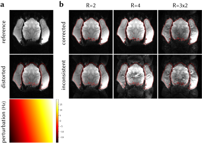 |
Highly accelerated fMRI of awake behaving non-human primates via model-based dynamic off-resonance correction
Mo Shahdloo1, Daniel Papp2, Urs Schüffelgen1, Karla L. Miller2, Matthew Rushworth1, and Mark Chiew2
1Wellcome Centre for Integrative Neuroimaging, Department of Experimental Psychology, University of Oxford, Oxford, United Kingdom, 2Wellcome Centre for Integrative Neuroimaging, FMRIB, Nuffield Department of Clinical Neurosciences, University of Oxford, Oxford, United Kingdom
Dynamic B0 field inhomogeneities due to vigorous body motion - after mechanical head stabilisation - introduce severe artefacts in accelerated fMRI of awake behaving non-human primates (NHPs) by invalidating the calibration data used for unaliasing reconstruction. Here, we propose a method to estimate dynamic field perturbations via model-based frame-to-frame comparison of EPI reference navigators. These estimates can be used to improve reconstruction quality by matching each data frame to the calibration data, and simultaneously correcting the geometric distortions. The proposed method successfully estimates field perturbations and improves reconstruction quality in accelerated NHP fMRI, without the need for sequence modification or extra acquisitions.
|
|||
2597.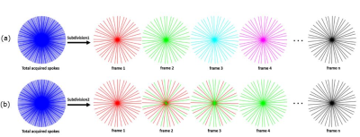 |
Efficient DCE-MR Image reconstruction with feasible temporal resolution in L+S Decomposition model
Faisal Najeeb1, Jichang Zhang2, Xinpei Wang2, Chengbo Wang2, Hammad Omer1, Penny Gowland3, Sue Francis3, and Paul Glover3
1MIPRG,Comsats University, Islamabad, Pakistan, 2SPMIC, The University of Nottingham Ningbo China, Ningbo, China, 3SPMIC, The University of Nottingham, Nottingham, United Kingdom
In this work, temporal shift-windowing function is integrated into Low rank plus sparse (L+S) decomposition to fix the certain motion phase with a high temporal resolution and reconstruction efficiency for free-breathing golden angle radial DCE-MRI of liver. A smooth weighting curve based on a sigmoid function is used to achieve a smooth transition for the spokes between the desired phase and other motion phases. Furthermore, the Fast Iterative Shrinkage-thresholding Algorithm (FISTA) was implemented to solve the L+S optimization problem which enables faster convergence. Results of the proposed method are compared with RACER-GRASP.
|
|||
 |
2598.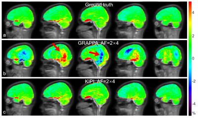 |
Accelerated Chemical Exchange Saturation Transfer Acquisition by Joint K-space and Image-space Parallel Imaging (KIPI)
Zu Tao1, Sun Yi2, Wu Dan1, and Zhang Yi1
1Key Laboratory for Biomedical Engineering of Ministry of Education, Department of Biomedical Engineering, College of Biomedical Engineering & Instrument Science, Zhejiang University, Hangzhou, China, 2MR Collaboration, Siemens Healthcare Ltd., Shanghai, China
The clinical use of chemical exchange saturation transfer (CEST) imaging is limited by its relatively long scan time, because it typically acquires multiple saturation image frames. Here, a novel auto-calibrated reconstruction method by joint k-space and image-space parallel imaging (KIPI) is proposed for faster CEST acquisition. By under-sampling CEST image frames with variable acceleration factors, KIPI allows an acceleration factor of up to 8-fold for acquiring source images, yielding a net speed-up of 6-fold in scan time, and produces image quality close to that of the ground truth.
|
||
2599. |
MEDI-d: Downsampled Morphological Priors for Shadow Reduction in Quantitative Susceptibility Mapping
Alexandra Grace Roberts1,2, Pascal Spincemaille2, Thanh Nguyen2, and Yi Wang2,3
1Electrical and Computer Engineering, Cornell University, Ithaca, NY, United States, 2Radiology, Weill Cornell, New York, NY, United States, 3Biomedical Engineering, Cornell University, Ithaca, NY, United States
Morphology Enabled Dipole Inversion (MEDI) is an iterative reconstruction algorithm for Quantitative Susceptibility Mapping (QSM) that is effective in suppressing streaking artifacts by exploiting the magnitude image as a morphological prior. However, contiguous areas of dipole incompatibility (such as noise) induce shadow artifacts whose spatial frequency components are not adequately regularized by the gradient based regularization in MEDI. Here, we show the feasibility of adding a downsampled morphological prior to suppress these shadow artifacts.
|
|||
2600.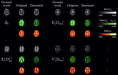 |
Enhancing diffusion tensor distribution imaging via denoising of tensor-valued diffusion MRI data
Jan Martin1, Patrik Brynolfsson2,3, Michael Uder4, Frederik Bernd Laun4, Daniel Topgaard1, and Alexis Reymbaut2
1Physical Chemistry, Lund University, Lund, Sweden, 2Random Walk Imaging AB, Lund, Sweden, 3NONPI Medical AB, Umeå, Sweden, 4Institute of Radiology, University Hospital Erlangen, Friedrich-Alexander-Universität Erlangen-Nürnberg (FAU), Erlangen, Germany
Diffusion tensor distribution imaging (DTD) is a versatile technique enabling to retrieve nonparametric intra-voxel diffusion tensor distributions from tensor-valued diffusion-encoded data. While DTD owes its versatility to the minimal set of assumptions on which it relies, such minimal constraints induce a high sensitivity to noise hindering DTD's potential clinical translation. In this work, we demonstrate within a brain-like numerical phantom that generalized singular-value shrinkage (GSVS) denoising of the data prior to DTD analysis drastically improves DTD's accuracy, mitigating the aforementioned issue.
|
|||
2601.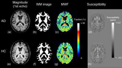 |
Simultaneous Myelin Water, Magnetic Susceptibility, and Morphometry Analyses Using Magnetization-prepared Multiple Spoiled Gradient Echo
Hirohito Kan1,2, Yuto Uchida3, Yoshino Ueki4, Satoshi Tsubokura5, Hiroshi Kunitomo5, Harumasa Kasai5, Noriyuki Matsukawa3, and Yuta Shibamoto2
1Department of Integrated Health Sciences, Nagoya University Graduate School of Medicine, Nagoya, Japan, 2Department of Radiology, Nagoya City University Graduate School of Medical Sciences, Nagoya, Japan, 3Department of Neurology, Nagoya City University Graduate School of Medical Sciences, Nagoya, Japan, 4Department of Rehabilitation Medicine, Nagoya City University Graduate School of Medical Sciences, Nagoya, Japan, 5Department of Radiology, Nagoya City University Hospital, Nagoya, Japan
This study confirmed the feasibility of the simultaneous acquisition and voxel-based myelin water fraction (MWF), quantitative susceptibility mapping (QSM), and morphometry (VBM) analysis using magnetization-prepared multiple spoiled gradient echo (MP-mSPGR) sequence throughout comparisons with patients with Alzheimer’s disease and healthy control. As a result, the voxel-based MWF could detect demyelination in patients with AD. In contrast, there was not a significant change in susceptibility. This result suggested that the MWF depended on only the myelin content. The VBM analysis delineated the atrophy pattern of AD. The MP-mSPGR sequence is feasible for simultaneous MWF, QSM, and VBM analyses.
|
|||
2602.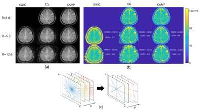 |
The impact of undersampling on the accuracy of the T2 maps reconstructed using CAMP
Nahla M H Elsaid1, Nadine L Dispenza2, R Todd Constable1,3, Hemant D Tagare1,2, and Gigi Galiana1
1Radiology and Biomedical Imaging, Yale School of Medicine, New Haven, CT, United States, 2Department of Biomedical Engineering, Yale University, New Haven, CT, United States, 3Neurosurgery, Yale University, New Haven, CT, United States Constrained alternating minimization for parameter mapping (CAMP) improves the image quality in highly accelerated parameter mapping by incorporating a linear constraint that relates consecutive images.
|
|||
2603.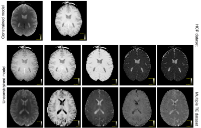 |
Extracting information from diffusion MRI models to visualize the adequacy of acquisition protocols
Samuel St-Jean1,2, Filip Szczepankiewicz2, Christian Beaulieu1, and Markus Nilsson2
1University of Alberta, Edmonton, AB, Canada, 2Clinical Sciences Lund, Lund University, Lund, Sweden
Parameter degeneracies and numerical fitting issues are the bane of diffusion MRI modelling. In this work, we present a framework using convex optimization to fit diffusion MRI models efficiently similarly to magnetic resonance fingerprinting. We also show how the singular value decomposition of these models can help to visualize how well the parameter space of a given model is sampled by a given acquisition protocol. Results on b-tensor encoded datasets and datasets leveraging multiple echo times literally show how these additional measurement dimensions disentangle model parameters better compared to traditional sequences.
|
|||
2604.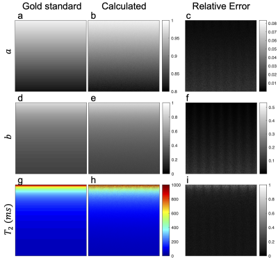 |
Towards analytical $$$T_2$$$ mapping using the bSSFP elliptical signal model
Yiyun Dong1, Qing-San Xiang2,3, and Michael Nicholas Hoff4
1Physics, University of Washington, Seattle, WA, United States, 2Physics, University of British Columbia, Vancouver, BC, Canada, 3Radiology, University of British Columbia, Vancouver, BC, Canada, 4Radiology, University of Washington, Seattle, WA, United States
bSSFP imaging enjoys widespread clinical use due to its high time efficiency and fluid-tissue contrast. Previous work with the elliptic signal model (ESM) and geometric solution (GS) inspire further analytical parameter quantification. Here the first exact solution for relaxation parameter T2 mapping is proposed based on the geometric properties of the ESM. Coupled with artifact-free images and field maps, a comprehensive imaging resource is generated. The analytic solution for source ESM parameters is proven to be relatively robust to variations in noise levels, T2 values and band proximity, and hence inspires a novel solution for T2 computation.
|
|||
2605.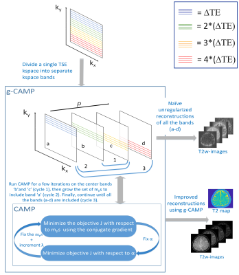 |
Quantitative T2 mapping from a single-contrast TSE scan using g-CAMP
Nahla M H Elsaid1, Nadine L Dispenza2, R Todd Constable1,3, Hemant D Tagare1,2, and Gigi Galiana1
1Radiology and Biomedical Imaging, Yale School of Medicine, New Haven, CT, United States, 2Department of Biomedical Engineering, Yale University, New Haven, CT, United States, 3Neurosurgery, Yale University, New Haven, CT, United States
This work presents quantitative T2-maps and a full T2w-image series generated from an ordinary single contrast T2w-dataset using the growing Constrained Alternating Minimization for Parameter mapping (g-CAMP) reconstruction method. Simulated data were used to study the accuracy of this approach under various echo spacings and with added noise, and the method is also demonstrated in an experimental T2w-dataset. This could ultimately lead to retrospective parameter mapping using data from standard single-contrast acquisitions.
|
|||
2606.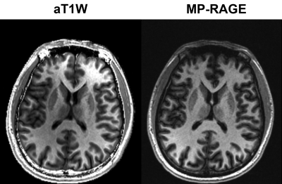 |
Augmented T1 weighted (aT1W) contrast imaging
Yongquan Ye1, Jingyuan Lv1, Yichen Hu1, Zhongqi Zhang2, Jian Xu1, and Weiguo Zhang1
1UIH America, Inc., Houston, TX, United States, 2United Imaging Healthcare, Shanghai, China
In this work, we propose a robust and effective method of an accentuated T1 weighted (aT1W) method. By dividing T1W images by proton density weighted (PDW) images, both obtained using GRE but with different flip angles, the resultant aT1W images offer improved level of T1 contrast. By adopting the multi-dimensional integration (MDI) technique, aT1W images have high signal-to-noise ratio (SNR) despite the use of division operation.
|
|||
2607.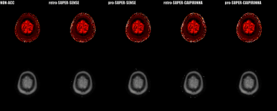 |
Accelerating 3D variable-flip-angle T1 mapping: a prospective study based on SUPER-CAIPIRINHA
Fan Yang1, Jian Zhang2, Guobin Li2, Jiayu Zhu2, Xin Tang1, and Chenxi Hu1
1Institute of Medical Imaging Technology, School of Biomedical Engineering, Shanghai Jiao Tong University, Shanghai, China, 2United Imaging Healthcare Co., Ltd, Shanghai, China
Three-dimensional variable-flip-angle (VFA) T1 mapping is a valuable T1 quantification method subject to a long scan time due to acquisition of multiple 3D images. SUPER (Shift Undersampling improves Parametric-mapping Efficiency and Resolution) is a recently developed method providing fast blockwise reconstruction for accelerated parametric mapping. Here we develop a combination of SUPER and 3D CAIPIRINHA to achieve 5-fold acceleration with 5 flip angles, validated with both retrospective and prospective reconstruction. The results suggest that the proposed method is highly accurate for accelerating 3D VFA T1 mapping, reducing the whole-brain scan time from 6 to 1.5 minutes.
|
|||
2608.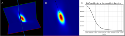 |
Discriminative Ensemble Average Propagator radial profiles along fixels of the centrum semiovale
Gabrielle Grenier1, François Rheault1,2, and Maxime Descoteaux1
1Sherbrooke Connectivity Imaging Lab (SCIL), Université de Sherbrooke, Sherbrooke, QC, Canada, 2Electrical Engineering, Vanderbilt University, Nashville, TN, United States
The Ensemble Average Propagator (EAP) encapsulates the angular and radial information of the diffusion magnetic resonance imaging (MRI) process and can highlight complex characteristics of the underlying tissue microstructure. Here, by sampling EAP values along different radii of the 3-way crossing of the centrum semiovale, we discriminate the orientations of the corticospinal tract from the arcuate fasciculus and the corpus callosum. This process finds microstructure signatures of fiber elements (fixels) of interest. Better tractometry as well as better local direction selection in tractography algorithms could be achieved with this information.
|
|||
2609.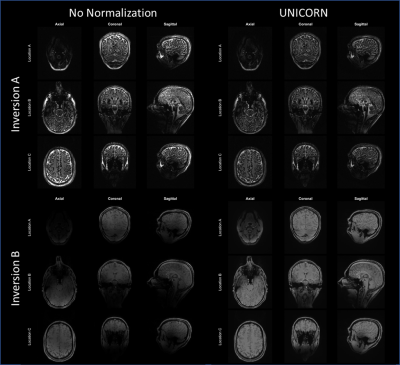 |
Uniform Combined Reconstruction for Improving Receive Intensity Homogeneity of N-dimensional 7T MRI
Venkata Veerendranadh Chebrolu1, Xiaodong Zhong2, Patrick Liebig3, and Robin Heidemann3
1Siemens Medical Solutions USA, Inc., Rochester, MN, United States, 2Siemens Medical Solutions USA, Inc., Los Angeles, CA, United States, 3Siemens Healthcare GmbH, Erlangen, Germany
Uniform combined reconstruction (UNICORN) was recently proposed and used to improve receive uniformity of 7T musculoskeletal and brain MRI data from two-dimensional (2D) sequences. In this work, we extend the UNICORN algorithm to improve receive uniformity of two-, three-, or more-dimensional (N-dimensional or ND) MRI. We also demonstrate UNICORN results using L1-based (computationally efficient compared to singular value decomposition or SVD) optimal combination of the multi-channel data and compare the results with the previously used SVD-based combination.
|
|||
2610.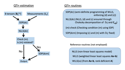 |
QTI+: a constrained estimation framework for q-space trajectory imaging
Magnus Herberthson1, Tom Dela Haije2, Deneb Boito3,4, Aasa Feragen5, Carl-Fredrik Westin6, and Evren Özarslan3,4
1Dept. of Mathematics, Linköping University, Linköping, Sweden, 2Dept.of Computer Science, University of Copenhagen, Copenhagen, Denmark, 3Dept. of Biomedical Engineering, Linköping University, Linköping, Sweden, 4Center for Medical Image Science and Visualization, Linköping University, Linköping, Sweden, 5Department of Applied Mathematics and Computer Science, Technical University of Denmark, Lyngby, Denmark, 6Laboratory for Mathematics in Imaging, Department of Radiology, Brigham and Women’s Hospital, Harvard Medical School, Boston, Boston, MA, United States
Q-space trajectory imaging (QTI) provides a means to estimate the moments for a diffusion tensor distribution (DTD) characterizing the tissue. Commonly employed estimation methods do not typically yield mathematically acceptable estimates, namely that the tensor distribution consists of symmetric positive semidefinite tensors and that the second cumulant represents a covariance. We introduce the QTI+ framework, which utilizes semi-definite programming (SDP) to address this issue. Simulations using a DTD taking the form of a non-central Wishart distribution as well as real data show a marked improvement in the technique's robustness to noise compared to more common (unconstrained) estimation methods.
|
|||
2611.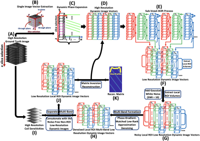 |
Complex-Valued Spatial-Temporal Super-Resolution Combined with Multi-Band Technique on MRI temperature mapping
Duohua Sun1, Jean-Philippe Galons2, Chidi Ugonna1, Silu Han1, and Nan-kuei Chen1
1Biomedical Engineering, The University of Arizona, Tucson, AZ, United States, 2Medical Imaging, The University of Arizona, Tucson, AZ, United States
We present an approach for improving the phase variation representation of complex-valued dynamic MRI temperature mapping. Our technique utilizes phase information to better recover signal loss caused by susceptibility gradients and generate finer representations of dynamic phases variation. Results from numerical and hybrid simulation show that promising improvements in image resolution, susceptibility artifact reduction and phase variation representation can be achieved using our complex-valued super-resolution MRI scheme.
|
|||
2612.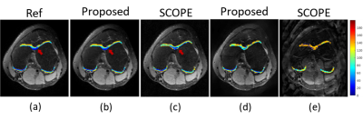 |
Deep Generalization of Signal Compensation for Fast Parameter Mapping in k-Space
Zhuo-Xu Cui1, Yuanyuan Liu2, Qingyong Zhu1, Jing Cheng2, and Dong Liang1,2
1Research center for Medical AI, Shenzhen Institutes of Advanced Technology, Chinese Academy of Sciences, Shenzhen, China, 2Paul C. Lauterbur Research Center for Biomedical Imaging, Shenzhen Institutes of Advanced Technology, Chinese Academy of Sciences, Shenzhen, China
Thanks to powerful clinical applications, magnetic resonance (MR) parameter mapping has received widespread attentions. This work shows that the physical decay about parameter maps can be implicitly absorbed into an annihilation relation from k-space measurement of weighted MR images. Routinely, this relation can be estimated via null-space decomposition of a structured matrix, but, which usually results in computational burden. To alleviate it, we propose to train a convolutional neural network (CNN) to estimate this annihilation relation from undersampled measurement to further realize k-space interpolation. Experiments reveal the effectiveness of the proposed method compared with other competing methods.
|
|||
2613.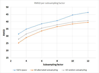 |
Faster and better HARDI using FSE and holistic reconstruction
Maarten Naeyaert1, Vladimir Golkov2, Daniel Cremers2, Jan Sijbers3, and Marleen Verhoye4
1Radiology, Universitair Ziekenhuis Brussel, Brussels, Belgium, 2Department of Computer Science, Technical University of Munich, Garching, Germany, 3Imec-Vision Lab, University of Antwerp, Wilrijk, Belgium, 4Bio-Imaging Lab, University of Antwerp, Wilrijk, Belgium
To accelerate the acquisition of HARDI data, compressed sensing can be used to subsample the data both in k-space and in q-space, using a holistic algorithm for the combined reconstruction. Fast spin echo (FSE) data has fewer deformation artefacts as EPI data, but often requires a multishot acquisition, making subsampling k-space more attractive. In this work FSE data was subsampled retrospectively to investigate different types of subsampling: subsampling q-space only, also using 1D k-space subsampling, or using q-space and alternated 1D k-space subsampling. The results show that for a given subsampling factor the alternated 1D k-space subsampling strategy performs best.
|
|||
2614.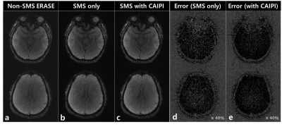 |
Simultaneous multi-slice 3D Spatiotemporal Encoding (SPEN) Imaging: Emulation study
Jaeyong Yu1,2, Sugil Kim3, Jae-Kyun Ryu4, and Jang-Yeon Park1,2,4
1Department of Biomedical Engineering, Sungkyunkwan University, Suwon, Korea, Republic of, 2Department of Intelligent Precision Healthcare Convergence, Sungkyunkwan University, Suwon, Korea, Republic of, 3Siemens Healthineers, Seoul, Korea, Republic of, 4Biomedical Institute for Convergence at SKKU, Sungkyunkwan University, Suwon, Korea, Republic of
Acceleration imaging techniques such as parallel imaging and simultaneous multi-slice (SMS) imaging have been developed to achieve shorter acquisition time and larger FOV and incorporated into various imaging techniques including ultrafast imaging techniques such as echo-planar imaging (EPI). Here, we propose a new way to accelerate 3D spatiotemporal encoding (SPEN) imaging by combining SMS with controlled aliasing for parallel imaging results in higher acceleration (CAIPIRINHA) and split slice generalized auto-calibrating partially parallel acquisitions (Split Slice-GRAPPA).
|
|||
2615.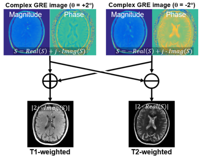 |
Simultaneous T1- and T2-weighted imaging using RF phase modulated gradient echo imaging
Daiki Tamada1 and Scott B. Reeder1,2,3,4,5
1Radiology, University of Wisconsin-Madison, Madison, WI, United States, 2Medical Physics, University of Wisconsin-Madison, Madison, WI, United States, 3Biomedical Engineering, University of Wisconsin-Madison, Madison, WI, United States, 4Medicine, University of Wisconsin-Madison, Madison, WI, United States, 5Emergency Medicine, University of Wisconsin-Madison, Madison, WI, United States
A novel method for simultaneous T1 and T2-weighted imaging using RF phase-modulated GRE with small RF phase increments is presented. Configuration theory approach was used to derive an equation for the steady-state GRE signal. The equation reveals that small RF phase increments provide separable T1 and T2-weighted contrast in the real and imaginary components of the signal. Simulation and phantom studies were performed for quantitative analysis of the proposed method. Brain in-vivo imaging was included to show clinical feasibility. The results suggested the proposed method enables faster imaging compared to conventional FSE imaging.
|
The International Society for Magnetic Resonance in Medicine is accredited by the Accreditation Council for Continuing Medical Education to provide continuing medical education for physicians.