Digital Posters
Signal Representations in Acquisition & Reconstruction
ISMRM & SMRT Annual Meeting • 15-20 May 2021

Digital Posters
Signal Representations in Acquisition & Reconstruction
| Concurrent 1 | 13:00 - 14:00 |
2616.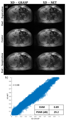 |
Fast Deep Learning Motion-Resolved Golden-Angle Radial MRI Reconstruction
Ramin Jafari1, Richard K G Do2, Yousef Mazaheri Tehrani1,2, Ty Cashen3, Sagar Mandava3, Maggie Fung3, Ersin Bayram3, and Ricardo Otazo1,2
1Medical Physics, Memorial Sloan Kettering Cancer Center, New York, NY, United States, 2Radiology, Memorial Sloan Kettering Cancer Center, New York, NY, United States, 3GE Healthcare, Waukesha, WI, United States
To use deep learning to reconstruct motion-resolved dynamic images from multicoil undersampled radial data without image quality degradation and 800-fold reduction in reconstruction time compared to the iterative XD-GRASP algorithm.
|
|||
2617.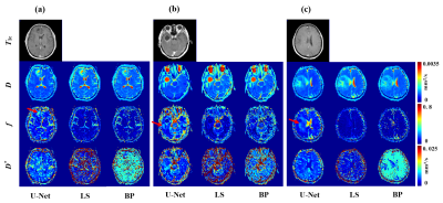 |
High Efficient Reconstruction Method for IVIM Imaging Based on Deep Neural Network and Synthetic Training Data and its Application in IVIM-DKI
Lu Wang1, Zhen Xing2, Jian Wu1, Qinqin Yang1, Congbo Cai1, Shuhui Cai 1, Zhong Chen1, and Dairong Cao2
1Department of Electronic Science, Xiamen University, Xiamen, Fujian, China, 2Department of Radiology, First Affiliated Hospital of Fujian Medical University, Fuzhou, Fujian, China
Intravoxel incoherent motion (IVIM) imaging is a non-invasive MR perfusion imaging that could prevent patients from the harm of exogenous reagent. Previous studies proved that the least square and Bayesian approaches are so far the best algorithms in IVIM fitting. However, they still suffer from time-consuming and high noise level. We proposed a deep neural network-based reconstruction method with synthetic training data for IVIM imaging and extended it to hybrid IVIM-DKI (diffusion kurtosis imaging) model fitting. Experimental results show that our method owns prominent performance in both image quality and accuracy of fitting results with a remarkably short reconstruction time.
|
|||
2618.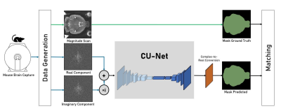 |
CU-Net: A Completely Complex U-Net for MR k-space Signal Processing
Dipika Sikka1,2, Noah Igra3,4, Sabrina Gjerswold-Sellec1, Cynthia Gao5, Ed Wu6, and Jia Guo7
1Department of Biomedical Engineering, Columbia University, New York, NY, United States, 2VantAI, New York, NY, United States, 3Department of Applied Mathematics, Columbia University, New York, NY, United States, 4Sackler School of Medicine, Tel Aviv University, Tel Aviv, Israel, 5Department of Computer Science, Columbia University, New York, NY, United States, 6Department of Electrical and Electronic Engineering, The University of Hong Kong, Pok Fu Lam, Hong Kong, China, 7Department of Psychiatry, Mortimer B. Zuckerman Mind Brain Behavior Institute, Columbia University, New York, NY, United States
While the application of deep learning in MR image analysis has gained significant popularity, using raw MR k-space data as part of deep learning analysis is an underexplored area. Here we develop a completely complex U-Net deep learning architecture, CU-Net, where we apply deep learning components and operations in the complex space. CU-Net leverages k-space MR signals while training a U-Net with Attention and Residual components, as opposed to using processed spatial (real) data, typically seen with MRI deep learning applications. As part of a proof-of-concept study, the complex networks demonstrated their utility and potential superiority over their spatial counterparts.
|
|||
2619.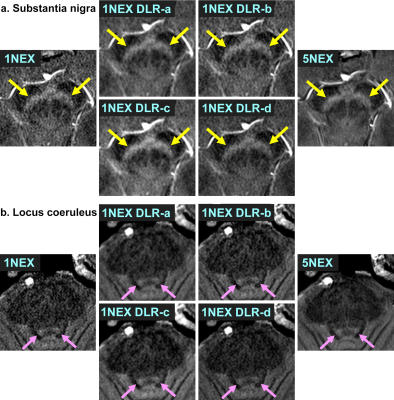 |
Neuromelanin-sensitive MRI using deep learning reconstruction (DLR) denoising: comparison of DLR patterns
Sonoko Oshima1, Yasutaka Fushimi1, Satoshi Nakajima1, Akihiko Sakata1, Takuya Hinoda1, Sayo Otani1, Krishna Pandu Wicaksono1, Hiroshi Tagawa1, Yang Wang1, Yuichiro Sano2, Rimika Imai2, Masahito Nambu2, Koji Fujimoto3, Hitomi
Numamoto4, Kanae Kawai Miyake4, Tsuneo Saga4, and Yuji Nakamoto1
1Department of Diagnostic Radiology and Nuclear Medicine, Graduate School of Medicine, Kyoto University, Kyoto, Japan, 2Canon Medical Systems Corporation, Otawara, Japan, 3Department of Real World Data Research and Development, Graduate School of Medicine, Kyoto University, Kyoto, Japan, 4Department of Advanced Medical Imaging Research, Graduate School of Medicine, Kyoto University, Kyoto, Japan
We applied four patterns of deep learning reconstruction (DLR) denoising methods to 1 NEX neuromelanin-sensitive MR images. DLR with denoising intensity coefficient of 1.0 and edge enhancement off provided the best image quality among the four types of DLR, and it was significantly better than or as good as 5 NEX images. ROC analyses using images with all DLR patterns showed good AUCs for diagnosis of Parkinson’s disease. This DLR denoising method can improve image quality of neuromelanin-sensitive MRI with good diagnostic ability to differentiate patients with Parkinson’s disease from healthy controls.
|
|||
2620.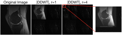 |
Utilizing the Wavelet Transform's Structure in Compressed Sensing
Nicholas Dwork1, Daniel O'Connor2, Corey A. Baron3, Ethan M. I. Johnson4, John M. Pauly5, and Peder E.Z. Larson6
1Radiology and Biomedical Imaging, UCSF, San Francisco, CA, United States, 2Mathematics and Statistics, University of San Francisco, San Francisco, CA, United States, 3Robarts Research, Western University, London, ON, Canada, 4Biomedical Engineering, Northwestern University, Evanston, IL, United States, 5Electrical Engineering, Stanford University, Stanford, CA, United States, 6Radiology and Biomedical Imaging, University of California in San Francisco, San Francisco, CA, United States
In this work, we present a modification of the standard implementation of compressed sensing that takes advantage of the structure of the Daubechies wavelet transform. By doing so, we show that we retain additional detail in the reconstructed images when few data samples are used.
|
|||
2621.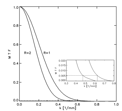 |
Application of compressed sensing in High Spectral and Spatial resolution (HiSS) MRI – evaluation of effective resolution
Milica Medved1, Marco Vicari2, and Gregory S Karczmar1
1Department of Radiology, The University of Chicago, Chicago, IL, United States, 2Fraunhofer MEVIS, Bremen, Germany
Compressed sensing (CS) was evaluated as an acceleration technique for high spectral and spatial resolution (HiSS) MRI, at acceleration factors up to R=10. Effective spatial resolution was maintained in the readout direction, and decreased with R in the phase encoding direction, although acceleration factors of up to R = 4 are realistic. Noise amplification was not observed. CS could improve diagnostic utility of HiSS MRI in breast by allowing longer echo trains and thus heavier T2* weighting in a fewer number of k-space lines. CS could also facilitate use of HiSS MRI in geometrically constrained applications, such as prostate MRI.
|
|||
2622.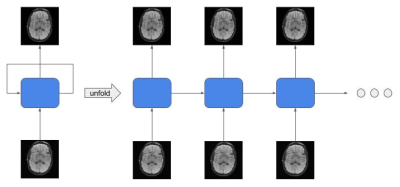 |
Variational Feedback Network for Accelerated MRI Reconstruction
Pak Lun Kevin Ding1, Riti Paul1, Baoxin Li1, Ameet C. Patel2, and Yuxiang Zhou2
1CIDSE, Arizona State University, Tempe, AZ, United States, 2RADIOLOGY, Mayo Clinic College of Medicine, Tempe, AZ, United States
Conventional Magnetic Resonance Imaging (MRI) is a prolonged procedure. Therefore, it’s beneficial to reduce scan time as it improves patient experience and reduces scanning cost. While many approaches have been proposed for obtaining high quality reconstruction images using under-sampled k-space data, deep learning has started to show promising results when compared with conventional methods. In this paper, we propose a Variational Feedback Network (VFN) for accelerated MRI reconstruction. Specifically, we extend the previously proposed variational network with recurrent neural network (RNN). Quantitative and qualitative evaluations demonstrate that our proposed model performs superiorly against other compared methods on MRI reconstruction.
|
|||
2623.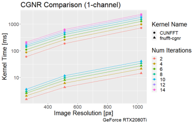 |
Ultrafast Non-uniform Fast Fourier Transform for real-time radial acquisitions.
Falk Christian Mayer1,2, Peter Bachert1,2, Mark E Ladd1,2,3, and Benjamin Knowles1
1Medical Physics in Radiology, German Cancer Research Center (DKFZ), Heidelberg, Germany, 2Department of Physics and Astronomy, Heidelberg University, Heidelberg, Germany, 3Faculty of Medicine, Heidelberg University, Heidelberg, Germany
A GPU-based Fast non-Uniform Fast Fourier Transform (NUFFT) was implemented, which uses optimized libraries and algorithms such as stencils and managed memory to perform highly efficient transformations from the image to the k-space domain and vice-versa. In testing, the transform execution time was measured to be approximately 1ms for 32 channel data and a 256x256 grid size. A conjugate gradient based solution to the inverse NUFFT was also implemented, in which a solution was given within approximately 10ms. The proposed implementation has valuable applications for non-Cartesian real-time imaging.
|
|||
2624.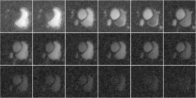 |
Low Rank Plus Joint Sparse Reconstruction for Hyperpolarized MRI
Nicholas Dwork1
1Radiology and Biomedical Imaging, UCSF, San Francisco, CA, United States
We present a model based reconstruction algorithm with low-rank and joint sparse regularization terms for use with hyperpolarized MRI. Results show improved details.
|
|||
2625.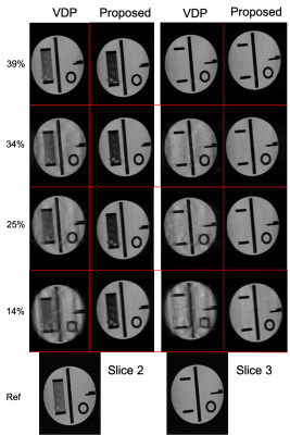 |
Optimised sampling for low-dimensional compressed sensing
Joshua Michael McAteer1, Olivier Mougin1, James Harkin2, Paul Glover1, and Penny Gowland1
1Physics, University of Nottingham, Nottingham, United Kingdom, 2Medicine, University of Nottingham, Nottingham, United Kingdom
Optimising compressed sensing sampling can yield a significant increase in measured and observed image quality over heuristic sampling methods. Using an example image, such as a previous acquired scan of the same anatomy, a bespoke sampling pattern can be designed that optimally samples the data. This increase in image quality allows for greater acceleration or better SNR with the same imaging time over standard methods such as variable density Poisson disc sampling. This has been tested on a 0.5T upright MR scanner in phantoms.
|
|||
2626.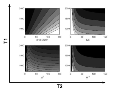 |
Phase-cycled balanced TFE disentangled using configuration states: Multi-purpose imaging for the MRI-Linac workflow
Astrid van Lier1, Yulia Shcherbakova1, and Cornelis van den Berg1
1UMC Utrecht, Utrecht, Netherlands Phase cycled bGRE images disentangled into configuration modes can be used to generate alternative signal contrasts for for example for radiotherapy delineation purposes while simultaneously geometrical errors due to B0 inhomogeneity can be quantified. Numerical simulations show the change in modulation for different configurations states. In vivo experiments on the male pelvis are used to illustrate the changed image contrast and obtained B0 map against the reference method. |
|||
2627.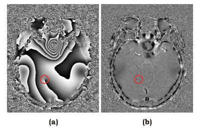 |
Background Correction with Phase Diffusor (BACOPSOR) for Susceptibility Weighted Imaging
Qing-San Xiang1,2
1Radiology, University of British Columbia, Vancouver, BC, Canada, 2Physics and Astronomy, University of British Columbia, Vancouver, BC, Canada
Susceptibility Weighted Imaging (SWI) has many applications. One crucial step of SWI is background phase error removal that typically involves smoothing either before or after phase unwrapping. Both approaches may have to face difficulties when large isolated phase loops (around poles or singularities) are present in the image. In this work, background correction with phase diffusor (BACOPSOR) is introduced. It is straightforward to implement and has a desirable immunity to phase loops. Application of BACOPSOR to SWI has been demonstrated with in vivo data.
|
|||
2628.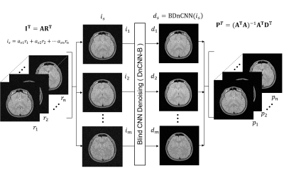 |
Parallelized Blind MR Image Denoising using Deep Convolutional Neural Network
satoshi ITO1, taro SUGAI1, kohei TAKANO1, and shohei OUCHI1
1Utsunomiya University, Utsunomiya, Japan
To improve the denoising performance of a convolutional neural network (CNN), a parallelized blind image denoising (ParBID) was proposed and demonstrated. ParBID procedure is similar to SENSE technique, 1) linear combination of adjacent 2D sliced noisy images, 2) blind noise level CNN denoising, and 3) separation of linearly combined and denoised images by solving linear equation. Experimental studies showed that the PSNR and the SSIM were improved for all noise levels, from 2.5% to 7.5%. ParBID showed that the greatest PSNR improvements were obtained when three slice images were used for linear image combination.
|
|||
2629.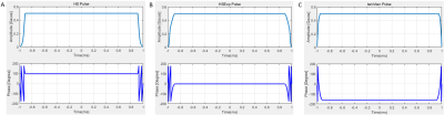 |
The optimization of three adiabatic pulses with constant amplitude spin-lock
Yuxin Yang1, Xi Xu1, Yuanyuan Liu1, Yanjie Zhu1,2, Dong Liang1,2,3, and Hairong Zheng1,2
1Shenzhen Institute of Advanced Technology,Chinese Academy of Sciences, Shenzhen, China, 2Shenzhen College of Advanced Technology, University of Chinese Academy of Sciences, Shenzhen, China, Shenzhen, China, 3Research Centre for Medical AI, Shenzhen Institutes of Advanced Technology, Chinese Academy of Science, Shenzhen, China, Shenzhen, China
An optimization aimed at shortening pulse durations was carried out for three types of adiabatic spin-lock pulses by means of Bloch simulation. The variance of a part of the trajectory of Mz with respect to a range of off-resonance values was calculated to find the optimal pulse parameters and decent T1ρ-weighted imaging and T1ρ mapping results were obtained.
|
|||
2630.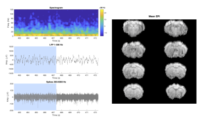 |
A Singular Value Shrinkage Approach to Remove Artifacts from Neuro-electrophysiology Data Recorded During fMRI at 16.4T
Corey Edward Cruttenden1, Wei Zhu1, Yi Zhang1, Xiao-Hong Zhu1, Rajesh Rajamani2, and Wei Chen1
1Center for Magnetic Resonance Research, University of Minnesota, Minneapolis, MN, United States, 2Mechanical Engineering, University of Minnesota, Minneapolis, MN, United States
Acquiring neuro-electrophysiology signal simultaneously with fMRI is hindered by electromagnetic field interactions that generate artifacts, including fMRI gradient induced artifacts in the neuro-electrophysiology data. This abstract presents a novel method using a separation boundary on the singular value decomposition of the first difference of artifact-contaminated data to accurately reconstruct clean neural signals. The separation boundary can be estimated from a brief baseline recording period followed by simultaneous fMRI and neuro-electrophysiological data acquisition. The method is successfully demonstrated on neural recording data acquired simultaneously with time-series echo planar imaging at 16.4T.
|
|||
2631.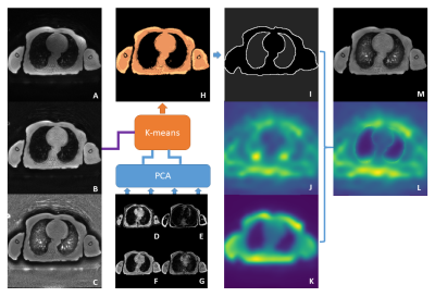 |
Hybrid bias correction of thoracic zero echo time (ZTE) images
Chang Sun1, Roido Manavaki1, Jason Tarkin2, Christopher Wall2, James HF Rudd2, Fiona J Gilbert1, and Martin J Graves1
1Department of Radiology, University of Cambridge, Cambridge, United Kingdom, 2Division of Cardiovascular Medicine, University of Cambridge, Cambridge, United Kingdom
Bias correction in the thoracic region is challenging due to the low proton density in the lung. Traditional retrospective bias correction techniques, such as surface fitting method and the histogram-based method, suffer from over suppression in the lung regions or increased noise in the tissue. We propose a hybrid bias correction method that combines the advantages of the surface fitting and the histogram-based methods. The hybrid method normalized the signal intensity in lung and reduced the signal variation in tissue.
|
|||
2632.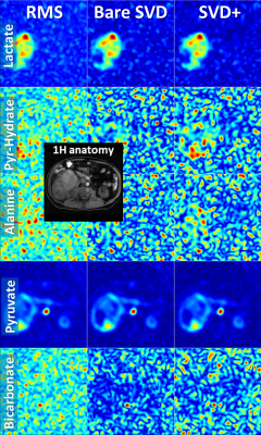 |
SVD-Based Multi-Channel-Receive-Coil Combination for 13C Metabolic Imaging
Rolf F Schulte1, Mary A McLean2, Joshua D Kaggie2, Stephan Ursprung2, Ramona Woitek2, Ferdia A Gallagher2, Esben S S Hansen3, Nikolaj Bogh3, and Christoffer Laustsen3
1GE Healthcare, Munich, Germany, 2Department of Radiology, University of Cambridge, Cambridge, United Kingdom, 3MR Research Centre, University of Aarhus, Aarhus, Denmark
Multi-channel receive coils can improve coverage for metabolic imaging with hyperpolarised 13C compounds. Combining different receive channels in an SNR-optimal way is challenging due to the difficulties in determining sensitivity maps. The main aim of this work was to implement and optimise a Singular-Value-Decomposition (SVD) based sensitivity map extraction from metabolic images with a single spectral point per metabolite and to investigate its performance in SNR-limited metabolic imaging experiments.
|
|||
2633.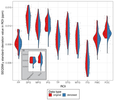 |
Linked Independent Component Analysis for Denoising multi-centre 7T MRI data
Catarina Rua1,2, Alberto Llera3,4, Olivier Mougin5, Mauro Costagli6, Renat Yakupov7, Richard Bowtell5, James B Rowe1,8, and Christopher T Rodgers9,10
1Department of Clinical Neurosciences and University of Cambridge Centre for Parkinson-plus, University of Cambridge, Cambridge, United Kingdom, 2Wolfson Brain Imaging Centre, University of Cambridge, cambridge, United Kingdom, 3Cognitive Neuroscience, Radboud University Medical Centre, Nijemen, Netherlands, 4Donders Institute, Centre for Cognitive Neuroimaging, Radboud University, Nijmegen, Netherlands, 5Sir Peter Mansfield Imaging Centre, School of Physics and Astronomy, University of Nottingham, Nottingham, United Kingdom, 6IMAGO7 Foundation, Pisa, Italy, 7German Centre for Neurodegenerative Diseases (DZNE), Magdeburg, Germany, 8Medical Research Council Cognition and Brain Sciences Unit, University of Cambridge, Cambridge, United Kingdom, 9Wolfson Brain Imaging Centre, University of Cambridge, Cambridge, United Kingdom, 10Department of Clinical Neurosciences, University of Cambridge, Cambridge, United Kingdom
Multi-site studies are an attractive option at ultra-high field (7T) as it is possible to pool larger number of datasets of healthy and patients increasing the statistical power of neuroimaging studies. However, differences on scanner hardware and software increase variability in the measurements obtained on across imaging sites.In this study we have piloted the application of a multimodal ICA approach for denoising scanner effects across two different 7T MRI scanner platforms.
|
The International Society for Magnetic Resonance in Medicine is accredited by the Accreditation Council for Continuing Medical Education to provide continuing medical education for physicians.