Digital Posters
Modelling Signals Throughout the Body
ISMRM & SMRT Annual Meeting • 15-20 May 2021

Digital Posters
Modelling Signals Throughout the Body
| Concurrent 1 | 15:00 - 16:00 |
2850.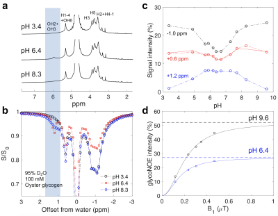 |
Mechanism and quantitative assessment of saturation transfer for water-based detection of the aliphatic protons in carbohydrate polymers
Yang Zhou1, Peter van Zijl2,3, Jiadi Xu2,3, and Nirbhay N. Yadav2,3
1Institute of Biomedical and Health Engineering, Shenzhen Institutes of Advanced Technology, Shenzhen, China, 2The Russell H. Morgan Department of Radiology, The Johns Hopkins University School of Medicine, Baltimore, MD, United States, 3F.M. Kirby Research Center for Functional Brain Imaging, Kennedy Krieger Institute, Baltimore, MD, United States
A three-pool relayed NOE (rNOE) model and its theoretical solution is formulated to describe the rNOE-based signal in CEST experiments and the analytical solution for rNOE transfer presented. We show that experimental data for glycogen solutions in D2O and H2O could be analyzed successfully using this analytical theory, which was further validated using numerical simulations with the Bloch equations. The study increases the understanding of the aliphatic components of Z-spectra of sugar polymers as well as of other macromolecules such as proteins and lipids.
|
|||
2851.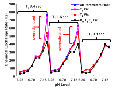 |
Improving the Bloch Fitting Method for the Analysis of acidoCEST MRI
Tianzhe Li1, Aikaterini Kotrotsou1, Shu Zhang1, Kyle Jones1, and Mark Pagel1
1Cancer Systems Imaging, UT MD Anderson Cancer Center, Houston, TX, United States
With the expansion of clinical CEST MRI, the analysis methods for Z-spectra need further validation and optimization. Fitting CEST spectra with the modified Bloch-McConnell equations provides the gold standard for chemical exchange rate (kex) quantification, but the current direct-fitting process requires fitting eight parameters. In this study, we have optimized the Bloch-fitting process and incorporated experimentally measured information. We tested the performance of the optimized algorithm using iopamidol phantoms of various pH levels and discovered that including experimentally determined T1, T2 and B0 information can increase the accuracy of the kex fitting results.
|
|||
| 2852. | Time resynchronization of data obtained during cardiovascular MRI combined with catheterization using biophysical cardiac modeling
Maria Gusseva1,2, Daniel Alexander Castellanos3, Mohamed Abdelghafar Hussein4,5, Joshua Greer 4, Gerald Greil4, Surendranath Veeram Reddy4, Dominique Chapelle1,2, Tarique Hussain4, and Radomir Chabiniok1,2,4,6
1Inria, Palaiseau, France, 2LMS, Ecole Polytechnique, Palaiseau, France, 3Department of Cardiology, Boston Children’s Hospital, Boston, MA, United States, 4Division of Pediatric Cardiology, UT Southwestern Medical Center Dallas, Dallas, TX, United States, 5Pediatric department, Kafrelsheikh University, Kafr Elsheikh, Egypt, 6Department of Mathematics, Czech Technical University in Prague, Faculty of Nuclear Sciences and Physical Engineering, Prague, Czech Republic
Time resynchronization of data is often needed when the analysis is based on combining several cardiovascular MRI (CMR) sequences and/or when CMR is combined with a different modality such as pressure catheter during interventional CMR (iCMR). In the present work we propose using patient-specific biophysical model to resynchronize the intraventricular pressure and volume data. The model-driven resynchronization strategy generates pressure-volume (P-V) loops, which can be then used for clinical interpretation. Moreover, the patient-specific model provides additional mechanical indicators of patient's physiology. This framework has the perspective to contribute to planning of complex surgical interventions.
|
|||
2853.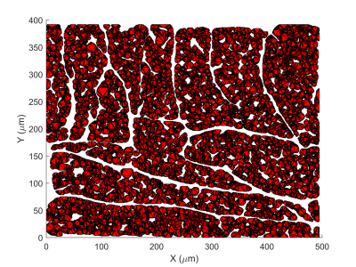 |
Realistic diffusion tensor cardiovascular magnetic resonance simulations in a histology-based substrate: The effect of membrane permeability
Jan N Rose1, Ignasi Alemany1, Andrew D Scott2,3, and Denis J Doorly1
1Department of Aeronautics, Imperial College London, London, United Kingdom, 2Cardiovascular Magnetic Resonance unit, Royal Brompton Hospital, London, United Kingdom, 3National Heart and Lung Institute, Imperial College London, London, United Kingdom
Computational simulations and specifically random walks provide a unique opportunity to investigate how several confounding factors might affect the diffusion tensor cardiovascular magnetic resonance (DT-CMR) parameters. We investigate the effect of membrane permeability and diffusion time on the fractional anisotropy (FA) and mean diffusivity in a realistic histology-based 3D substrate considering two common sequence types. Comparing the results from permeable with that from impermeable membranes helps to considerably reduce FA towards levels expected from in-vivo DT-CMR. We further reveal that FA is unaffected by changes in diffusion times above 500ms. A similar, albeit weaker effect is observed regarding MD.
|
|||
2854.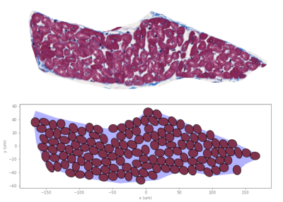 |
Packing hierarchical structures in myocardial tissue to synthesise a realistic substrate
Jan N Rose1, Andrew D Scott2,3, and Denis J Doorly1
1Department of Aeronautics, Imperial College London, London, United Kingdom, 2Cardiovascular Magnetic Resonance unit, Royal Brompton Hospital, London, United Kingdom, 3National Heart and Lung Institute, Imperial College London, London, United Kingdom
In order to facilitate parameter studies of diffusion tensor cardiovascular magnetic resonance through simulations, there is a need to synthesise realistic microstructural substrates. We developed an algorithm to pack arbitrary shapes (polygons) into an arbitrarily shaped target region. We pack cardiomyocytes into the myocardial cell groupings known as sheetlets. In turn, these sheetlets are packed into a voxel. Our method produces a substrate that is comparable to histology in both extra-cellular volume fraction and distribution of extra-cellular space. Future work aims to expand capabilities and extend packing from 2D to 3D.
|
|||
2855.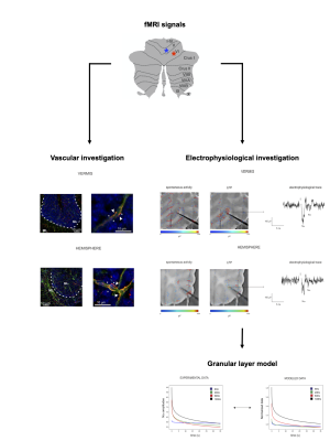 |
Neurovascular coupling in the cerebellum: reconstructing the neurophysiological basis of different cerebellar fMRI responses.
Anita Monteverdi1,2, Giuseppe Gagliano2, Stefano Casali2, Fulvia Palesi1,2, Claudia AM Gandini Wheeler-Kingshott 1,2,3, Lisa Mapelli2, and Egidio D'Angelo1,2
1Brain Connectivity Center Research Department, IRCCS Mondino Foundation, Pavia, Italy, 2Brain and Behavioral Sciences, University of Pavia, Pavia, Italy, 3Queen Square MS Centre, Department of Neuroinflammation, UCL Queen Square Institute of Neurology, Faculty of Brain Sciences, University College London, London, United Kingdom
Neuroimaging studies in humans showed different patterns of BOLD responses in cerebellar vermis and cortical regions when performing tasks at different grip forces. In this study we investigated the basis of different cerebellar fMRI responses ex-vivo. Capillary dilation and neuronal activity were recorded in the vermis and hemisphere of mouse cerebellar slices, highlighting a region and frequency dependency of neurovascular responses in the granular layer. The correlation between neuronal activity and vessel diameter changes was explored through a computational model, which supported a central role played by the NMDAR-NO pathway in shaping the vasodilation time course.
|
|||
2856.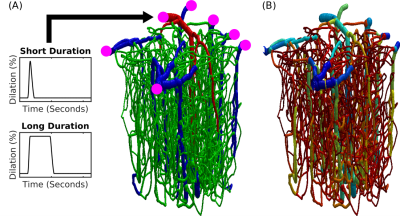 |
Simulations of the BOLD Non-Linearity Based on a Viscoelastic Model for Capillary and Vein Compliance
Joerg Peter Pfannmoeller1, Grant Addison Hartung1, Xiaojun Cheng2, Avery Berman1, David Boas2, and Jonathan Rizzo Polimeni1
1Athinoula A. Martinos Center for Biomedical Imaging, Massachusetts General Hospital, Charlestown, MA, United States, 2Neurophotonics Center, Boston University, Boston, MA, United States
The stimulus duration depended nonlinearity of the BOLD response is difficult to model, as it often requires stimulus-specific parameter values. Here we have extended an existing framework of BOLD response modeling based on biophysical simulations of realistic microvascular anatomy and dynamics to incorporate viscoelastic properties of individual blood vessels and tissue. We then applied this model to examine BOLD response nonlinearities to long-duration stimuli. We find that this model largely captures the differences in BOLD responses observed to short- and long-duration stimuli.
|
|||
2857.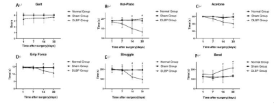 |
Established a rat model of discogenic low back pain for evaluating the paravertebral muscle functional magnetic resonance changes
Luo Bao fa1, Huang Yi long1, Yang Kai wen1, Nie Li sha2, and He Bo1
1The First Affiliated Hospital of Kunming Medical University, kunming, China, 2GE Healthcare, MR Research China, Beijing, Beijing, China
In this study, a discogenic low back pain (DLBP) model was established by puncture of rat intervertebral disc under the guidance of X-ray, and functional magnetic resonance imaging (fMRI) was performed on paravertebral muscles of DLBP rats to explore the feasibility of the DLBP model and the changes of T2 value and R2*value of paravertebral muscles in the early stage of DLBP. The conclusion is that it is feasible to construct DLBP rat model by X-ray guided puncture of intervertebral disc, and the T2 value changes earlier than R2*in the early stage of DLBP.
|
|||
2858.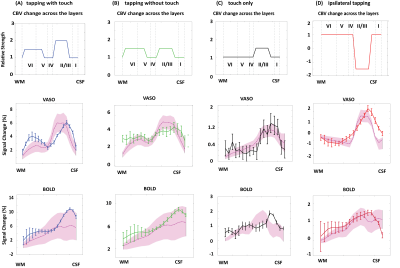 |
Modelling Depth-Dependent VASO and BOLD Signal Changes in Human Primary Motor Cortex
Atena Akbari1, Saskia Bollmann1, and Markus Barth1,2,3
1Centre for Advanced Imaging, The University of Queensland, Brisbane, Australia, 2School of Information Technology and Electrical Engineering, Brisbane, Australia, 3ARC Training Centre for Innovation in Biomedical Imaging Technology, Brisbane, Australia
Depth-dependent high resolution fMRI studies in animals and humans hold promise to reveal the input-output and feedforward-feedback connections across cortical layers. However, our understanding of the underling physiology is limited. In this work, using a cortical vascular model we simulated BOLD and VASO signal changes in human primary motor cortex.
|
|||
2859.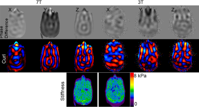 |
A Comparison on the Estimated Stiffness and Signal-to-Noise Ratio of Magnetic Resonance Elastography Images Acquired at 3T and 7T
Yuan Le1, Andrew J. Fagan1, Jun Chen1, Eric G. Stinson2, Joel P. Felmlee1, Matthew C. Murphy1, Kevin J. Glaser1, Arvin Arani1, Phillip J. Rossman1, Stephan Kannengiesser3, Bradley D. Bolster, Jr.2, John Huston, III1, and Richard L. Ehman1
1Radiology, Mayo Clinic, Rochester, MN, United States, 2Siemens Medical Solutions USA, Inc., Malvern, PA, United States, 3Siemens Healthcare GmbH, Erlangen, Germany
This study aimed to compare MRE image quality and stiffness measurements performed at 3T and 7T. It was found that in PVC phantom images the estimated stiffness values were very close between images acquired at 7T and 3T; the signal-to-noise ratio (SNR) at 7T was much higher than that at 3T, and the octahedral shear strain based SNR more than doubled at 7T. These results indicate potential of obtaining high resolution MRE images without affecting the stiffness measurement at 7T.
|
|||
2860.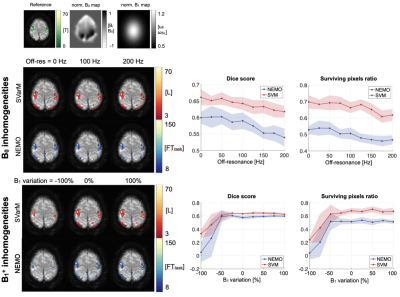 |
Towards direct neuronal-current MRI: a novel statistical processing technique for measurements in the presence of system imperfections.
Chiara Coletti1, Sebastian Domsch1, Frans Vos1, and Sebastian Weingärtner1
1Imaging Physics, TU Delft, Delft, Netherlands
Functional imaging based on detection of neuronal currents using spin-lock prepared MRI may overcome limitations inherent to haemodynamic fMRI with BOLD. However, in-vivo application is hindered by system imperfections. In this work, the effects of B0 and B1+ inhomogeneities and background noise on neuro-current MRI signals are quantified. Furthermore, a new statistical data-processing technique based on the analysis of signal variability, SVarM, is proposed for neuro-current MRI time series. SVarM achieves overall higher sensitivity than existing data-processing methods and is shown to be more robust in the presence of system imperfections.
|
|||
2861.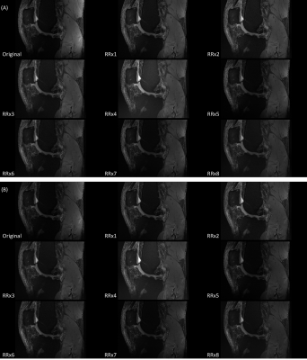 |
UNIform COmbined RecoNstruction (UNICORN) for 7T Clinical Fat-Suppressed TSE Imaging of the Human Knee
Xiaowei Zou1, Venkata V Chebrolu2, and Nakul Gupta3
1Siemens Medical Solutions USA Inc., Houston, TX, United States, 2Siemens Medical Solutions USA Inc., Rochester, MN, United States, 3Department of Radiology, Houston Methodist Research Institute, Houston, TX, United States
Fat-suppressed turbo-spin-echo (TSE) imaging is very important to visualize knee pathology such as cartilage defects in clinical routine. Ultra-high-field 7T magnetic resonance imaging (MRI) provides higher signal-to-noise-ratio (SNR) and contrast-to-noise-ratio (CNR) than 3T and 1.5T MRI, enabling better visualization of fine anatomical structures and physiological effects. However, imaging at 7T has higher receive and transmit non-uniformity that may degrade its clinical value. Recently, a UNIform COmbined RecoNstruction (UNICORN) algorithm was proposed for reducing the receive non-uniformity. The purpose of this preliminary work is to quantitatively and qualitatively evaluate and optimize UNICORN performance for 7T clinical TSE with fat suppression.
|
|||
2862.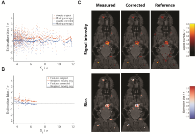 |
Correcting Signal Intensity Bias in 19F MR Imaging of Inflammation by Statistical Modelling
Ludger Starke1, Thoralf Niendorf1, and Sonia Waiczies1
1Berlin Ultrahigh Field Facility (B.U.F.F.), Max Delbrück Center for Molecular Medicine in the Helmholtz Association, Berlin, Germany
Labeling cells with 19F nanoparticles (NPs) continues to elicit interest for non-invasive localization of inflammation and monitoring immune cell therapy. Systematic overestimation in low SNR MRI of 19F-NPs has been previously described which needs to be corrected for valid quantitative conclusions. We develop a statistical model which successfully compensates this bias and demonstrate its efficacy for the correct estimation of signal intensities on neuroinflammation data acquired in a mouse model of multiple sclerosis. The correction only relies on the image data itself and promises to be a valuable contribution to the development of reliable quantitative 19F MRI.
|
|||
2863. |
Automated 3D modeling and analysis of cerebral small vessels with MR angiography at 7 Tesla
Zhixin Li1,2,3, Yue Wu1,2,3, Dongbiao Sun1,2,3, Jing An4, Qingle Kong5, Rong Xue1,2,3, Yan Zhuo1,2,3, and Zihao Zhang1,2,3
1State Key Laboratory of Brain and Cognitive Science, Institute of Biophysics, Chinese Academy of Sciences, Beijing, China, 2The Innovation Center of Excellence on Brain Science, Chinese Academy of Sciences, Beijing, China, 3University of Chinese Academy of Sciences, Beijing, China, 4Siemens Shenzhen Magnetic Resonance Ltd., Shenzhen, China, 5MR Collaboration, Siemens Healthcare Ltd, Beijing, China
Due to the possible interaction between cerebral small vessel disease (CSVD) and a variety of brain diseases or pathological changes, people's interest in small vessel pathology was growing. With the increased imaging resolution at ultra-high field MRI, imaging cerebral small vessels become feasible with TOF-MRA sequence. In this study, we introduced an automated vascular segmentation and tracing method based on deep learning, machine learning and multi filtering. Our method performed well for the multistage branching of cerebral vessels, and could quantitatively evaluate the vasculature. The technique was potentially useful for the clinical studies of CSVD.
|
|||
2864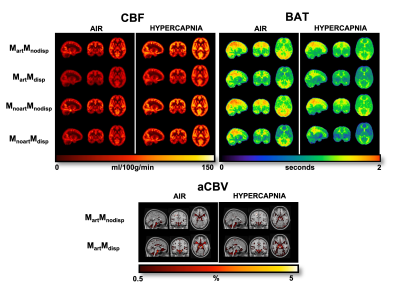 |
Impact of ASL modelling strategies on cerebral blood flow and reactivity assessment Video Permission Withheld
Joana Pinto1, Nicholas P. Blockley2, James W. Harkin3, and Daniel P. Bulte1
1Institute of Biomedical Engineering, Department of Engineering Science, University of Oxford, Oxford, United Kingdom, 2School of Life Sciences, University of Nottingham, Nottingham, United Kingdom, 3Respiratory Medicine Department, School of Medicine, University of Nottingham, Nottingham, United Kingdom
Despite most ASL cerebrovascular reactivity (CVR) studies being based on single-PLD approaches, it has been shown that the vascular spatiotemporal dynamics of cerebral blood flow (CBF) are altered during hypercapnia. This ultimately leads to inaccuracies in some ASL modelling assumptions, compromising CVR assessment. In this work, we test several multiple-PLD ASL modelling strategies and assess their impact on CBF dynamics and CVR assessment. In particular, the individual and combined impact of estimating dispersion effects and the macrovascular signal are evaluated in terms of quantification of several haemodynamic parameters using multiple-PLD ASL data during two different conditions (resting-state and hypercapnia).
|
|||
2865.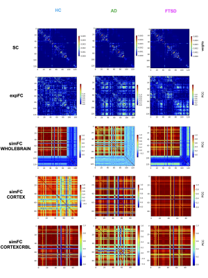 |
Improving the predictive power of The Virtual Brain in healthy and neurodegenerative diseases with cerebro-cerebellar loops integration.
Anita Monteverdi1,2, Fulvia Palesi1,2, Claudia AM Gandini Wheeler-Kingshott 1,2,3, and Egidio D'Angelo1,2
1Brain Connectivity Center Research Department, IRCCS Mondino Foundation, Pavia, Italy, 2Brain and Behavioral Sciences, University of Pavia, Pavia, Italy, 3NMR Research Unit, Queen Square MS Centre, Department of Neuroinflammation, UCL Queen Square Institute of Neurology, Faculty of Brain Sciences, UCL, London, United Kingdom
The Virtual Brain (TVB) has been recently developed to simulate whole-brain dynamics in healthy and pathological conditions, including only cerebral nodes. However, it has been demonstrated that the integration of cerebro-cerebellar connections in TVB can improve its predictive power. Thus, in this study we integrated cerebro-cerebellar connectivity in TVB simulation exploring its impact on prediction of brain dynamics in three different conditions: healthy, Alzheimer’s disease and Frontotemporal Spectrum Disorder. Notably we demonstrated that cerebro-cerebellar circuits integration in the TVB further improve its predictive power, both in healthy and pathological states.
|
|||
2866. |
Hemodynamic simulations reveal changes in ascending venules leads to enhanced venous CBV response to arterial dilation.
Grant Hartung1, Joerg Pfannmoeller1, Avery J. L. Berman1, and Jonathan R. Polimeni1
1Athinoula A. Martinos Center for Biomedical Imaging, Boston, MA, United States
Here we extend a recent approach employing biophysical simulations of realistic cortical microvascular anatomy and dynamics to test whether the topology of the microvasculature impact the BOLD response. We create 3D vascular anatomical networks with varying ratios of arteries to venules and simulated the effects on cerebral blood flow and volume responses. We observed a difference in the cerebral blood volume response after we varied the ratio of diving arteries to ascending veins.
|
|||
2867.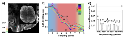 |
Pre-processing of high-resolution gradient-echo images for laminar fMRI applications
Patricia Pais-Roldan1, Seong Dae Yun1, and Jon N Shah1
1Forschungszentrum Juelich, Juelich, Germany
Due to their rapid acquisition and high contrast-to-noise-ratio, MRI sequences based on gradient-echo (GE) schemes, e.g., gradient-echo echo-planar-imaging (GE-EPI), are the most commonly used method in human functional MRI. However, their enhanced sensitivity to veins restricts their use in laminar fMRI due to poor signal localization, i.e., venous bias. Here, we investigated the spatial specificity of high-resolution GE signals, pre-processed with ten different approaches. Removal of motion parameters, physiological signals and non-GM tissue contributions, as well as regression of the pre-processed phase image from the magnitude image, significantly increased the spatial specificity of GE-fMRI in resting-state and task paradigms.
|
|||
2868.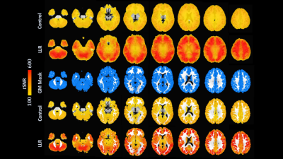 |
Improved signal integrity in multi-echo fMRI through locally low-rank tensor regularization
Nolan K Meyer1, Daehun Kang2, MyungHo In2, John Huston2, Yunhong Shu2, Matt A Bernstein2, and Joshua D Trzasko2
1Mayo Clinic Graduate School of Biomedical Sciences, Rochester, MN, United States, 2Radiology, Mayo Clinic, Rochester, MN, United States
Locally low-rank (LLR) regularization is extended to a multi-echo tensor framework for resting-state fMRI, building on previous generalizations of LLR frameworks. We demonstrate substantial increases in temporal SNR with improved robustness in mapping default mode, auditory, and sensorimotor resting-state connectivity networks in a preliminary seed-based analysis.
|
|||
2869.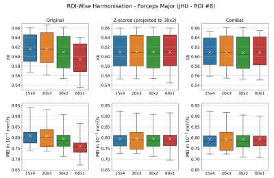 |
Comparison of Region-Wise and Voxel-Wise Diffusion Signal Harmonisation via Z-scoring and ComBat
Stefan Winzeck1,2, Maíra Siqueira Pinto3, Virginia F. J. Newcombe2, Ben Glocker1, David K. Menon2, and Marta M. Correia4
1BioMedIA, Department of Computing, Imperial College London, London, United Kingdom, 2Division of Anaesthesia, Department of Medicine, University of Cambridge, Cambridge, United Kingdom, 3Universitair Ziekenhuis Antwerpen, Antwerp, Belgium, 4MRC Cognition and Brain Sciences Unit, University of Cambridge, Cambridge, United Kingdom
This study compares the impact of voxel-wise versus region-wise harmonisation of diffusion-weighted imaging (DWI) signal via Z-scoring and ComBat. Both approaches and algorithms were shown equally effective in significantly reducing the variation in regionally observed FA and MD in a multi-acquisition DWI dataset.
|
The International Society for Magnetic Resonance in Medicine is accredited by the Accreditation Council for Continuing Medical Education to provide continuing medical education for physicians.