Digital Posters
GI & Pancreas Cancers
ISMRM & SMRT Annual Meeting • 15-20 May 2021

| Concurrent 5 | 17:00 - 18:00 |
2318.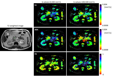 |
The reduction in the contribution of a fast diffusing component for pancreatic malignancy detection – A preliminary study
Debbie Anaby1, Maria Raitses-Gurevich2, Sara Apter1,3, Yael Inbar1,3, Hadassa Degani4, and Talia Golan2,3
1Diagnostic Imaging Department, Sheba Medical Center, Ramat Gan, Israel, 2Department of Oncology, Sheba Medical Center, Ramat Gan, Israel, 3Sackler School of Medicine, Tel Aviv University, Tel Aviv, Israel, 4Life Sciences, Weizmann Institute of Science, Rehovot, Israel
Early detection of pancreatic cancer is challenging but essential to improve its poor prognosis. This preliminary study utilized DT-MRI in pancreas in a clinical setting. DT-MRI was based on two pairs of b-values (0,500 s/mm2 and 100,500 s/mm2), such that the existence of a fast diffusing component was observed. We found that pancreatic tumors show similar values of parametric DTI measures (λ1 and MD-ADC) when using both b-value pairs, suggesting that the fast diffusing component is reduced compared with healthy or cystic pancreatic regions. We conclude that DT-MRI of the pancreas may assist in the detection of pancreatic tumors.
|
|||
2319. |
Predicting fibrosis grades of pancreatic ductal adenocarcinoma using intravoxel incoherent motion diffusion-weighted imaging
Qi Liu1, Wei Xing1, Jilei Zhang2, JingGang Zhang1, Jie Chen1, and Bei Li1
1Department of Radiology, The Third Affiliated Hospital of Soochow University, Changzhou, China, 2Clinical Science, Philips Healthcare, Shanghai, China
This study explored the feasibility of evaluating fibrosis of patients with PDAC and correlate it with histopathological features using IVIM-DWI compared with DWI. No statistically significant differences were found in ADC and D* values between the high- and low-fibrosis groups. A significant negative correlation between D values and fibrosis and a significant positive correlation between f values and fibrosis were observed. D and ƒ values derived from the IVIM model had high sensitivity and diagnostic performance for grading fibrosis in PDAC compared with the conventional DWI model. IVIM-DWI could serve as an imaging biomarker for predicting the fibrosis grade of PDAC.
|
|||
2320. |
A study of pancreatic cancers based on integrated 18F-FDG PET/MRI scans in diagnosis of malignant tumors from benign lesions
Gang FENG1, Mingxiang Sun1, Liling Peng1, Zhaoting Meng1, and Xin Gao1
1Shanghai Universal Medical Imaging Diagnostic Center, Shanghai, China
To explore the value of assessing pancreatic cancers with integrated positron emission tomography (PET)/magnetic resonance imaging (MRI), fluorine-18 fluorodeoxyglucose (FDG) PET and diffusion weighted imaging (DWI) were applied. Integrated PET/MR is a promising and powerful tool for pancreatic tumor detection and assessment. It is relatively advantageous to combine PET and DWI together to discriminate malignant tumors from benign lesions.
|
|||
2321. |
Monitoring tumor microenvironment in a mouse model of pancreatic ductal adenocarcinoma using MRI during early stages of tumor development.
Ravneet Vohra1, Yak-Nam Wang2, Helena Son3, Stephanie Totten2, and Donghoon Lee1
1Radiology, University of Washington, Seattle, WA, United States, 2Applied Physics Laboratory, University of Washington, Seattle, WA, United States, 3Gastroenterology, University of Washington, Seattle, WA, United States
Pancreatic cancer is expected to become the second leading cause of the cancer related deaths in the USA by 2020. A genetically engineered mouse model (KPC) offers an alternative to transplantation models for preclinical therapeutic evaluation as it expresses mutations similar to human pancreatic cell. Magnetic resonance imaging can be used to monitor tumor microenvironment in variety of tumors. The goal of this study was to monitor tumor microenvironment in KPC mouse model during the early stages of tumor development.
|
|||
2322.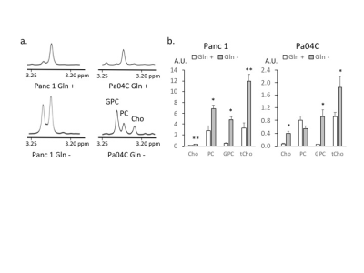 |
Glutamine depletion alters choline metabolism and reduces survival of pancreatic cancer cells
Noriko Mori1, Balaji Krishnamachary1, Yelena Mironchik1, and Zaver M. Bhujwalla1,2,3
1The Russell H. Morgan Department of Radiology and Radiological Science, The Johns Hopkins University, Baltimore, MD, United States, 2The Sidney Kimmel Comprehensive Cancer Center, The Johns Hopkins University, Baltimore, MD, United States, 3Department of Radiation Oncology and Molecular Radiation Sciences, The Johns Hopkins University School of Medicine, Baltimore, MD, United States
Pancreatic ductal adenocarcinoma (PDAC) has poor prognosis due to a combination of late-stage diagnosis and limited response to radiation and chemotherapy. Effective treatments for PDAC are urgently needed. Mutant Kras has been reported as a major cause of glutamine (Gln) addiction in PDAC. Here we investigated the effects of Gln depletion on PDAC cell survival and metabolism. Our data identified Gln as critical for PDAC cell viability. Gln depletion significantly altered choline metabolism in the cell lines investigated.
|
|||
2323.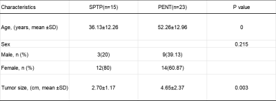 |
Differentiation of solid pseudopapillary tumor and neuroendocrine tumor of pancreas using enhanced T2 * weighted angiography
Longshuang Wang1, Yi Wang2, Ailian Liu3, and Qinhe Zhang3
1School of Medical Imaging,Dalian Medical University,Dalian, China, Dalian Liaoning, China, 2Department of Radiology, Dalian Friendship Hospital, Dalian, China, Dalian Liaoning, China, 3Department of Radiology, the First Affiliated Hospital of Dalian Medical University, Dalian Liaoning, China
It is difficult to differentiate the solid pseudopapillary tumor of the pancreas (SPTP) from pancreatic neuroendocrine tumors (PNET), due to their similar imaging characteristics. Enhanced T2 * weighted angiography (ESWAN) technique can simultaneously obtain amplitude, phase, R2 * and T2 * values, which can quantitatively assess the magnetic sensitivity characteristics of tissues. There was significant difference in R2 * (AUC: 0.707; sensitivity: 91.3%; specificity: 46.7%)and T2*(AUC: 0.829; sensitivity: 73.9%; specificity: 86.7%)between SPTP and PNET. Therefore, ESWAN may be an effective method to identify SPTP and PNET.
|
|||
2324.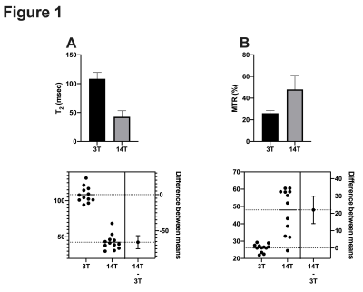 |
Comparison of MR values at 3T and 14T in a mouse model of pancreatic ductal adenocarcinoma.
Ravneet Vohra1, Yak-Nam Wang2, Helena Son3, Stephanie Totten2, and Donghoon Lee1
1Radiology, University of Washington, Seattle, WA, United States, 2Applied Physics Laboratory, University of Washington, Seattle, WA, United States, 3Gastroenterology, University of Washington, Seattle, WA, United States
Pancreatic cancer is expected to become the second leading cause of the cancer related deaths in the USA by 2020. The goal of all pre-clinical magnetic resonance imaging (MRI) studies is to translate the pre-clinical MR values to the clinical scanners and ultimately to human population. MRI has been proven to be extremely useful in clinical trials for monitoring tumor development and assessing therapeutic effects of novel therapeutic strategies. The aim of this study was to measure (T2) relaxation time and magnetization ratio (MTR) in a mouse model of pancreatic ductal adenocarcinoma (PDA) at pre-clinical (14T) and clinical (3T) scanner.
|
|||
2325.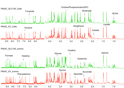 |
Glutamine transporter downregulation mediates metabolic reprogramming in pancreatic tumors
Raj Kumar Sharma1, Balaji Krishnamachary1, Ishwarya Sivakumar1, Yelena Mironchik1, Marie-France Penet1, Paul T Winnard Jr.1, Santosh K. Bharti1, and Zaver M. Bhujwalla1
1Division of Cancer Imaging Research, The Russell H. Morgan Department of Radiology and Radiological Science, The Johns Hopkins University School of Medicine, Baltimore, MD, United States
Pancreatic cancer cells are glutamine dependent for growth and proliferation. The glutamine transporter SLC1A5 is upregulated in pancreatic cancer and is being actively investigated as a pharmacological target. We genetically engineered human pancreatic cancer cells to express SLC1A5 shRNA to downregulate SLC1A5. 1H MRS was used to analyze metabolic differences in SLC1A5 downregulated Pa04C_SLC1A5 pancreatic cancer cells and tumors compared to empty vector cells and tumors. SLC1A5 downregulation resulted in a significantly lower glutamine/glutamate ratio in Pa04C_SLC1A5 cells. In Pa04C_SLC1A5 tumors we observed a significant reduction of several metabolites highlighting the metabolic reprogramming that occurred in tumors with SLC1A5 downregulation.
|
|||
2326.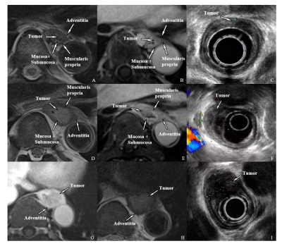 |
MRI Role in Evaluation of T staging and Lymphatic metastases for Esophageal Cancer
Yingyu Lin1, Mengzhu Wang2, and Shi-Ting Feng1
1The First Affiliated Hospital, Sun Yat-sen University, Guangzhou, China, 2MR Scientific Marketing, Siemens Healthcare, Guangzhou, China
Esophageal cancer ranks seventh in terms of incidence and sixth in mortality among all cancers. Accurate staging of esophageal cancer helps to choose appropriate therapeutic strategy. Current imaging modalities used for evaluating esophageal cancer have their own disadvantages. Therefore, we compared MRI, which is non-invasive, non-radiating and of high soft-tissue resolution, with EUS in preoperative T staging and with PET/CT in preoperative lymphatic metastases of esophageal cancer. In this study, MRI showed higher sensitivity, specificity and accuracy in T staging, while MRI and PET/CT showed similar performance in lymph nodes evaluation.
|
|||
2327.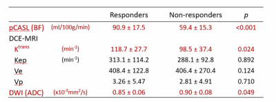 |
Pseudo continuous arterial spin labeling perfusion MRI for predicting tumor response to neoadjuvant chemotherapy in advanced rectal cancer
Yoshihiko Fukukura1, Yuichi Kumagae1, Koji Takumi1, Hiroaki Nagano1, Masanori Nakajo1, Kiyohisa Kamimura1, Takashi Iwanaga2, Yuta Akamine3, and Takashi Yoshiura1
1Kagoshima University Graduate School of Medical and Dental Sciences, Kagoshima, Japan, 2Kagoshima University Hospital, Kagoshima, Japan, 3Philips Japan, Minatoku, Japan
This study focused on the feasibility of pseudo continuous arterial spin labeling (pCASL) perfusion MRI as a non-invasive tool for predicting the response of locally advanced rectal cancer treated with neoadjuvant chemotherapy in comparison with dynamic contrast-enhanced MRI and diffusion-weighted imaging. Our results showed blood flow derived from pCASL was significantly higher in responders than in non-responders. These results suggested the promise of pCASL as a non-invasive alternative to dynamic contrast-enhanced MRI for predicting the treatment response to NAC in locally advanced rectal cancer.
|
|||
2328.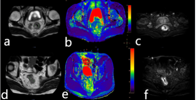 |
The value of quantitative T2-mapping in distinguishing rectal tubular adenocarcinoma from non-tubular adenocarcinoma
Yuhui Liu1, AIlian Liu1, Anliang Chen1, Jiazheng Wang2, Geli Hu2, Wan Dong1, Qingwei Song1, Mingxiao Wang1, Xinru Zhang1, and Xinao Wang1
1The First Affiliated Hospital of Dalian Medical University, Dalian, China, Dalian, China, 2Philips Healthcare,Beijing,China, Beijing, China
This study aims to evaluate the value of quantitative T2-mapping in distinguish rectal tubular adenocarcinoma from non-tubular adenocarcinoma, which can provide rich information and help clinical decision-making. The results showed that the quantitative value of T2mapping of rectal tubular adenocarcinoma was significantly different from that of rectal non-tubular adenocarcinoma. Therefore, quantitative T2 mapping can be used as a non-invasive method for diagnosis rectal tubular adenocarcinoma and non-tubular adenocarcinoma.
|
|||
2329.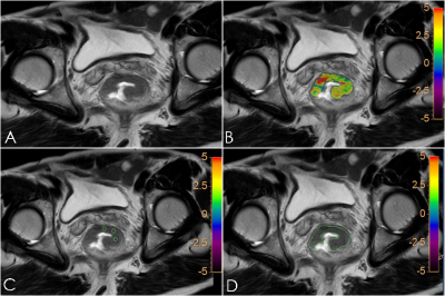 |
The measurements of APTw in Rectal Cancer: the impact of ROI methods on APTw values and interobserver variability
Xiaoyao Lei1, Yaxin Niu1, Longchao Li1, Min Tang1, Yanrong Yang1, Xiaoling Zhang1, Xin Zhang1, Juan Li1, and Xiuzheng Yue2
1Shaanxi Provincial People's Hospital, Xi-an, China, 2Philips Healthcare, Beijing, China
The study aimed to explore the impact of three methods of delineating region of interest (ROI) methods on the measurement of tumor amide proton transfer-weighted (APTw) values. The result showed that whole volume ROI method was reproducible and could reflect the pathophysiological condition of the tumor.
|
|||
2330.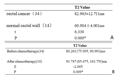 |
Feasibility of quantitative T2 mapping imaging in the diagnosis and chemotherapy response tracking of rectal cancer
Yunxing Tang1, Ailian Liu1, Jiazheng Wang2, Zhiwei Shen2, Yunsong Liu1, Yuhui Liu1, Anliang Chen1, and QingWei Song1
1The First Affiliated Hospital of Dalian Medical University, Dalian, China, 2Philips Healthcare, Beijing, China
Clinical diagnosis and real-time evaluation of chemotherapy response for rectal cancer remain challenging due to the lack of effective non-invasive examinational means. The quantitative T2 mapping technique is an effective method for MRI relaxometry in tissues. In this study, we demonstrated that T2 values were significantly higher in rectal cancer than those in the healthy rectal wall, and significantly increased after chemotherapy. Therefore, quantitative T2 mapping might be a promising non-invasive method in the diagnosis and chemotherapy response tracking of rectal cancer.
|
|||
2331.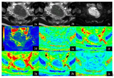 |
The combination of T2 mapping and diffusion kurtosis imaging (DKI) enhances the diagnosis of rectal cancer with and without vascular invasion
Deshuo Dong1, Ailian liu1, Jiazheng Wang2, Peng Sun2, Anliang Chen1, Wan Dong1, Yuhui Liu1, Qingwei Song1, and Renwang Pu1
1Radiology, The First Affiliated Hospital of Dalian Medical University, Dalian, China, 2Philips Healthcare, Beijing, China
Rectal cancer with and without vascular invasion differ greatly in terms of biological behavior, treatment, and prognosis. The T2 mapping and DKI allows non-invasive evaluation of tissue composition. In this study, we investigate the performance of the combination of T2 mapping and DKI on differentiating rectal cancer with and without vascular invasion. The results indicate a better differential diagnosis is achieved by the joint use of T2 mapping and DKI.
|
|||
2332.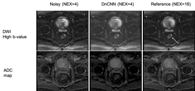 |
Deep Learning Denoising to Accelerate Diffusion-Weighted Imaging of Rectal Cancer
Mohaddese Mohammadi1, Elena Kayee1, Youngwook Kee1, Jennifer Golia Pernicka 2, Iva Petkovska2, and Ricardo Otazo 2
1Medical physics, Memorial Sloan Kettering Cancer Center, New York, NY, United States, 2Memorial Sloan Kettering Cancer Center, New York, NY, United States
Deep learning denoising using a convolutional neural network (DnCNN) is proposed to accelerate the acquisition of diffusion-weighted imaging (DWI) data in patients with rectal tumors. 4-fold acceleration was achieved by reducing the number of averages from 16 to 4 and applying the DnCNN to denoise the data. The DnCNN was trained using pairs of noisy (1 average) and reference (16 averages) images from 92 patients. The trained network was then tested with the data from 6 patients and the results were evaluated qualitatively by radiologists. DnCNN represents a powerful approach to improve efficiency of DWI in patients with rectal cancer.
|
|||
2333.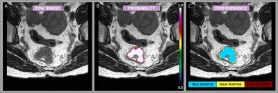 |
Deep Learning Segmentation of Rectal Cancer on MRI
Endre Grøvik1,2, Darvin Yi3, Franziska Knuth4, Sebastian Meltzer5, Anne Negård5, and Kathrine Røe Redalen4
1Department for Diagnostic Physics, Oslo University Hospital, Oslo, Norway, 2Faculty of Health Sciences, University of South-Eastern Norway, Drammen, Norway, 3University of Illinois at Chicago, Chicago, IL, United States, 4Norwegian University of Science and Technology, Trondheim, Norway, 5Akershus University Hospital, Lørenskog, Norway
Treatment of rectal cancer often requires repeated identification of the tumor volume by means of manual delineation by expert radiologists or oncologists. This is a tedious and time-consuming task, particularly with the growing use of multi-sequence 3D imaging. In this work, we have implemented a deep neural network for automatic detection and segmentation of rectal cancer. Our model demonstrates high detection and segmentation performance, equivalent to that of an expert reader, thus illustrating the potential use of deep learning-based segmentation in a clinically relevant setting.
|
|||
2334.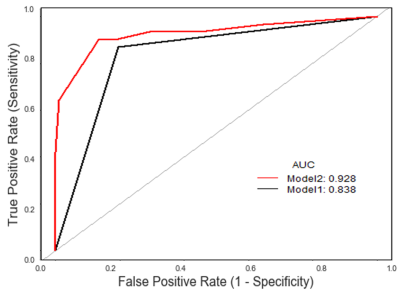 |
The effects of different manual segmentation on High-Resolution T2WI Based-Radiomics in the Preoperative T Staging of Rectal Cancer
Haidi LU1, Fu Shen1, Yuwei Xia2, and Jianping Lu1
1Changhai Hospital, Shanghai, China, 2Huiying Medical Technology Co., Ltd., Beijing, China
Manual delineation of volume of interest (VOI) is widely used in current radiomics analysis, suffering from high variability. The purpose of our study was to investigate the effects of delineation of VOIs on radiomics analysis for the preoperative T staging based on high-resolution T2WI. The result demonstrated that differences in delineation of VOIs affected radiomics analysis, the minimum contour method has better stability in extracting features and the maximum contour method has better diagnostic efficiency in patients with rectal cancer.
|
|||
2335.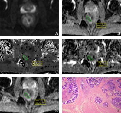 |
The Value of Multiparametric Diffusion-Weighted Imaging in the Preoperative T Staging of Rectal Cancer
Fu Shen1 and Shaotinng Zhang1
1Changhai Hospital, Shanghai, China
This study aimed to explore the value of MP-DWI in the preoperative T staging of rectal cancer.
|
|||
2336.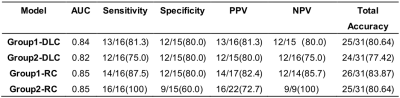 |
AI Methods for Predicting Sensitivity of Total Neoadjuvant Treatment (TNT) in Rectal Cancer Based on Multiparameter MRI and Clinical Data
Ganlu Ouyang1, Zhebin Chen2,3, Jitao Zhou1, Meng Dou2,3, Xu Luo2,3, Han Wen2,3, Yu Yao2,3, and Xin Wang1
1Department of Radiation Oncology, Cancer Center, West China Hospital, Sichuan University, Chengdu, China, 2Chengdu Institute of Computer Application, Chinese Academy of Sciences, Chengdu, China, 3University of Chinese Academy of Sciences, Beijing, China
This work aimed for developing a model for predicting sensitivity and response of total neoadjuvant treatment (TNT) for locally advanced rectal cancer (LARC) based on baseline magnetic resonance imaging (MRI) and clinical data by artificial intelligence method. The results showed that the models for predicting high sensitivity and pCR built with radiomics features achieved the mean area under the ROC curve (AUC) of 0.85 respectively, while the other built with deep-learning (DL) method yielded the mean AUC of 0.82 and 0.84 respectively. The models of two methods for predicting high sensitivity and pCR may be valuable in clinical practice.
|
|||
2337. |
Multiparametric MRI of rectal cancer patients for early detection of therapeutic efficacy of neoadjuvant radio-chemotherapy
Martin Buechert1 and Arnd-Oliver Schäfer2
1Medizinische Fakultät, Universität Freiburg, Freiburg, Deutschland, Radiologische Klinik, Medizinphysik, Universitätsklinikum Freiburg, Freiburg, Germany, 2Klinik für Radiologie, Klinikum St. Georg Leipzig, Leipzig, Germany
Significant changes of tumor properties during and after neoadjuvant radiochemo therapy are observed in DCE- as well as in DWI-MRI. However heterogneiity of the patient cohort impede more clear results. To overcome this an increase in patient numbers may allow to gain significant results for sharper definied sub groups. This will include combining single results of DCE- and DWI.MRI data in a more integral approach to increase the predictive value.
|
The International Society for Magnetic Resonance in Medicine is accredited by the Accreditation Council for Continuing Medical Education to provide continuing medical education for physicians.