Digital Posters
Cancer: Clinical
ISMRM & SMRT Annual Meeting • 15-20 May 2021

| Concurrent 2 | 13:00 - 14:00 |
0939.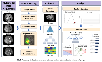 |
Radiomics Based Classification of Ependymoma and High Grade Glioma Using Multimodal MRI
Apoorva Safai1, Sumeet Shinde1, Manali Jadhav1, Tanay Chougule1, Abhilasha Indoria2, Manoj Kumar2, Vani Santosh2, Shumyla Jabeen2, Manish Beniwal2, Subhash Konar2, Jitender Saini2, and Madhura Ingalhalikar1
1Symbiosis International University, Pune, India, 2National Institute of Mental Health and Neurosciences, Bangalore, India
Adult Supratentorial Extraventricular Ependymoma (STEE) are rare neoplasms that are often misdiagnosed as high-grade gliomas (HGG) due to their similar radiological manifestation on MRI. However, the pathogenesis and treatment plan of ependymoma differs significantly from gliomas, and hence an early and accurate diagnosis is crucial. We propose a novel machine learning based diagnostic model that can accurately distinguish adult STEE from HGG subtypes using quantitative radiomic signatures from a multi-model MRI data. First order and texture based radiomic features, particularly from FLAIR, T2 and ADC, can capture intricate pathological variations and aid in accurate and differential diagnosis of STEE tumors.
|
|||
0940.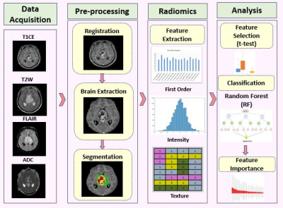 |
Creating a radiomic signature for H3K27M mutation in midline glioma on multimodal MRI
Manali Balasaheb Jadhav1, Richa Singh Chauhan2, Priyanka Tupe Waghmare3, Archit Rajan4, Abhilasha Indoria2, Jitender Saini5, Vani Santosh6, Madhura Ingalhalikar4, and Subhas Konar7
1Symbiosis Center for Medical Image Analysis, Pune, India, 2Radiology, National Institute of Mental Health and Neuroscieces, Bengaluru, India, 3Symbiosis Institute on Technology, Pune, India, 4Symbiosis Centre for Medical Image Analysis, Pune, India, 5Radiology, National Institute of Mental Health and Neuroscieces, Pune, India, 6Neuropathology, National Institute of Mental Health and Neuroscieces, Bengaluru, India, 7Neurosurgery, National Institute of Mental Health and Neuroscieces, Bengaluru, India
H3K27M mutation in diffuse midline glioma is an independent predictor of overall survival however has a very poor prognosis. Identification of the mutation using conventional radiological analysis is complicated while the deep location of the tumors in the brain makes biopsy challenging with substantial risk of morbidity. To alleviate these issues, our work employs radiomics based machine learning framework to predict the H3K27M mutation from multi-modal MRI on 46 subjects. Results revealed 91% cross validation accuracy illustrating its future potential in clinical use.
|
|||
0941.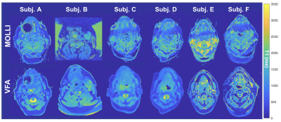 |
Using Variable Flip Angle (VFA) and Modified Look Locker Inversion Recovery (MOLLI) T1 Mapping in Clinical OE-MRI
Emma Bluemke1, Ambre Bertrand1, Kwun-Ye Chu2,3, Nigar Syed2, Andrew Murchison3,4, Tessa Greenhalgh3,5, Brian Burns6, Martin Craig7, Nia Taylor3, Ketan Shah2,3, Fergus Gleeson2,3, and Daniel Bulte1
1University of Oxford, Oxford, United Kingdom, 2MRC Oxford Institute for Radiation Oncology, Department of Oncology, University of Oxford, Oxford, United Kingdom, 3Radiotherapy Department, Oxford University Hospitals NHS, Oxford, United Kingdom, 4Nuffield Department of Medicine, University of Oxford, Oxford, United Kingdom, 5University Hospital Southampton NHS FT, Southhampton, United Kingdom, 6GE Healthcare, Menlo Park, CA, United States, 7University of Nottingham, Nottingham, United Kingdom
As clinical studies adopt Oxygen-Enhanced MRI to assess its feasibility in human tumours, it is important for researchers to report any practical complications experienced. We report our practical experience with using both MOLLI and VFA T1 mapping for a recently completed OE-MRI clinical oncology study with the aim that sharing this is helpful to researchers planning to use OE-MRI in clinical oncology. Specifically, we report 4 elements: (1) difference in estimated T1 from each method used (MOLLI and VFA), (2) standard deviation within tumour ROIs, (3) OE-MRI response resulting from either method, and (4) artefacts and practical difficulties encountered.
|
|||
0942.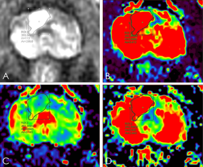 |
Assessment the Preponderant Diagnostic Performances of Oligometastatic Prostate Cancer Using DCE-MRI of Tofts Model
SHUANG MENG1, Ailian Liu1, Lihua Chen1, Qinhe Zhang1, Qingwei Song1, and Yunsong Liu1
1The First Affiliated Hospital of Dalian Medical University, Dalian, China
It remains a challenge to diagnose the oligometastatic prostate cancer (PCa) due to the ambiguous definition of oligometastatic PCa. Previous studies had shown that dynamic contrast enhanced (DCE) - MRI imaging could be used to assess tumor aggressiveness. This study indicated that transfer constant (Ktrans) had the potential to assess the aggressiveness of PCa. And Ktrans combined with clinical characteristics (such as age and prostate specific antigen) had the higher diagnostic efficiency for oligometastatic and widely metastasis PCa.
|
|||
0943.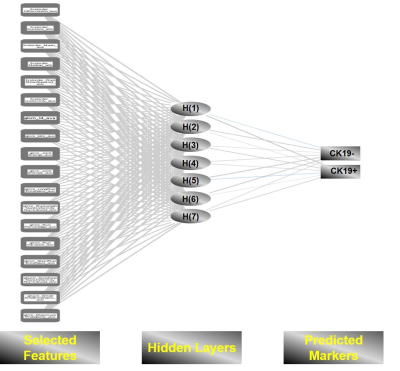 |
Evaluating the cytokeratin 19 (CK-19) status via neural network model established by the SWI-derived radiomics features
Zhijun Geng1, Yunfei Zhang2, Chuanmiao Xie1, and Yongming Dai2
1Sun Yat-sen University Cancer Center, Guangzhou, China, 2Central Research Institute, United Imaging Healthcare, Shanghai, China
Susceptibility Weighted Imaging (SWI) has shown tremendous clinical significance for identifying the hepatic micro-structural abnormalities such as micro-bleeding, vascularity, nodule and so forth. Recent studies concluded that cytokeratin 19 (CK-19) is an important marker for prognostic prediction of hepatocellular carcinoma (HCC). We hypothesized that the neural network model can be established by means of extracting high throughput radiomics features from SWI images for noninvasively evaluating the CK-19 status with high accuracy. The results demonstrated that such deep learning based neural network model yielded excellent diagnostic performance for predicting the CK-19 status.
|
|||
0944. |
Improved gastric T2WI imaging by an TSE corrected HASTE sequence: a comparation study with conventional HASTE, TSE and BLADE-TSE sequences on 3T
Xiaosheng Xu1, Qinglei Shi2, Weishuai Wang3, Jia Wei4, Li Yang4, Qian Xu4, and Gaofeng Shi4
1The Fourth Hospital of Hebei Medical University, Shijiazhuang, China, 2MR Scientific Marketing,Siemens Healthcare, Beijing, China, 3MR Scientific Marketing,Siemens Healthcare, JINAN, China, 4The Fourth Hospital of Hebei Medical University, shijiazhuang, China
In this study, we evaluated the scan time and image quality of a serious of gastric T2WI imaging, including conventional TSE with cartesian acquisition (TSE), BLADE-TSE, conventional HASTE sequence and a new TSE corrected HASTE sequence (TSE-HASTE). We found that the HASTE sequences can provide comparable image quality to TSE sequences, while is insensitive to motion and susceptibility artifacts. For patients with poor respiratory movement, HASTE sequence may be used as a good replacement to the TSE sequences, and TSE-HASTE sequence may provide a better image contrast compared to conventional HASTE.
|
|||
0945.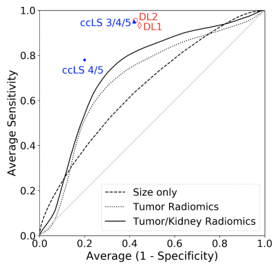 |
Deep learning prediction for clear cell renal carcinoma cancer compared with human and radiomics analysis
Junyu Guo1, Lauren Hinojosa1, Yin Xi1, Keith Husley1, and Ivan Pedrosa1
1Radiology, UT southwestern medical center, Dallas, TX, United States
Clear cell renal carcinoma cancer (ccRCC) is the most aggressive subtype among small renal masses. ccRCC identification can help in decision making between active surveillance and definitive intervention. Recently, a clear cell likelihood score (ccLS) using subjective interpretation of multiparametric MRI by radiologists was proposed. In this study, we investigate whether radiomics and deep learning (DL) technique can facilitate the prediction of ccRCC using T2-weighted images only. We compared the results of two different approaches, radiomics and DL, with the reported ccLS performance. Our results demonstrate that both radiomics and deep learning may provide useful information for identification of ccRCC.
|
|||
0946. |
The value of preoperative prediction of ki67 and P53 expression in DCE-MRI texture analysis of rectal cancer
Yuhui Liu1, Ailian Liu1, and Mingxiao Wang1
1The First Affiliated Hospital of Dalian Medical University, Dalian, China, Dalian, China This study aims to analyze the intrinsic relationship between imaging indicators and tumor biological behavior, and improve the clinical value of DCE-MRI.The correlation between DCE-MRI quantitative parameter values (Ktrans, Ve, Kep) and molecular biomarkers (Ki67,P53) was compared and explored in patients with rectal cancer.
|
|||
0947. |
Diagnostic performance of machine learning-based MRI for posterior fossa tumors: a meta-analysis
Chen Chen1, Fabao Gao1, and Xiaoyue Zhou2
1Department of Radiology, West China Hospital, Chengdu, China, 2MR Collaboration, Siemens Healthineers Ltd., Shanghai, China
Because of variations in severity and treatment methods of pilocytic astrocytoma, medulloblastoma, and ependymoma, accurate and specific diagnoses of the tumors are critical. Non-invasive diagnosis of posterior fossa tumors based on machine learning-based magnetic resonance imaging are being reported. However, conventional MRI, diffusion MRI, MR perfusion, and magnetic resonance spectroscopy have variable diagnostic values. We present here a meta-analysis of all the relevant published studies and conducted a large sample-size assessment concerning the diagnostic performance and potential covariates that could influence the diagnostic performance of machine learning.
|
|||
0948. |
Automatic segmentation of glioma based on MRI K-space data
Yikang Li1,2,3, Zhan li Hu1,2,3, Sen Jia1,2,3, Wenjing Xu4, Zongyang Li1,2,3, Hairong Zheng1,2,3, Xin Liu1,2,3, and Na Zhang1,2,3
1Paul C. Lauterbur Research Center for Biomedical Imaging, Shenzhen Institutes of Advanced Technology, Chinese Academy of Sciences, Shenzhen, China, 2Key Laboratory for Magnetic Resonance and Multimodality Imaging of Guangdong Province, Shenzhen Institutes of Advanced Technology, Chinese Academy of Sciences, Shenzhen, China, 3CAS key laboratory of health informatics, Shenzhen Institutes of Advanced Technology, Chinese Academy of Sciences, Shenzhen, China, 4Faculty of Information Technology, Beijing University of Technology, Beijng, China
Nowadays, Magnetic Resonance Imaging (MRI) plays a pivotal role in gliomas diagnosis, analysis, and surgery planning. Nevertheless, the accuracy of MRI segmentation is enormously restricted by the quality of images. Therefore, we demonstrate a new method that can directly make segmentations from K space data. And the results show that our method achieves the state of the art.
|
|||
0949.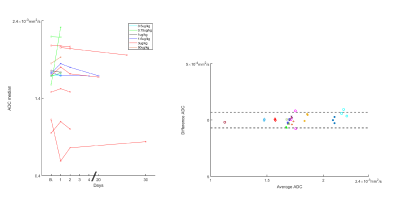 |
ADC Decreases in Solid Tumors Following Monotherapy With PEGylated Recombinant Human Hyaluronidase: Results From Early-Phase Clinical Trials
Andres Mauricio Arias-Lorza1 and Natarajan Raghunand1
1Moffitt Cancer Center, Tampa, FL, United States
In early-phase clinical trials, we observed a general decrease in tumor ADC following administration of PEGylated Recombinant Human Hyaluronidase PH20 (PEGPH20) as monotherapy. This decrease was related with reduction of tumor hyaluronan measured in biopsy samples. Reduction in ADC is suggestive of a decrease in tumor water content following hyaluronan depletion by PEGPH20.
|
|||
0950.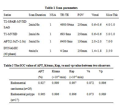 |
Differential diagnosis of Endometrial carcinoma and polyps using Amide proton transfer-weighted imaging and permeability analysis
Ye Li1, Xulun Lu1, Shifeng Tian1, Jiazheng Wang2, Zhiwei Shen2, Qingwei Song1, and Ailian Liu1
1The First Affiliated Hospital of Dalian Medical University, Dalian, China, 2Philips Healthcare, Beijin, China
The accurate differential diagnosis of endometrial carcinoma and endometrial polyps always met many challenges. In this study, we evaluated the capability of differentiation of endometrial carcinoma from endometrial polyps with APT signal intensity (SI) and permeability parameters. The diagnostic efficiency is the highest using APT combined with Kep. APT and permeability parameters are potentially a promising and valuable method in differentiation of endometrial carcinoma from endometrial polyps.
|
|||
0951.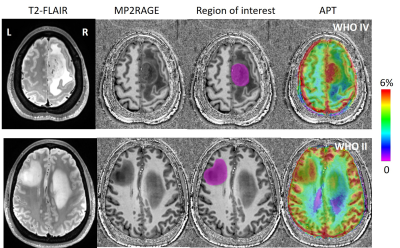 |
Preoperative discrimination between the low-grade glioma and high-grade glioma and early exploration of metastatic margin with APT imaging at 7T
Yifan Yuan1, Qi Yue1, Xiang Zou1, Jiajun Cai1, Ying-Hua Chu2, Yi-Cheng Hsu2, Patrick Alexander Liebig3, Hui Zhang4, He Wang4, Liang Chen1, and Ying Mao1
1Department of Neurosurgery, Huashan Hospital, Fudan University, Shanghai, China, 2MR Collaboration, Siemens Healthcare Ltd., Shanghai, China, 3Siemens Healthcare GmbH, Erlangen, Germany, 4Institute of Science and Technology for Brain-Inspired Intelligence, Fudan University, Shanghai, China
Preoperative assessment of histological grades and possible margin of glioma are challenges to neurosurgeons, which may also make a huge difference in therapeutic strategies. In this study, we aim to establish a grade-discrimination criterion via APT and explore the possible metastatic margin of diffuse glioma at 7T. Eleven patients underwent structural imaging and APT CEST imaging. Results of the APT were merged with the T1 image, which showed a promising indication of preoperative histological grades. Meanwhile, with the early exploration of metastatic margin, the APT CEST at 7 Tesla may significantly facilitate the surgical strategy and surgical excision extension.
|
|||
0952.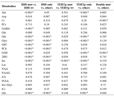 |
The Effect of Cramer-Rao Lower Bound Thresholds on Classification of IDH and TERTp Mutation Status in Gliomas using 1H-MRS
Abdullah Bas1, Banu Sacli-Bilmez1, Ayca Ersen Danyeli2,3, Cengiz Yakicier3,4, M.Necmettin Pamir3,5, Koray Ozduman3,5, Alp Dincer3,6, and Esin Ozturk-Isik1,3
1Institute of Biomedical Engineering, Bogazici University, İstanbul, Turkey, 2Department of Medical Pathology, Acibadem Mehmet Ali Aydinlar University, Istanbul, Turkey, 3Center for Neuroradiological Applications and Reseach, Acibadem Mehmet Ali Aydinlar University, Istanbul, Turkey, 4Department of Molecular Biology and Genetics, Acibadem Mehmet Ali Aydinlar University, Istanbul, Turkey, 5Department of Neurosurgery, Acibadem Mehmet Ali Aydinlar University, Istanbul, Turkey, 6Department of Radiology, Acıbadem Mehmet Ali Aydinlar University, Istanbul, Turkey
Cramer-Rao lower bound (CRLB) is commonly employed as an exclusion criterion for bad quality MR spectra. This study aims to investigate the CRLB differences of the metabolites between isocitrate dehydrogenase (IDH) and telomerase reverse transcriptase promoter (TERTp) mutational subgroups in gliomas, and to assess the effects of CRLB threshold on their classifications. GPC, PCh, 2HG, and Ins were more reliably detected in IDH-mutant gliomas. Ins had higher CRLB values in TERTp-mutant than TERTp-wildtype gliomas. Different CRLB thresholds followed by zero-imputing had a small impact on the classification accuracies, but affected the choice of best features and classification algorithms.
|
|||
0953.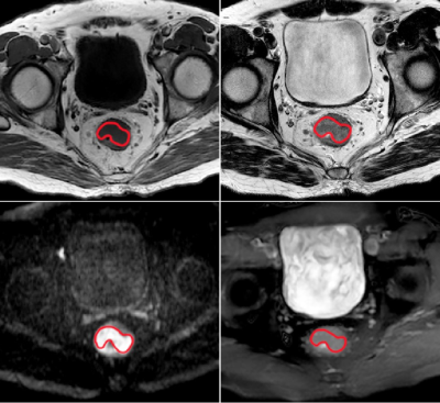 |
Correlation between Amide Proton Transfer Imaging and Pathological Staging of Rectal Cancer
Honglei Hu1, Xixi Zhao1, Qiming Wei2, Chuyao Chen1, Yuewei Huang1, Yingjie Mei3, and Yikai Xu1
1Medical Imaging Center, Nanfang Hospital, Southern Medical University, Guangzhou, China, 2Intervention, Traditional Chinese Medicine Hospital of Guangdong Province, Guangzhou, China, 3Philips Healthcare, Guangzhou, China
Rectal cancer is the third deadliest cancer in the world. The choice of its clinical treatment plan is closely related to clinical staging, which mainly depends on preoperative MR staging. Clinical work experience has found that the MR staging obtained by the existing MR technology is often less precise and often higher than the postoperative pathological staging. In order to solve this problem, our research has found that Amide Proton Transfer(ATP) imaging has a strong correlation with postoperative pathological staging, which can help clinicians to make a more accurate diagnosis of rectal cancer staging before surgery.
|
|||
0954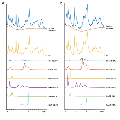 |
Correlations of Single Voxel 1H-MRS Findings with Tumor Biology in Meningiomas. Video Permission Withheld
Banu Sacli-Bilmez1, Ayca Ersen Danyeli2,3, Murat Şakir Ekşi4, Kübra Tan5, Ozge Can6, Cengiz Yakicier7, M.Necmettin Pamir3,4, Alp Dincer3,8, Koray Özduman3,4, and Esin Ozturk-Isik1
1Institute of Biomedical Engineering, Bogazici University, Istanbul, Turkey, 2Department of Medical Pathology, Acibadem Mehmet Ali Aydinlar University, Istanbul, Turkey, 3Center for Neuroradiological Applications and Reseach, Acıbadem Mehmet Ali Aydinlar University, Istanbul, Turkey, 4Department of Neurosurgery, Acibadem Mehmet Ali Aydinlar University, Istanbul, Turkey, 5Health Institutes of Turkey, Istanbul, Turkey, 6Department of Medical Engineering, Acibadem Mehmet Ali Aydinlar University, Istanbul, Turkey, 7Department of Molecular Biology and Genetics, Acibadem Mehmet Ali Aydinlar University, Istanbul, Turkey, 8Department of Radiology, Acibadem Mehmet Ali Aydinlar University, Istanbul, Turkey
Meningiomas are a heterogeneous group of tumors that arise at the pachymeninges and tend to invade surrounding bone and rarely the brain. In this work, we studied the single-voxel 1H-MRS correlates of aggressive biology (measured by WHO grade), peritumoral edema, brain invasion, bone invasion, skull base location in meningiomas. Higher Glyc and Ins+Glyc levels were observed in aggressive (WHO grade II or III) meningiomas. Higher Glu, GPC, and Glx levels were observed in hyperostotic tumors. The classification accuracies were 81.28% for tumor grade, 73.18% for hyperostosis detection, and 76.88% for skull base localization of meningiomas.
|
|||
0955.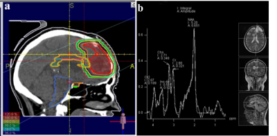 |
Early detection of radiation-induced injury and prediction of cognitive deficit by MRS metabolites in radiotherapy of low grade glioma
Zahra Alirezaei1,2, Mohammadreza Nazemzadeh3, Masoud Hasanpour3, Alireza Amouheidari4, and Sajad Iraji3
1Isfahan University of Medical Sciences, Isfahan, Iran (Islamic Republic of), 2Bushehr University of Medical Sciences, Bushehr, Iran (Islamic Republic of), 3Tehran University of Medical Sciences, Tehran, Iran (Islamic Republic of), 4Milad Hospital, Isfahan, Iran (Islamic Republic of)
To compare the sensitivity of alteration in MRS metabolites and MoCA and ACE-R cognitive tests in early and early-delayed post-radiation phases in detection of radiation-induced injury of Low Grade Glioam patients. The MRS metabolites, the ACE-R and MoCA, and the dosimetric parameters in the corpus callosum were analyzed during RT and up to 6-month post-RT for 10 LGG patients. NAA/Cr and Cho/Cr declined significantly at least 3 months before detecting alterations in ACE and MoCA cognitive tests. Therefore, the MRS-based biomarkers may be more sensitive than the state of the art cognitive test tools in prediction of post-radiation cognitive impairments.
|
|||
0956.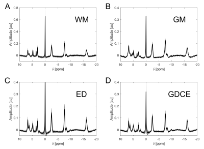 |
31P spectral profiles in brain tissues of volunteers and glioma patients at 7T
Andreas Korzowski1, Nina Weckesser2, Vanessa L Franke1, Heinz-Peter Schlemmer2, Mark E Ladd1, Peter Bachert1, and Daniel Paech2
1Medical Physics in Radiology, German Cancer Research Center (DKFZ), Heidelberg, Germany, 2Radiology, German Cancer Research Center (DKFZ), Heidelberg, Germany
The low spatial resolution of 31P MRSI of the human brain leads to a weighted mixture of specific spectral profiles from different tissue types in localized 31P spectra of individual voxels. Application of high-resolution 31P MRSI to volunteers and glioma patients at ultra-high B0 yields a possibility to approach brain tissue-specific profiles, which could aid interpretation of observations from 31P MRSI. The presented high-quality 31P brain spectra from individual tissue types obtained at B0 = 7T illustrate clear differences not only between healthy and tumor tissues, but also between different compartments within diseased tissue, i.e. contrast-enhanced regions and edema.
|
|||
0957.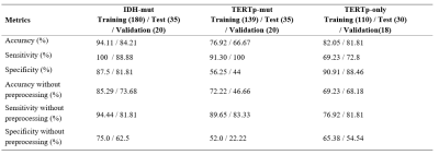 |
1D-CNN for the Detection of IDH and TERTp Mutations in Diffuse Gliomas using Proton Magnetic Resonance Spectroscopy
Abdullah BAS1, Banu Sacli-Bilmez1, Ayca Ersen Danyeli2,3, Cengiz Yakicier3,4, M.Necmettin Pamir3,5, Koray Ozduman3,5, Alp Dincer3,6, and Esin Ozturk-Isik1,3
1Institute of Biomedical Engineering, Bogazici University, Istanbul, Turkey, 2Department of Medical Pathology, Acibadem Mehmet Ali Aydinlar University, Istanbul, Turkey, 3Center for Neuroradiological Applications and Reseach, Acibadem Mehmet Ali Aydinlar University, Istanbul, Turkey, 4Department of Molecular Biology and Genetics, Acibadem Mehmet Ali Aydinlar University, Istanbul, Turkey, 5Department of Neurosurgery, Acibadem Mehmet Ali Aydinlar University, Istanbul, Turkey, 6Department of Radiology, Acıbadem Mehmet Ali Aydinlar University, Istanbul, Turkey
Isocitrate dehydrogenase (IDH) and telomerase reverse transcriptase promoter (TERTp) mutations affect the clinical behavior and survival rate of diffuse gliomas. The detection of these mutations preoperatively is very critical for treatment planning. In this study, three different one dimensional convolutional neural network (1D-CNN) models were designed to identify IDH mutant (IDH-mut), TERTp mutant (TERTp-mut), and TERTp-only (IDH-wild type and TERTp-mut) gliomas based on proton magnetic-resonance spectroscopy (1H-MRS). The 1D-CNN models could identify IDH-mut, TERTp-mut, and TERTp-only gliomas with 94.11%, 76.92%, and 82.05% accuracies, respectively. This study showed the potential of deep-learning in predicting especially IDH-mutations in gliomas using 1H-MRS data.
|
|||
0958.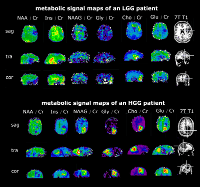 |
Metabolic profiles of glioma grade and IDH mutation status using high resolution 7T 3D-FID-MRSI: Preliminary results
Cornelius Cadrien1,2, Sukrit Sharma1, Philipp Lazen1, Julia Furtner3, Alexandra Lipka1,4, Eva Hečková1, Lukas Hingerl1, Stanislav Motyka1, Stephan Gruber1, Bernhard Strasser1, Barbara Kiesel2, Mario Mischkulnig2, Matthias Preusser5,
Thomas Roetzer6, Adelheid Wöhrer6, Michael Weber7, Christian Dorfer2, Karl Rössler2, Siegfried Trattnig1,4, Wolfgang Bogner1, Georg Widhalm2, and Gilbert Hangel1,2
1High-field MR Center, Department of Biomedical Imaging and Image-guided Therapy, Medical University of Vienna, Vienna, Austria, 2Department of Neurosurgery, Medical University of Vienna, Vienna, Austria, 3Division of Neuroradiology and Musculoskeletal Radiology, Department of Biomedical Imaging and Image-guided Therapy, Medical University of Vienna, Vienna, Austria, 4Christian Doppler Laboratory for Clinical Molecular MR Imaging, Vienna, Austria, 5Division of Oncology, Department of Inner Medicine I, Medical University of Vienna, Vienna, Austria, 6Division of Neuropathology and Neurochemistry, Department of Neurology, Medical University of Vienna, Vienna, Austria, 7Division of Medical Imaging and Nuclear Medicine, Medical University of Vienna, Vienna, Austria
We measured 38 HGG and LGG patients with a 15 min MR spectroscopic imaging sequence with an isotropic voxel size of 3.4 mm, covering a 64×64×39 matrix. After post-processing the measured spectra were analyzed statistically. Clinically segmented tumor regions showed statistically significant differences in metabolic ratios compared to normal-appearing white matter. Further analysis of metabolic hotspots with respect to tumor grade and IDH mutation status yielded no significant differences beyond myo-Inositol decreasing with tumor grade.
|
The International Society for Magnetic Resonance in Medicine is accredited by the Accreditation Council for Continuing Medical Education to provide continuing medical education for physicians.