Digital Posters
Spectroscopy: Acq/Recon/Analysis
ISMRM & SMRT Annual Meeting • 15-20 May 2021

Digital Posters
Spectroscopy: Acq/Recon/Analysis
| Concurrent 2 | 15:00 - 16:00 |
1983.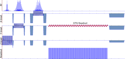 |
Ultrahigh-field echo-planar spectroscopic imaging with semi-adiabatic spatial-spectral pulses
Gaurav Verma1, Rebecca Emily Feldman2, and Priti Balchandani1
1Radiology, Icahn School of Medicine at Mount Sinai, New York, NY, United States, 2Medical Physics, University of British Columbia, Kelowna, BC, Canada
An echo-planar spectroscopic imaging sequence has been demonstrated incorporating semi-adiabatic band-limited refocusing (SABRE) pulses. The sequence simultaneously addresses two major obstacles to the implementation of high-resolution spectroscopic imaging at ultrahigh field: presence of B1 field inhomogeneity and aliasing due to bandwidth limitation in the spectral-spatial readout. Both bi-polar and flyback variants of the EPSI readout have been implemented in a user-selectable manner. An accompanying Matlab-based reconstruction has been developed which performs extraction and complete processing from raw data including eddy current compensation, phase reversal of even echoes in the bi-polar readout and removal of spurious points acquired during gradient switching.
|
|||
1984.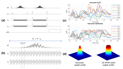 |
Double Spin Echo Spectroscopic Imaging Using Optimized 3D Spectral-Spatial Pulses for Brain Studies at 10.5T
Xiaoxuan He1, Edward J. Auerbach1, Michael Garwood1, Naoharu Kobyashi1, Alireza Sadeghi‐Tarakameh1, Yigitcan Eryaman1, Xiaoping Wu1, and Gregory J. Metzger1
1Center for Magnetic Resonance Research, University of Minnesota, Minneapolis, MN, United States
A parallel transmit optimized 3D spatial-spectral pulse is developed for spectroscopic imaging for brain studies at 10.5T with reduced SAR, intrinsic water suppression and field inhomogeneity mitigation. Phantom studies are used to compare the new method with a conventional approach.
|
|||
1985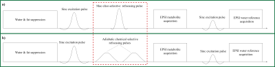 |
Simultaneous Water and Lipid Suppression Using Chemical Selective Adiabatic Refocusing Pulses Echo Planar Spectroscopic Imaging (EPSI) at 7T Video Permission Withheld
Guodong Weng1, Sulaiman Sheriff2, Claus Kiefer1, Irena Zubak3, Andrew A Maudsley2, and Johannes Slotboom1
1Institute for Diagnostic and Interventional Neuroradiology, Support Center for Advanced Neuroimaging (SCAN), University of Bern, Bern, Switzerland, 2Department of Radiology, University of Miami School of Medicine, Miami, FL, United States, 3Inselspital Bern and University Hospital, Bern, Switzerland
To tackle the chemical shift displacement artifacts (CSDA), B1-inhomoheneity, water and fat suppression as well as specific absorption-rate (SAR) limitation at 7T spectroscopy, an EPSI-variant sequence is developed in this study. The conventional slice-selective refocusing pulse is replaced by a spectral-selective adiabatic 2π-pulse pair. The results show a 77% reduction of CSDA, homogeneous refocusing map, water suppression factor ≥ 1000 within an acceptable SAR both in vitro and in vivo measurement. The scan time for the whole brain is within 8 minutes. This new EPSI-variant sequence shows its high application potential clinical routine.
|
|||
1986.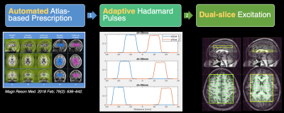 |
Atlas-based adaptive Hadamard-encoded Acquisition for Multiband 2D MRSI at 3T
Huawei Liu1, Adam Autry1, Duan Xu1, Peder Larson1, and Yan Li1
1Department of Radiology and Biomedical Imaging, University of California, San Francisco, San Francisco, CA, United States
Proton MR spectroscopy has been widely used for non-invasive assessment of brain metabolites. Metabolic alterations in the motor and sensory cortex, cingulate cortex and subcortical structures have been associated with pathology. It is desirable to acquire MRSI in these regions simultaneously with short scan time and sufficient SNR. We have developed a novel technique for simultaneous multiband 2D MRSI acquisition, which combines automated atlas-based prescription and adaptive Hadamard pulses. This technique helps to reduce prescription time, improve reproducibility and increase SNR efficiency.
|
|||
1987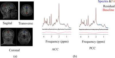 |
Potential of Dual-SPECIAL sequence in revealing interregional differences in human brain metabolite concentrations Video Permission Withheld
Masoumeh Dehghani1,2 and Jamie Near1,2
1McGill university, Montreal, QC, Canada, 2Centre d'Imagerie Cérébrale, Douglas Mental Health University, Montreal, QC, Canada
Previously, we demonstrated the feasibility of performing simultaneous MRS localization in two human brain regions using the dual-SPECIAL technique. Based on the SPin ECho, full Intensity Acquired Localized sequence, this approach yields simultaneous acquisition from two regions in combination with Hadamard encoding, reducing the acquisition time by half compared with serial acquisition. Here, we demonstrate that MRS acquisition using dual-Special sequence reveals significant differences in the ratio of tCho/tCr between anterior and posterior regions and higher concentration of several metabolites in regions with higher gray matter fraction.
|
|||
1988.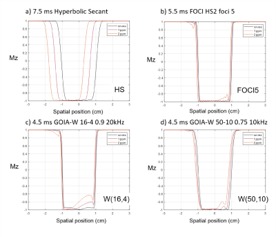 |
GOIA-WURST optimisation for ultra-high field single-voxel MRS at short-TE
Adam Berrington1, Joseph S Gillen2,3, and Vincent Boer4
1Sir Peter Mansfield Imaging Centre, School of Physics and Astronomy, University of Nottingham, Nottingham, United Kingdom, 2Russell H. Morgan Department of Radiology and Radiological Science, Johns Hopkins University School of Medicine, Baltimore, United Kingdom, 3F.M. Kirby Research Centre, Kennedy Krieger Institute, Baltimore, MD, United States, 4Danish Research Centre for Magnetic Resonance,Centre for Functional and Diagnostic Imaging and Research, Copenhagen University Hospital, Hvidovre, Denmark
Gradient modulated pulses allow for accurate slice localisation in single-voxel MRS. However, they suffer from ‘smearing’ artifacts at off-resonance leading to poor performance across the inversion band, particularly when B1 field strength is low. An optimized GOIA-WURST pulse shape was found by simulating changes in the modulation parameters to improve the off-resonance profile at 7T for B1 of 15 μT. When used in a semi-LASER localisation, the optimised GOIA(50,10) pulses resulted in a minimum TE of 23 ms which led to upright peak shapes for J-coupled multiplets in phantom.
|
|||
1989.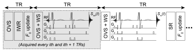 |
Spectral registration for real-time frequency correction of single-voxel GABA-edited MRS data: Proof of concept
Mark Mikkelsen1,2, Steve C. N. Hui1,2, and Richard A. E. Edden1,2
1Russell H. Morgan Department of Radiology and Radiological Science, The Johns Hopkins University School of Medicine, Baltimore, MD, United States, 2F. M. Kirby Research Center for Functional Brain Imaging, Kennedy Krieger Institute, Baltimore, MD, United States
Offsets in B0 field frequency have a detrimental impact on the SNR, linewidth, and editing efficiency of edited 1H MRS data. We have previously described an interleaved water referencing (IWR) method for interspersed acquisition of the unsuppressed water signal to correct frequency drift at regular intervals. Here, we add to IWR a spectral registration (SR) approach for updating the center frequency every TR. Proof-of-concept results from phantom and in vivo experiments demonstrate that SR can be successfully combined with IWR for fully prospective correction of frequency offsets across the whole edited MRS scan.
|
|||
1990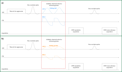 |
SLOW: Whole Brain Spectral Editing EPSI Based Technique using Chemical Selective Adiabatic 2π-Refocusing Pulses applied to 2HG and GABA Editing Video Permission Withheld
Guodong Weng1, Claus Kiefer1, Irena Zubak2, and Johannes Slotboom1
1Institute for Diagnostic and Interventional Neuroradiology, Support Center for Advanced Neuroimaging (SCAN), University of Bern, Bern, Switzerland, 2Department of Neurosurgery, Inselspital Bern and University Hospital, Bern, Switzerland
To overcome the B1+ inhomogeneities and SAR limitation for using MRSI-editing at UHT (≥7T), a new spectral editing scheme was developed within an EPSI sequence for whole brain MRSI. The slice-selective refocusing pulse was replaced by a chemical-selective adiabatic 2π-refocusing pulse pair with varying passbands. The results show an excellent glutamate, 2HG and GABA-editing pattern with in vitro and in vivo measurements. This study has shown the excellent performance of the proposed new spectral editing scheme, and proved that it could be an alternative method for MEGA editing.
|
|||
1991.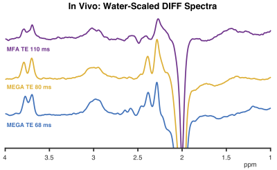 |
Long-TE mixed flip angle editing of GABA
Sofie Tapper1,2, Muhammad G. Saleh3, Helge J. Zöllner1,2, Steve C.N. Hui1,2, and Richard A.E. Edden1,2
1Russell H. Morgan Department of Radiology and Radiological Science, Johns Hopkins University School of Medicine, Baltimore, MD, United States, 2F. M. Kirby Research Center for Functional Brain Imaging, Kennedy Krieger Institute, Baltimore, MD, United States, 3Department of Diagnostic Radiology and Nuclear Medicine, University of Maryland School of Medicine, Baltimore, MD, United States
The aim of this work was to investigate an alternative MRS approach for GABA editing. By performing a range of simulations, we found a long-TE scheme that reduces the influence of co-edited macromolecular signal without lower GABA losses than long-TE MEGA-PRESS. We termed this scheme ‘Mixed flip angle’ (MFA) editing, and validated the scheme with phantom measurements and a proof-of-principle in vivo measurement. Preliminary results show that longer-TE editing of GABA has the potential to increase selectivity by allowing for longer editing pulse duration, avoiding the need for symmetrical suppression of MM signals.
|
|||
1992.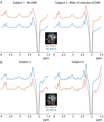 |
Improved prospective frequency correction for macromolecule-suppressed GABA editing with metabolite cycling at 3T
Kimberly Chan1, Andreas Hock2, Richard Edden3,4, Erin MacMillan5, and Anke Henning1,6
1The University of Texas Southwestern, Dallas, TX, United States, 2MR Clinical Science, Philips Health Systems, Horgen, Switzerland, 3Russell H. Morgan Department of Radiology and Radiological Science, The Johns Hopkins University School of Medicine, Baltimore, MD, United States, 4. M. Kirby Research Center for Functional Brain Imaging, Kennedy Krieger Institute, Baltimore, MD, United States, 5UBC MRI Research Centre, University of British Columbia, Vancouver, BC, Canada, 6Max Planck Institute for Biological Cybernetics, Tübingen, Germany
Macromolecule-suppressed GABA-editing with symmetrical suppression is often preferred over conventional GABA-editing due to its greater specificity. However, this pulse sequence is more sensitive to magnetic field instabilities than conventional GABA-editing. This leads to macromolecule contamination in the edited GABA signal. Here, we combine metabolite cycling with J-difference (MC-MEGA) editing to allow for prospective volume-localized frequency correction at each repetition time without the acquisition of additional water reference transients. We show here that prospective MC-MEGA reduces B0 field instability relative to intermittent prospective frequency correction with water suppressed (WS) MEGA and reduces macromolecule contamination and subtraction artifacts.
|
|||
1993.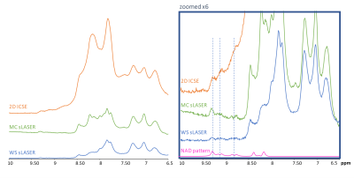 |
Detection and quantification of NAD+ in the human brain at 3 T: Comparison of three different localization techniques
Martyna Dziadosz1, Maike Hoefemann1, André Döring2, Malgorzata Marjanska3, Edward Auerbach3, and Roland Kreis1
1Departments of Radiology and Biomedical Research, University of Bern, Bern, Switzerland, 2Cardiff University Brain Research Imaging Centre (CUBRIC), School of Psychology, Cardiff University, Cardiff, United Kingdom, 3Department of Radiology, Center for Magnetic Resonance Research, University of Minnesota, Minnesota, MN, United States
The detection and quantification of Nicotinamide adenine dinucleotide (NAD+) was demonstrated to be challenging in 1H Magnetic Resonance Spectroscopy, due to its low concentration and reported polarization exchange with water. Frequency-selective excitation with slice-selective refocusing was proposed to prevent saturation-transfer from water. In this study we compare three techniques to access NAD+ quantification – standard WS semiLASER and two nWS (MC semiLASER and 2D I-CSE). NAD+ was detected with all techniques with a limited visibility for the WS semiLASER. While utilizing nWS concept allows to detect NAD+ at the visibility of 66%.
|
|||
1994.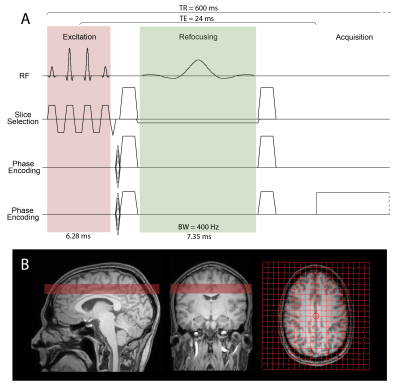 |
Mapping of downfield resonances in the human brain using 1H-MRSI with binomial spectral-spatial excitation and selective refocusing
Michal Považan1, Michael Schär1, Joseph S Gillen1,2, and Peter B Barker1,2
1Department of Radiology and Radiological Science, The Johns Hopkins University School of Medicine, Baltimore, MD, United States, 2Kennedy Krieger Institute, F. M. Kirby Research Center for Functional Brain Imaging, Baltimore, MD, United States
Water suppression employed in 1H-MR spectroscopy affects signals of exchangeable protons that resonate downfield from water. We have developed a novel method without water suppression for mapping these resonances on a 3T scanner using 2D 1H-MRSI in combination with binomial spectral spatial excitation and selective refocusing. Acquired data were consistent across all scanned subjects and within the whole FOV, with spectral patterns in agreement with previous single voxel studies.
|
|||
1995.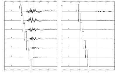 |
Effect of digitization in gradient modulated adiabatic pulses with a spatial offset.
Jan Willem van der Veen1 and Jun Shen1
1Magnetic Resonance Spectroscopy Core, NIH, NIMH, Bethesda, MD, United States
Spectroscopy on high field clinical scanners with limited RF amplitude suffer from large chemical shift artifacts with conventional pulses. Latest developments in adiabatic pulses with modulated gradients like GOIA-WURST pulses offer wide bandwidth at low RF amplitude. The modulated gradient however requires spatial offsets to be added to the phase modulation of the RF pulse. The additional phase offset may cause error due to insufficient digitization of the rapidly changing RF phase. We examined this effect on the voxel profile for two popular pulse parameter sets.
|
|||
1996.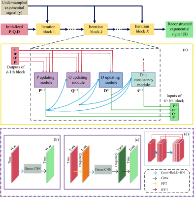 |
Accelerate Magnetic Resonance Spectroscopy with Deep Low Rank Hankel Matrix
Yihui Huang1, Jinkui Zhao1, Zi Wang1, Di Guo2, and Xiaobo Qu1
1Department of Electronic Science, National Institute for Data Science in Health and Medicine, Xiamen University, Xiamen, China, 2School of Computer and Information Engineering, Xiamen University of Technology, Xiamen, China
Nuclear Magnetic Resonance (NMR) spectroscopy is regarded as an important tool in bio-engineering while often suffers from its time-consuming acquisition. Non-Uniformly Sampling (NUS) method can speed up the acquisition, but the missing FID signals need to be reconstructed with proper method.. In this work, we proposed a deep learning reconstruction method based on unrolling the iterative process of a state-of-the-art model-based low rank Hankel matrix method. Experimental results show that the proposed method provides a better approximation of low rank and preserves the low-intensity signals much better.
|
|||
1997.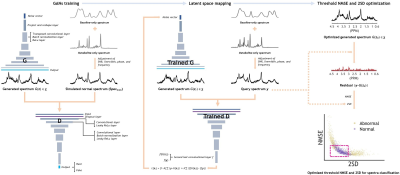 |
Unsupervised anomaly detection using generative adversarial networks in 1H-MRS of the brain
Joon Jang1, Hyeong Hun Lee1, Ji-Ae Park2, and Hyeonjin Kim3,4
1Department of Biomedical Sciences, Seoul National University, Seoul, Korea, Republic of, 2Division of Applied RI, Korea Institute of Radiological & Medical Science, Seoul, Korea, Republic of, 3Department of Medical Sciences, Seoul National University, Seoul, Korea, Republic of, 4Department of Radiology, Seoul National University Hospital, Seoul, Korea, Republic of
The applicability of generative adversarial networks (GANs) capable of unsupervised anomaly detection (AnoGAN) was investigated in the management of quality of 1H-MRS human brain spectra. The AnoGAN showed potential in the detection of the spectra with poor SNR or abnormal NAA levels. Despite the fact that those spectra contaminated with ghost, residual water or lipid have never been involved in the training or optimization of the AnoGAN, they were successfully filtered out depending on the intensity of the artifacts. Our unsupervised learning-based approach could be an option in the spectral quality management in addition to the previous supervised learning-based approaches.
|
|||
1998.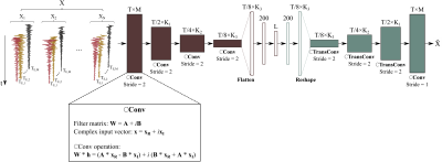 |
SNR-Enhancing Reconstruction for Multi-TE MRSI Using a Learned Nonlinear Low-dimensional Model
Yahang Li1,2, Zepeng Wang1,2, and Fan Lam1,2
1Department of Bioengineering, University of Illinois Urbana-Champaign, Urbana, IL, United States, 2Beckman Institute for Advanced Science and Technology, University of Illinois Urbana-Champaign, Urbana, IL, United States
We report a new method for SNR-enhancing reconstruction of multi-TE MRSI data. Specifically, we designed a deep complex convolutional autoencoder (DCCAE) to learn a nonlinear low-dimensional model of the high-dimensional multi-TE spectra which allowed for effective separation of molecular signals and noise. A constrained reconstruction formulation is used to incorporate the learned model for denoising spatial-temporal reconstruction. The performance of the learned model and the proposed reconstruction method have been evaluated using both simulation and experimental multi-TE $$$^1$$$H-MRSI data. Results obtained demonstrate superior denoising performance achieved by the proposed method over alternative spatial-spectrally constrained denoising strategies.
|
|||
1999.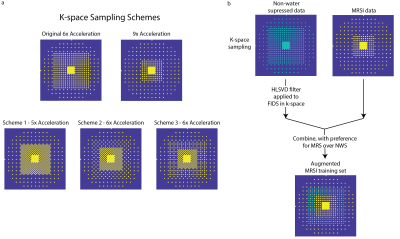 |
Highly accelerated variable-density MultiNet CAIPIRINHA for 1H MRSI and augmented MRSI neural network training
Kimberly Chan1 and Anke Henning1,2
1The University of Texas Southwestern, Dallas, TX, United States, 2Max Planck Institute for Biological Cybernetics, Tübingen, Germany
It has previously been shown that neural networks combined with variable k-space undersampling (MultiNet GRAPPA) is superior to a conventional GRAPPA reconstruction and is feasible at 7T. Here, MultiNet reconstruction of several new CAIPIRINHA-based variable-density k-space undersampling schemes is investigated. A new approach to train the neural networks (NN) by augmenting the MRSI data with the non-water suppressed (NWS) data to provide additional self-calibration training data is also introduced and evaluated. In this study, both are shown here to reduce lipid artifacts and improve metabolic maps at high acceleration factors relative to those previously proposed for MultiNet GRAPPA.
|
|||
2000.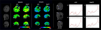 |
k-Space-based Coil Combination via Geometric Deep Learning for Reconstruction of non-Cartesian MR Spectroscopic Imaging Data
Stanislav Motyka1, Lukas Hingerl1, Bernhard Strasser1, Gilbert Hangel1, Eva Heckova1, Asan Agibetov2, Georg Dorffner2, and Wolfgang Bogner1
1Department of Biomedical Imaging and Image-guided Therapy, Medical University of Vienna, Wien, Austria, 2Section for Artificial Intelligence and Decision Support (CeMSIIS), Medical University of Vienna, Wien, Austria
A new coil combination method of non-Cartesian kspace MRSI data based on Geometric deep learning is introduced and compared to the conventional image-based coil combination. MRSI data were represented as a graph and a shallow neural network was used to solve the coil combination task. The training data were based on in vivo data and the performance of the network was tested on volunteer data, whose data were never shown to the network. The results were similar to conventional image-domain based coil combination. Thus, a highly accelerated online reconstruction is feasible with this method.
|
|||
2001. |
Magnetic Resonance Imaging and Spectroscopy in Late-Onset GM2-Gangliosidosis
Olivia E Rowe1, Rangaprakash Deshpande1, Akila Weerasekera1, Christopher Stephen2,3, Robert L Barry1,4, Florian Eichler3,5, and Eva-Maria Ratai1
1Athinoula A. Martinos Center for Biomedical Imaging, Department of Radiology, Massachusetts General Hospital and Harvard Medical School, Charlestown, MA, United States, 2Movement Disorders Division and Ataxia Center, Department of Neurology, Massachusetts General Hospital and Harvard Medical School, Boston, MA, United States, 3Center for Rare Neurological Diseases, Department of Neurology, Massachusetts General Hospital and Harvard Medical School, Boston, MA, United States, 4Harvard-Massachusetts Institute of Technology Health Sciences & Technology, Cambridge, MA, United States, 5Leukodystrophy Clinic, Department of Neurology, Massachusetts General Hospital and Harvard Medical School, Boston, MA, United States
Late-onset GM2-Gangliosidoses (LOGG) are rare lysosomal storage disorders with slowly progressing neurological symptoms that encompass late-onset Tay-Sachs disease (LOTS) and Sandhoff disease (LOSD). We performed sMRI-MRS to discern how cerebellar aberrations may relate to underlying metabolic abnormalities, and their relationships to clinical presentations. Results revealed that structural and metabolic differences between LOTS patients and controls were more prominent than the collective group of LOGG patients vs. controls. Significant clinical associations with imaging-markers also differed when LOTS patients were analyzed separately versus in conjunction with LOSD patients. While LOSD could not be accurately assessed, it appeared to be distinct from LOTS.
|
|||
2002.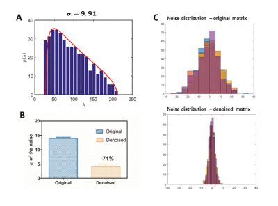 |
The impact of Marchencko-Pasteur principal component analysis denoising on high-resolution MR spectroscopic imaging in the rat brain at 9.4T.
Dunja Simicic1,2,3, Jessie Julie Mosso1,2,3, Thanh Phong Lê3,4, Ruud B. van Heeswijk5, Ileana Ozana Jelescu1,2, and Cristina Cudalbu1,2
1CIBM Center for Biomedical Imaging, Lausanne, Switzerland, 2Animal Imaging and Technology, EPFL, Lausanne, Switzerland, 3Laboratory of Functional and Metabolic Imaging, EPFL, Lausanne, Switzerland, 4Geneva School of Health Sciences, HES-SO University of Applied Sciences and Arts Western Switzerland, Geneva, Switzerland, 5Department of Radiology, Lausanne University Hospital (CHUV) and University of Lausanne (UNIL), Lausanne, Switzerland
MRSI is a powerful tool for the non-invasive simultaneous mapping of metabolic profiles at multiple spatial positions. This method is highly challenging due to low concentration of metabolites, long measurement times, low SNR, hardware limitations and need for advanced pulse sequences. Denoising based on singular value decomposition has been previously used, but determination of the appropriate thresholds that separate the noise from the signal is problematic leading to possible loss of spatial resolution. Aim of the present study was to implement an improved denoising technique (Marchenko-Pastur principal component analysis) on high resolution MRSI data acquired at 9.4T in the rat-brain.
|
The International Society for Magnetic Resonance in Medicine is accredited by the Accreditation Council for Continuing Medical Education to provide continuing medical education for physicians.