Digital Posters
Molecular Imaging & X-Nuclei: Developments
ISMRM & SMRT Annual Meeting • 15-20 May 2021

Digital Posters
Molecular Imaging & X-Nuclei: Developments
| Concurrent 2 | 13:00 - 14:00 |
1804.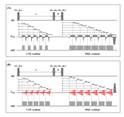 |
Triple-quantum-filtered Sodium MRI at 7T: Optimization of the Enhanced SISTINA Sequence Using FLORET k-space Trajectories
Qingping Chen1,2,3, Wieland A. Worthoff1, and N. Jon Shah1,2,4,5
1Institute of Neuroscience and Medicine 4, INM-4, Forschungszentrum Jülich, Germany, 2Department of Neurology, RWTH Aachen University, Aachen, Germany, 3Department of Biomedical Engineering, The University of Melbourne, Parkville, Australia, 4Institute of Neuroscience and Medicine 11, INM-11, JARA, Forschungszentrum Jülich, Germany, 5JARA - BRAIN - Translational Medicine, Aachen, Germany
It has been reported that enhanced SISTINA, one of the triple-quantum-filtered sodium MRI techniques, can selectively measure the distribution of restricted sodium, which may be sensitive to metabolic cellular dysfunction. In this work, the enhanced SISTINA sequence was optimized for 7T using FLORET, which has high k-space filling efficiency and good potential for undersampling. The optimized enhanced SISTINA sequence greatly improves the UTE image quality, while maintaining the multiple-quantum-filtered image performance and introducing incoherent randomness to the raw data. We believe the optimized enhanced SISTINA has promising potential for clinical applications with faster scans using compressed-sensing reconstruction in future.
|
|||
1805.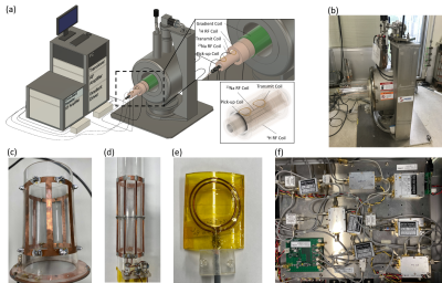 |
Development of an add-on 23Na MRI platform for an existing 1H MRI scanner using a crossband repeater: Proof-of-concept
Michiru Kajiwara1, Tomoyuki Haishi2,3, and Yasuhiko Terada1
1Institute of Applied Physics, University of Tsukuba, Tsukuba, Japan, 2Department of Radiological Sciences, School of Health Sciences at Narita, International University of Health and Welfar, Narita, Japan, 3MRTechnology Inc., Tsukuba, Japan
23Na MRI provides novel information on energy metabolism, and there are growing expectations for its application in clinical MRI. However, implementation of 23Na MRI for existing 1H MRI systems requires expensive dedicated transceivers and RF coils for sodium imaging. Here, we have developed an add-on crossband repeater system that can acquire 23Na MRI signals simply by inserting it into the magnet’s bore of an existing 1H MRI, and proved this concept by phantom and in vivo mouse experiments. This add-on platform is applicable to other 1H MRI systems and will enhance the feasibility of 23Na MRI in clinical practice.
|
|||
 |
1806.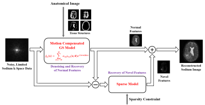 |
High-Resolution Sodium Imaging Using Anatomical and Sparsity Constraints for Denoising and Recovery of Novel Features
Yibo Zhao1,2, Rong Guo1,2, Yudu Li1,2, Keith R. Thulborn3, and Zhi-Pei Liang1,2
1Department of Electrical and Computer Engineering, University of Illinois, Urbana-Champaign, Urbana, IL, United States, 2Beckman Institute for Advanced Science and Technology, University of Illinois, Urbana-Champaign, Urbana, IL, United States, 3Center for Magnetic Resonance Research, University of Illinois at Chicago, Chicago, IL, United States
Quantitative sodium MRI is a unique tool for assessing tissue viability noninvasively. A major obstacle to widespread clinical applications of sodium MRI is low sensitivity, which leads to low spatial resolution even with long scan times. We present a novel method for reconstruction of high-resolution sodium maps from noisy, limited k-space data. The proposed method has been validated using simulated and experimental data, producing high-SNR and high-resolution tissue sodium concentration maps. These high-resolution maps were also shown to improve the detection of tumor responses to radiation therapy.
|
||
1807.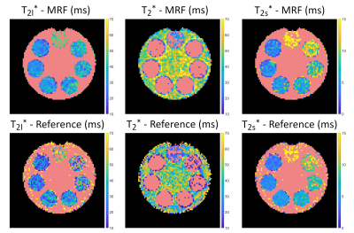 |
Three Dimensional Sodium Magnetic Resonance Fingerprinting using Irreducible Spherical Tensor Operator Simulations
Fabian J. Kratzer1, Sebastian Flassbeck1,2,3, Sebastian Schmitter1,4, Tobias Wilferth5, Arthur W. Magill1, Benjamin R. Knowles1, Tanja Platt1, Peter Bachert1, Mark E. Ladd1, and Armin M. Nagel1,5
1Medical Physics in Radiology, German Cancer Research Center (DKFZ), Heidelberg, Germany, 2Center for Advanded Imaging Innovation and Research, New York University, New York, NY, United States, 3Center for Biomedical Imaging, Dept. of Radiology, New York University, New York, NY, United States, 4Physikalisch Technische Bundesanstalt (PTB), Braunschweig and Berlin, Germany, 5Institute of Radiology, University Hospital Erlangen, Friedrich-Alexander-Universität Erlangen (FAU), Erlangen, Germany
Sodium relaxation times have been shown to be altered in several diseases. Hence, 2D sodium relaxometry using Magnetic Resonance Fingerprinting (23Na-MRF) was demonstrated recently as a proof of concept. In this work an extension to a 3D sequence is presented. Furthermore, a more complex signal model based on irreducible spherical tensor operators was investigated. The feasibility of simultaneous 3D quantification of T1, T2s*, T2l*, T2* and ΔB0 was demonstrated in phantom measurements.
|
|||
1808.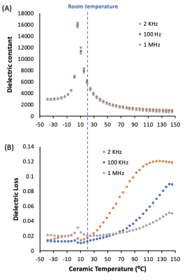 |
New tuHDC BST-BT Ceramics with Optimal Permittivity Greatly Improve B1 efficiency and SNR at Room Temperature for 17O MRSI Application at 10.5T
Hannes Michel Wiesner1, Xiao-Hong Zhu1, Maryam Sarkarat2, Xin Li1, Matt Waks1, Michael T. Lanagan2, Qing X. Yang3, and Wei Chen1
1CMRR, Department of Radiology, University of Minnesota, Minneapolis, MN, United States, 2Department of Engineering, Pennsylvania State College of Engineering, University Park, PA, United States, 3CNMRR, Department of Neurosurgery, Penn State University, Hershey, PA, United States
Recently we reported that the ceramics made of composite BST (Ba0.6Sr0.4TiO3) material have an extremely high tunability of relative permittivity when changing the ceramic temperature; and by cooling the ceramics to 14-15 °C, they provided the best improvement for 17O MRSI application at 10.5T, resulting in an optimal permittivity of ~6000. In this study, we developed new ceramic disks made of mixed Ba0.6Sr0.4TiO3 and BaTiO3 (BST-BT) materials to achieve the optimal permittivity at room temperature. The results indicate that unprecedented performance is possible for 17O MRSI application at 10.5T without the need of a temperature controller.
|
|||
1809.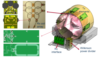 |
A Dedicated RF Coil System for 19F MRI of Myocardial Infarction at a 3 T Clinical MRI System
Ali Caglar Özen1,2, Felix Spreter1, Timo Heidt3, Constantin von zur Mühlen3, and Michael Bock1
1Deptartment of Radiology, Medical Physics, University Medical Center Freiburg, University of Freiburg, Freiburg, Germany, 2German Consortium for Translational Cancer Research Partner Site Freiburg, German Cancer Research Center (DKFZ), Heidelberg, Germany, 3Department of Cardiology and Angiology I, UHZ, University Medical Center and Faculty of Medicine, University of Freiburg, Freiburg, Germany
Fluorine magnetic resonance imaging is a non-invasive background-free MRI approach to image infiltrating monocytes. The transfer of 19F MRI into large animals is needed for both method validation and the development of clinical applications. In this study, we have developed an 8 transmit/ 8 receive coil array tuned to 115.9 MHz (at 3 T) and developed a detachable 19F coil system for cardiac and thoracic MRI in pigs.
|
|||
1810.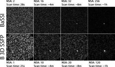 |
Fast Fluorine Spectroscopic Imaging with pseudo-spiral k-space sampling
Muhammed Yildirim1,2, Patrick Scholtz2, Markus Schütz2, Xenia Kovalyk2, Edwin Heijman2,3, Rolf Lamerichs3, Holger Grüll2, and Esin Ozturk Isik1
1Computational Imaging Lab, Institute Of Biomedical Engineering, Bogazici University, Istanbul, Turkey, 2Institute of Diagnostic and Interventional Radiology, Faculty of Medicine and University Hospital of Cologne, University of Cologne, Cologne, Germany, 3Philip Research Labs, Eindhoven, Netherlands
19F-MRI constitutes a viable platform for quantitative theranostics. However, being impaired by physiological absence, and complex NMR spectra of perfluorocarbons, for 19F-MR it is essential to achieve high imaging efficiency. This work introduces a novel spectroscopic imaging sequence, BaSSI, that resides on a balanced psuedo-spiral acquisition scheme and compares against 3D balanced UTE SSFP imaging, that has become the golden standard of fluorine imaging. The work, optimizes both sequences to their fullest, and tests their performance using miniscule samples of PFOB.
|
|||
1811.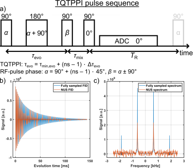 |
Comparison of low rank compressed sensing with non-uniform undersampled non-linear FID fitting for time efficient 23Na TQTPPI measurements
Simon Reichert1, Dennis Kleimaier1, and Lothar Schad1
1Computer Assisted Clinical Medicine, Medical Faculty Mannheim, Heidelberg University, Mannheim, Germany
This study demonstrates the feasibility of low rank compressed sensing and non-uniform undersampled non-linear FID fitting for time efficient sodium TQTPPI measurements. In simulations, undersampling factors of up to 5 resulted in less than 10% deviation from the ground truth for all parameters. This accuracy was confirmed in measurement data of agarose and protein samples. Hence, a measurement time reduction up to a factor of 5 is possible without significantly reducing the accuracy of the fit parameters. Thus, CS allows for time efficient TQTPPI measurements to investigate cellular processes as well as isolated sodium protein interactions.
|
|||
1812.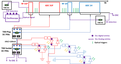 |
Interleaved 31P MRS and 1H dual-echo GRE B0 map pulse sequence for 7T Terra scanners
Jabrane Karkouri1 and Christopher T. Rodgers1
1Wolfson Brain Imaging Center, University of Cambridge, Cambridge, United Kingdom
In this abstract, we demonstrate a framework for interleaving 1H imaging and 31P spectroscopy on the Siemens 7T Terra platform. We show that this can be done without making hardware modifications and demonstrate the concept with interleaved B0 map acquisition with 31P-MRS on a phantom and the quadriceps of a healthy volunteer.
|
|||
1813. |
Three-Dimensional Sodium MRI Using A Rotation of Spiral Disc (RSD) Trajectory
Kwan-Jin Jung1 and Brad Sutton1,2
1Beckman Institute for Advanced Science and Technology, University of Illinois at Urbana-Champaign, Urbana, IL, United States, 2Electrical & Computer Engineering, University of Illinois at Urbana-Champaign, Urbana, IL, United States
Sodium MRI is challenging due to its low sensitivity and short T2 and hence a three-dimensional sequence with a spiral trajectory has been applied. The spiral trajectory was shortened in the TPI method by accelerating the span at the k-space origin using a radial start and then transiting into a spiral trajectory. This popular TPI method, however, requires a very high gradient slew rate when the 3D cone approaches the polar pole. This drawback has been resolved by rotating a two-dimensional disc filled with interleaved TPI trajectories. This method achieved the faster sweeping of TPI as well as a uniform gradient slew rate over the sphere sampling. We are reporting some artifacts in TPI-based scans not only in our study but also in other studies.
|
|||
1814.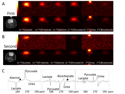 |
Conserving low amplitude resonances in tensor rank truncation for image enhancement of spectroscopic imaging data.
Alan J. Wright1, Richard Mair1,2,3, Anastasia Tsyben1, and Kevin M. Brindle1,3,4
1CRUK Cambridge Institute, University of Cambridge, Cambridge, United Kingdom, 2Department of Clinical Neurosciences, University of Cambridge, Cambridge, United Kingdom, 3Cancer Research UK Major Centre-Cambridge, University of Cambridge, Cambridge, United Kingdom, 4Department of Biochemistry, University of Cambridge, Cambridge, United Kingdom
Tensor decomposition can be used for denoising magnetic resonance spectroscopic imaging (MRSI) data by reconstructing the data from a small number of ranks. Selecting too low an order can remove data from the reconstruction and alter peak amplitudes. A condition for selection of rank order is proposed that removes only noise from the reconstructed data and, therefore, preserves signals from low amplitude resonances. Denoising algorithms that apply this condition are demonstrated for metabolic imaging data sets obtained with hyperpolarised [1-13C]pyruvate and [6,6-2H]glucose in preclinical cancer models, which improve signal-to-noise and reduce the Cramér-Rao lower bound errors in peak fitting.
|
|||
1815.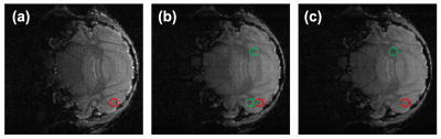 |
Variable Temporal Resolution Cartesian Sampling for Cell Tracking MRI
Mark Armstrong1, Felix Freppon2, Enrica Wilken2, Max Masthoff2, Cornelius Faber2, and Dan Xiao1
1University of Windsor, Windsor, ON, Canada, 2Universität Münster, Münster, Germany
Individual iron oxide nanoparticle labelled immune cells can be visualized with a dynamic MRI image series. Time-lapse MRI can provide insights in the studies of inflammatory diseases and metastasis of cancer. The MRI temporal resolution can be improved by undersampling k-space. However, the optimal acceleration factor cannot be easily determined prior to experiments. Strategies have been proposed for non-Cartesian flexible retrospective undersampling, which may not be readily applicable for most users. We propose a Cartesian sampling scheme where undersampling ratio and temporal resolution can be chosen retrospectively.
|
|||
1816.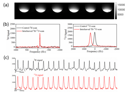 |
Single Loop Tri-frequency Surface Coil Design for 1H MRI and Interleaved Dynamic 2H and 17O MRS Applications at Ultrahigh Field of 16.4T
Parker John Bresnahan Jenkins1, Guangle Zhang1, Wei Zhu1, Xiao-Hong Zhu1, and Wei Chen1
1Center for Magnetic Resonance Research, University of Minnesota, Minneapolis, MN, United States
To capture anatomical information and dynamic changes of different metabolic activities in the brain or other organs using a singular RF coil, we developed a loop RF surface coil design with a wide tune-match isolation and active tune circuitry for proton ( 1H) MRI, and interleaved deuterium ( 2H) and oxygen ( 17O) MRS measurements at 16.4T. We obtained static 1H MRI and performed interleaved dynamic 2H and 17O measurements in phantom studies. Results showed proficient proton imaging and quasi-stable 2H and 17O signals with rapid acquisition, indicating the potential of the design for robust high temporal resolution metabolic and anatomical imaging applications.
|
|||
1817.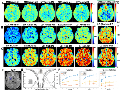 |
Parametric VARied Saturation (PaVARS) for enhanced CEST characterization of human brain at 3T
Zhensen Chen1, Yishi Wang2, Le He1, Hanyu Wei1, Yibing Chen3, and Xiaolei Song1
1Center for Biomedical Imaging Research, School of Medicine, Tsinghua University, Beijing, China, 2Philips Healthcare, Beijing, China, 3School of Information Science and Technology, Northwest University, Xi'an, China
Existing CEST methodologies suffer from low specificity in distinguishing various exchange species in tissue. To further utilize dependence of CEST spectra on saturation length (tsat), power (B1) as well as frequency, we setup a fast acquisition of Parametric VARied Saturation (PaVARS) in a 3T clinical scanner, by separating the seconds-long saturation preparation to 5-8 modules each followed by a low flip-angle readout. On phantoms of three metabolites and human brain, PaVARS enabled fast acquisition of multiple Zspectra, each weighted with different saturation length and power. PaVARS could be a ready-to-use, informative acquisition methods for better differentiation of endogeneous brain metabolites.
|
|||
1818.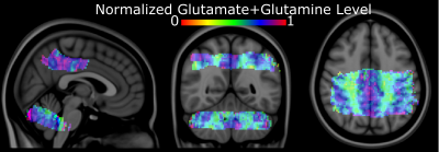 |
Towards a Probabilistic Neurochemical atlas via parcellated approach using ZOOM MRSI
Nicholas Farley1, Jaiyta Sood1, Antonia Susjnar2, Sean Lane3, Mark Chiew4, Michael Albert Thomas5, and Uzay Emir1,2
1School of Health Sciences, Purdue University, West Lafayette, IN, United States, 2Weldon School of Biomedical Engineering, Purdue University, West Lafayette, IN, United States, 3Department of Psychological Sciences, Purdue University, West Lafayette, IN, United States, 4Welcome Center for Integrative Neuroimaging, University of Oxford, Oxford, United Kingdom, 5Department of Radiology, University of California Los Angeles, Los Angeles, CA, United States
To create a probabilistic Neurochemical atlas of the human brain via parcellated approach using ZOOM MRSI, in this study, we aim to quantify the test-retest reliability of the ZOOM MRSI by assessing the stability of the molecular contrasts of the posterior cingulate and cerebellum across two independent scan sessions and by quantifying the intra- and inter-rater reliability of the ZOOM MRSI scan.
|
|||
1819.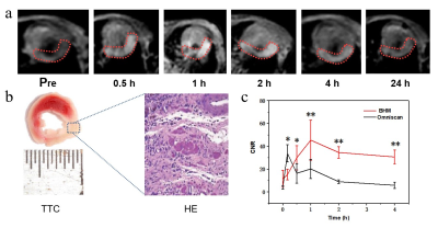 |
MRI contrast agent for myocardial infarction based on novel manganese-based nanoparticles
Chengbin He1, Xiaoxuan Zhou1, Cailing Pu1, Jingle Fei1, Yan Wu1, and Hongjie Hu1
1Department of Radiology, Sir Run Run Shaw Hospital (SRRSH), Zhejiang University School of Medicine, Hangzhou, China
Gadolinium is a commonly used contrast agent for myocardial infarction during CMR examinations, but its nephrotoxicity and short examination time window limited its application to some extent. We try to develop new contrast agents with superior imaging performance and good biocompatibility, which can achieve clinical translation.
|
|||
1820. |
MR detection of gas microbubbles via hyperCEST: a path toward a dual modality contrast agent
Christian T McHugh1, Phillip G Durham2, Michele Kelley1, Paul A Dayton3, and Rosa T Branca1
1Physics & Astronomy, University of North Carolina at Chapel Hill, Chapel Hill, NC, United States, 2Pharmacoengineering and Molecular Pharmaceutics, University of North Carolina at Chapel Hill, Chapel Hill, NC, United States, 3Biomedical Engineering, University of North Carolina at Chapel Hill, Chapel Hill, NC, United States
Gas microbubbles are capable of providing both ultrasound and MRI image contrast, but at differing concentration levels. In order for gas microbubbles to be detected by MRI at the clinical ultrasound dose, a more sensitive detection scheme must be employed. Here we demonstrate that hyperCEST is capable of detecting gas microbubbles at clinically relevant US concentrations, thus enabling their use as a dual modality contrast agent.
|
|||
1821. |
Kartogenin and synthetic melanin nanoparticle loaded hydrogel scaffold for cartilage regeneration and monitored by quantitative MRI
Chuyao Chen1, Shaoshan Huang2, Zelong Chen1, Chenggong Yan1, Yingjie Mei3, Yikai Xu1, and Rui Guo2
1Medical Imaging Center, Nanfang Hospital, Southern Medical University, Guangzhou, China, 2Key Laboratory of Biomaterials of Guangdong Higher Education Institutes, Guangdong Provincial Engineering and Technological Research Centre for Drug Carrier Development, Department of Biomedical Engineering, Jinan University, Guangzhou, China, 3Philips Healthcare, Guangzhou, China
We have developed a KGN/SMNP loaded cell-therapy theranostic hydrogel scaffold for cartilage repair. The multifunctional hydrogel system is expected to be a tissue engineering strategy for cartilage regeneration and non-invasive monitoring by multiparametric MR imaging.
|
|||
1822. |
Glycogen Synthesis and Glucose Utilization at 3T using Localized Spectroscopy and Chemical Shift Imaging
Shun Kishimoto1, Jeffrey R Brender1, Jeeva Munasinghe2, Martin Lizak2, Yu Saito1, Kota Yamashita1, Otowa Yasunori1, Kazu Yamamoto1, James Mitchell1, and Murali C. Krishna1
1NIH/NCI, Bethesda,, MD, United States, 2NIH/NINDS, Bethesda,, MD, United States
Aberrant glycogen metabolism has been implicated in vitro as a possible causative factor in multiple disorders. Progress has been stymied due to the rapid degradation of glycogen post mortem and the lack of methods for measuring glycogen metabolism in vivo. We show here that glucose metabolism and local synthesis of glycogen can be measured in mice by short echo PRESS following a single bolus of unlabeled glucose using a standard 3T preclinical MRI using low rank reconstruction. Imaging of glycogen synthesis was possible at 9.4T by CSI, where it was found in mice to be centered on the olfactory bulb
|
|||
1823 |
In vivo hijacking local activated macrophages for targeted theranostics of epilepsy with nanomedicines Video Permission Withheld
Lin Lin1,2, Xiaodan Chen3, and Pu-Yeh Wu4
1Fujian Medical University Union Hosptial, Fuzhou, China, 2Fudan University Huashan Hospital, Shanghai, China, 3Fujian Cancer Hospital, Fuzhou, China, 4GE Healthcare, Beijing, China
Epilepsy is closely linked to inflammation, oxidative stress, and hypoxia. The difficulty of accurate localization and targeted drug delivery to the lesion hinders the effective treatment. The local activated inflammatory cells offer a new opportunity for drug delivery to the lesion. Here, CD163 positive macrophages were first utilized as cell carriers after being hijacked by targeted albumin nanoparticles, which effectively delivered nanoparticles to the epileptic sites. Thus, accumulative nanoparticles enabled the visualization of the epileptogenic lesion though microenvironment-responsive MR-T1W imaging. Moreover, these manganese-based nanomaterials played a crucial role in protecting neurons from cell apoptosis mediated by oxidative stress and hypoxia.
|
The International Society for Magnetic Resonance in Medicine is accredited by the Accreditation Council for Continuing Medical Education to provide continuing medical education for physicians.