Digital Posters
Spectroscopy: Neuro II
ISMRM & SMRT Annual Meeting • 15-20 May 2021

| Concurrent 2 | 17:00 - 18:00 |
2219.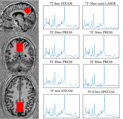 |
Assessing the agreement between 3T and 7T MRS measures of in-vivo neurochemistry.
Dana Goerzen1,2, Tiffany Bell3,4,5, Jamie Near1,2, and Ashley D Harris3,4,5
1McGill University, Montreal, QC, Canada, 2Centre d'Imagerie Cérébrale, Douglas Mental Health University Institute, Montreal, QC, Canada, 3Radiology, University of Calgary, Calgary, AB, Canada, 4Hotchkiss Brain Institution, Calgary, AB, Canada, 5Alberta Children's Hospital Research Institute, Calgary, AB, Canada
A key challenge in magnetic resonance spectroscopy in the human brain is assessing the reliability of metabolite concentration estimates, since the ground truth is generally unknown. This project assesses the correlations between 3T MRS metabolite measures against two “gold standard” 7T sequences as a means to assess which 3T sequences are the most reliable at detecting common metabolites of interest. This work will help researchers to make informed choices about which sequences to use for optimal detection of their metabolites of interest.
|
|||
2220.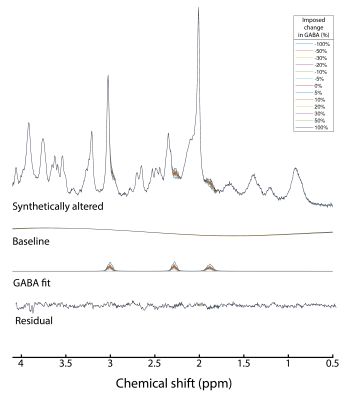 |
Quantification of GABA in ultra-short TE 7-T human MR spectra
Guglielmo Genovese1, Dinesh K. Deelchand1, Melissa Terpstra1, and Malgorzata Marjanska1
1Center for Magnetic Resonance Research (CMRR), Department of Radiology, University of Minnesota, Minneapolis, MN, United States
The goal of this abstract was to deepen the knowledge on the influence of LCModel fitting parameters and SNR on GABA quantification and on the sensitivity for detecting differences in GABA levels. To this aim, GABA concentrations were quantified using different LCModel approaches and different number of averages in synthetically altered spectra generated by adding synthetic GABA signal to in-vivo ultra-short TE, 7-T human spectra.
|
|||
2221. |
Improving the B0 homogeneity in 7 T MRSI applications using a 24-channel local array of shim coils
Philipp Lazen1, Sahar Nassirpour2, Paul Chang2, Lukas Hingerl1, Karl Rössler3, Siegfried Trattnig1,4, Wolfgang Bogner1, and Gilbert Hangel1,3
1Department of Biomedical Imaging and Image-guided Therapy, Medical University of Vienna, Vienna, Austria, 2MR Shim GmbH, Reutlingen, Germany, 3Department of Neurosurgery, Medical University of Vienna, Vienna, Austria, 4Christian Doppler Laboratory for Clinical Molecular MR Imaging, Vienna, Austria
A 24-channel array of local shim coils in a 7 T Siemens Terra Fit was tested on eight healthy volunteers. Significant improvements of the homogeneity of the static magnetic field could be achieved and were qualitatively shown and quantitatively analyzed. The standard deviation of the resonance frequency decreased from 42.5 Hz for the standard shim (up to second order spherical harmonics) to 34.5 Hz for the 24-channel local shim array. The full width at half maximum and the signal to noise ratio also improved with the better shim. Lastly, some preliminary MRSI results could be gathered.
|
|||
2222.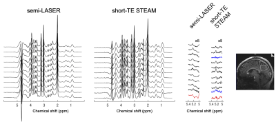 |
Comparison of Semi-LASER and Short-TE STEAM Pulse Sequences for Cerebral Glucose Quantification via Detecting H1-α-Glucose Peak in 1H MRS at 7 T
Hideto Kuribayashi1, Yuta Urushibata1, Thuy Ha Duy Dinh2, Hirohiko Imai3, Sinyeob Ahn4, Ravi Teja Seethamraju5, Tadashi Isa2, and Tomohisa Okada2
1Siemens Healthcare K.K., Tokyo, Japan, 2Human Brain Research Center, Kyoto University Graduate School of Medicine, Kyoto, Japan, 3Kyoto University Graduate School of Informatics, Kyoto, Japan, 4Siemens Medical Solutions, Berkeley, CA, United States, 5Siemens Medical Solutions, Boston, MA, United States
Single voxel MRS pulse sequences of semi-LASER and short-TE STEAM were compared for cerebral glucose quantification via detecting H1-α-glucose peak at 5.23 ppm. Young healthy non-fasted subjects were scanned on a whole-body 7T MRI. Spectra were analyzed on LCModel and in-house MATLAB software. Semi-LASER (32-ms TE) detected the H1-α-glucose peak at posterior cingulate cortex (27-mL volume) with higher SNR (9.1 vs 5.1) than short-TE STEAM (5-ms TE) in shorter scan time (11 vs. 12.5 minutes), respectively, and estimated glucose concentration to be 1.26 mM. More robust water suppression techniques are required to stabilize baselines around the peak.
|
|||
2223.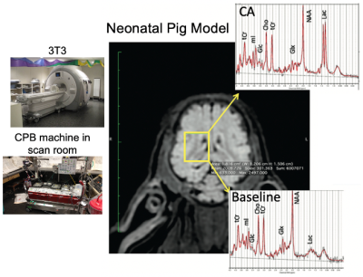 |
1H MRS of the Pediatric Brain Metabolism During Cardiopulmonary Bypass (CPB) Surgery
Daniel Spielman1, Phil Adamson2, Ralph Hurd1, Meng Gu1, Kirk Riemer3, Ryan Beckman3, Michael Ma3, Kenichi Okamura3, and Frank Hanley3
1Radiology, Stanford University, Stanford, CA, United States, 2Electrical Engineering, Stanford University, Stanford, CA, United States, 3Cardiothoracic Surgery, Stanford University, Stanford, CA, United States
We used single-voxel 1H MRS at 3T to study acute brain metabolic changes during cardiopulmonary bypass (CPB) surgery in a neonatal piglet model, with the overall goal of determining optimal surgical parameters for minimizing brain injury. The recent improvements in proton spectroscopy and MR scanner performance allowed robust measurement of dynamic brain water and metabolic changes during hypothermia and circulatory arrest. Key findings include a linear relationship between unsuppressed water and brain temperature, observation of the theoretically predicted NMR “powder pattern” from intravascular water during hypoxia, and quantitative measurements of glucose, lactate, and phosphocreatine metabolic rates vs temperature.
|
|||
2224.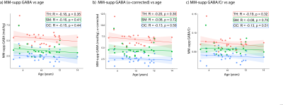 |
Macromolecule suppressed GABA levels show no relationship with age in children and adolescents
Tiffany Bell1,2,3, Mehak Stokoe1,2,3, and Ashley Harris1,2,3
1Department of Radiology, University of Calgary, Calgary, AB, Canada, 2Hotchkiss Brain Institute, University of Calgary, Calgary, AB, Canada, 3Alberta Children's Hospital Research Institute, University of Calgary, Calgary, AB, Canada
Studies have shown that GABA+ increases with age during development, however roughly 50% of this signal reflects macromolecules. It is often assumed that macromolecule levels are stable, but there has been no attempt to directly validate this in youth. Here, we use a sequence specifically designed to suppress the macromolecule signal that contaminates the measured GABA+ signal when using MEGA-PRESS. We show no relationship between macromolecule-suppressed GABA and age in youth aged 7-14 years. We suggest increases in macromolecule levels may drive previously shown relationships between age and GABA+ levels, highlighting the importance of controlling for macromolecules in MRS analyses.
|
|||
2225.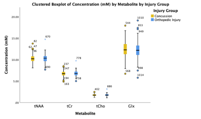 |
Exploring Brain Metabolites in Pediatric Concussion: An A-CAP Study
Parker L La1, Robyn Walker1, Julie M Joyce1, Tiffany Bell1, William Craig2, Quynh Doan3, Miriam Beauchamp4, Roger Zemek5, Pediatric Emergency Research Canada (PERC)6, Keith O Yeates7, and Ashley D Harris1
1Radiology, University of Calgary, Calgary, AB, Canada, 2Pediatrics, University of Alberta and Stollery Children's Hospital, Edmonton, AB, Canada, 3Pediatrics, University of British Columbia, Vancouver, BC, Canada, 4Psychology, University of Montreal and St Justine Hospital, Montreal, QC, Canada, 5Pediatrics and Emergency Medicine, University of Ottawa and Children's Hospital of Eastern Ontario, Ottawa, ON, Canada, 6Alberta Children's Hospital, Calgary, AB, Canada, 7Psychology, University of Calgary, Calgary, AB, Canada
Disruptions in brain metabolites following concussion are commonly reported in the literature. These studies often have different timings following injury, are limited to adults and have limited sample size. Using magnetic resonance spectroscopy (MRS), we show in the largest MRS in pediatric concussion study to date, that there are no group differences in metabolites between concussion and orthopedic injury at acute injury. Glx was associated with cognitive symptoms but no other metabolites showed a relationship with symptoms. No difference between groups may indicate that metabolic disturbance following injury may be a broad indicator of injury or alterations are regionally specific.
|
|||
2226.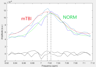 |
Measurement of local values of cerebral pH in mild traumatic brain injury using 1H MR spectroscopy
Peter Bulanov1, Andrei Manzhurtsev1,2, Petr Menshchikov3,4, Maxim Ublinskiy2,3, Ilya Melnikov2, Natalia Semenova1,2,3,5, and Tolib Akhadov2
1Lomonosov Moscow State University, Moscow, Russian Federation, 2Clinical and Research Institute of Emergency Pediatric Surgery and Traumatology, Moscow, Russian Federation, 3Emanuel Institute of Biochemical Physics of RAS, Moscow, Russian Federation, 4PHilips Healthcare, Russia, Moscow, Russian Federation, 5Semenov Institute of Chemical Physics of the Russian Academy of Sciences, Moscow, Russian Federation
The effect of mild traumatic brain injury (mTBI) on the values of the cerebral pH in the region of the posterior cingulate cortex was studied. pH was determined by analyzing the chemical shifts of mobile protons in the aromatic region of the spectrum of histidine, homocarnosine, resonating at ~7-8 ppm in PRESS spectra. According to our data, the pH value decreases by ~1% in posterior cingulate cortex after acute mTBI.
|
|||
2227.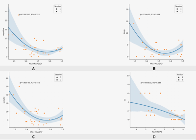 |
The Correlation of Neurometabolites on Presentation of Post-Concussion Phenotypes
Katherine Breedlove1, David R Howell2, Inga K Koerte3,4, Eduardo Coello4, Molly Charney4, Huijin Liao4, Corey Lanois5, and Alexander Lin4
1Radiology, Brigham and Women's Hospital, Boston, MA, United States, 2Childrens Hospital Colorado, Aurora, CO, United States, 3Ludwig-Maximilians-Universitat, Munich, Germany, 4Brigham and Women's Hospital, Boston, MA, United States, 5Boston Children's Hospital, Boston, MA, United States
A population of collegiate athletes was evaluated post-concussion for neurometabolic levels in the PCG by magnetic resonance spectroscopy (MRS) and Post-Concussion Symptom Scale (PCSS) following injury. Evaluations were conducted within 72 hours of diagnosed concussion with a follow-up two months after injury. Correlations between neurometabolic concentration and PCSS composite scores were explored. For the purpose of this work, focus was given to the concentrations and concentration ratios of total NAA (NAA+NAAG), total Glutamate (Glu+Gln), and Glutamate (Glu). The change in metabolites between the two time points was also computed.
|
|||
2228.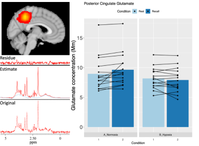 |
Hypoxia alters posterior cingulate cortex metabolism during a memory task: a 1H fMRS study
Matthew Rogan1,2,3, Joseph B R Smith1,2,3, Alexander Friend3,4, Jamie H Macdonald3,4, Sam J Oliver3,4, Gabriella M K Rossetti5, Mark Mikkelsen6,7, Richard A E Edden6,7, and Paul G Mullins1,2,3
1School of Psychology, Bangor University, Bangor, United Kingdom, 2The Bangor Imaging Unit, Bangor University, Bangor, United Kingdom, 3The Extremes Research Group, Bangor University, Bangor, United Kingdom, 4School of Sport Health and Exercise Sciences, Bangor University, Bangor, United Kingdom, 5Centre for Integrative Neuroscience and Neurodynamics, University of Reading, Reading, United Kingdom, 6Russell H. Morgan Department of Radiology and Radiological Science, The Johns Hopkins University School of Medicine, Baltimore, MD, United States, 7F. M. Kirby Research Center for Functional Brain Imaging, Kennedy Krieger Institute, Baltimore, MD, United States
Our group has previously shown that exposure to environmental hypoxia reduces regional CBF within the posterior nodes of the default mode network. Within the same region, hypoxia also reversed the task evoked BOLD response. However, it is unclear what neurometabolic signals underlie this peculiar hemodynamic response to hypoxia. To investigate we employed event related functional MRS to measure the dynamic changes in neurometabolites within the posterior cingulate during hypoxia. We found that hypoxia negates the functional increase in glutamate as seen in normoxia, and induced a regional reduction in glucose. This observation suggests that hypoxia may reduce regional oxidative metabolism.
|
|||
2229.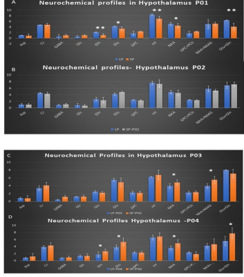 |
Photoperiodic Regulation of hypothalamic metabolism : a preliminary single voxel Magnetic Resonance Spectroscopy investigation at 3T
Nathalie Just1, Pierre-Marie Chevillard1, and Martine Migaud1
1NhyRVana, INRAE, Nouzilly, France
A proton MR Spectroscopy investigation of the sheep hypothalamus at 3T was proposed to compare the impact of photoperiodism on neurochemical profiles. Results showed transient metabolic changes as a function of time and significant differences between long days (LD) and short days (SD) demonstrating photoperiodic regulation of the metabolism of the hypothalamus.
|
|||
2230.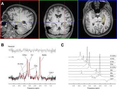 |
Hippocampal Single-Voxel MR Spectroscopy with Long Echo Time at 3 Tesla
Martin Gajdošík1,2, Karl Landheer1, Kelley M. Swanberg1, Fatemeh Adlparvar2, Guillaume Madelin2, Wolfgang Bogner3, Christoph Juchem1,4, and Ivan I. Kirov2,5,6
1Department of Biomedical Engineering, Columbia University, New York City, NY, United States, 2Bernard and Irene Schwartz Center for Biomedical Imaging, Department of Radiology, New York University Grossman School of Medicine, New York City, NY, United States, 3High-Field MR Center, Department of Biomedical Imaging and Image-Guided Therapy, Medical University of Vienna, Vienna, Austria, 4Department of Radiology, Columbia University Medical Center, New York City, NY, United States, 5Department of Neurology, New York University Grossman School of Medicine, New York City, NY, United States, 6Center for Advanced Imaging Innovation and Research, Department of Radiology, New York University Grossman School of Medicine, New York City, NY, United States
The hippocampus is one of the most challenging brain regions for proton MR spectroscopy (MRS) applications. Moreover, quantification of J-coupled species such as myo-inositol (m-Ins) and glutamate + glutamine (Glx) is affected by the presence of macromolecular background. Here we investigate the feasibility of reproducibly measuring their concentrations at long TE of 120 ms, using sLASER localization.
|
|||
2231.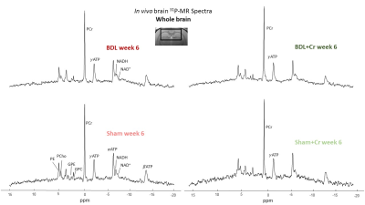 |
Protective role of Creatine in chronic hepatic encephalopathy developing brain: in vivo longitudinal 1H and 31P-MRS study
Dunja Simicic1,2,3, Katarzyna Pierzchala1,2, Olivier Braissant4, Stefanita-Octavian Mitrea1,2, Dario Sessa5, Valerie McLin5, and Cristina Cudalbu1,2
1CIBM Center for Biomedical Imaging, Lausanne, Switzerland, 2Animal Imaging and Technology, EPFL, Lausanne, Switzerland, 3Laboratory of Fuctional and Metabolic Imaging, EPFL, Lausanne, Switzerland, 4Service of Clinical Chemistry, University of Lausanne and University Hospital of Lausanne, Lausanne, Switzerland, 5Swiss Center for Liver Disease in Children, University Hospitals Geneva, Geneva, Switzerland
Type-C hepatic encephalopathy (CHE) is a complication of chronic liver disease (CLD). It is known that children are more affected by CLD than adult patients. Bile duct ligated rat (BDL) is a model of CLD-induced CHE. It was shown that rats having acquired CLD as pups display more profound neurometabolic disturbances than adults. Cr-treatment showed a positive effect in youngP21 BDL-rats resulting in less pronounced metabolic changes. Our aim was to test whether Cr-supplementation dampens the changes observed in CHE in a longitudinal model of CLD acquired in early childhood-P15 and if these changes are similar to those in P21-rats.
|
|||
2232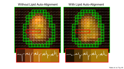 |
Fast lipid removal with multi-scale auto-alignment in full-FOV MRSI. Video Permission Withheld
Peter Adany1, In-Young Choi1,2, and Phil Lee1,3
1Hoglund Biomedical Imaging Center, University of Kansas Medical Center, Kansas City, KS, United States, 2Department of Neurology, University of Kansas Medical Center, Kansas City, KS, United States, 3Department of Radiology, University of Kansas Medical Center, Kansas City, KS, United States
The availability of robust lipid signal removal methods for whole slab MRSI opens up the possibility to measure signals from the edge of the brain, including cortical regions. We have developed a Fast LIpid signal Processing (FLIP) algorithm that effectively removes subcutaneous lipid signals. Recently, we have further developed our FLIP method to include a novel multi-scale auto-alignment feature at a sub-millimeter scale. This study demonstrates that the new alignment algorithm enables the detection of subtle displacement of subject head positions between scans and significantly improves the performance of lipid removal in full-FOV MRSI.
|
|||
2233.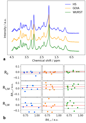 |
On the repeatability and reproducibility of SPECIAL-based in-vivo spectroscopy with different adiabatic inversion pulses
Layla Tabea Riemann1, Christoph Stefan Aigner1, Ralf Mekle2, Sebastian Schmitter1, Bernd Ittermann1, and Ariane Fillmer1
1Physikalisch-Technische Bundesanstalt (PTB), Braunschweig und Berlin, Germany, 2Center for Stroke Research Berlin, Charité Universitätsmedizin, Berlin, Germany
This work assesses the test-retest repeatability and reproducibility of spectral shapes and neurochemical profiles of the SPECIAL 1H MR spectroscopy sequence employing three different adiabatic pulses in-vivo at 7T: the standard hyperbolic secant pulse and two gradient-modulated pulses, namely a GOIA and a WURST pulse. Nine healthy volunteers were scanned four times each, with all three SPECIAL variants to establish three different repeatability or reproducibility measures and to evaluate the limits in the precision of the resulting metabolite quantification.
|
|||
2234.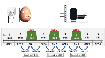 |
Polarity Dependent Modulation of the Motor Region Using tDCS: A Proton MR Spectroscopy Study
Rajakumar Nagarajan1, Anant Shinde2,3, Muhammed Enes Gunduz2,3, and Gottfried Schlaug2,3
1Human Magnetic Resonance Center, Institute for Applied Life Sciences, University of Massachusetts Amherst, Amherst, MA, United States, 2Biomedical Engineering, University of Massachusetts Amherst, Amherst, MA, United States, 3Baystate Medical Center, University of Massachusetts Medical School, Springfield, MA, United States
We studied the effects of motor region tDCS on metabolite levels in a spectroscopic voxel with repeated episodes of 5min of constant current dose levels anticipating stimulation-induced alterations in metabolite concentration at rest. The cumulative change in GABA from before the first tDCS stimulation epoch to after the last epoch showed a significant polarity effect. The first tDCS epoch revealed polarity dependent opposite trends in GABA and Glx from baseline (before stimulation) to post-stimulation. This study provides insights into how tDCS leads to polarity dependent modulation of cerebral metabolites and might provide a model for future MRS stimulation studies.
|
|||
2235.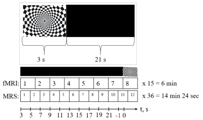 |
A time course of the glutamate+glutamine level in response to a short visual stimulus
Alexey Yakovlev1,2,3, Andrei Manzhurtsev1,3, Petr Menshchikov3,4, Maxim Ublinskiy3, Tolib Akhadov1, and Natalia Semenova1,2,3
1Clinical and Research Institute of Emergency Pediatric Surgery and Trauma, Moscow, Russian Federation, 2N.N.Semenov Federal research center of Chemical Physics Russian Academy of Sciences, Moscow, Russian Federation, 3N.M. Emanuel Institute of Biochemical Physics RAS, Moscow, Russian Federation, 4Philips Healthcare, Moscow, Russian Federation
Changes in the concentration of neurotransmitters in response to a short (or event-related) stimulus in not well investigated. Due to low neurotransmitter cycle rates, these changes can be related to neurotransmitter release from vesicles. In this work, we measured a time course of Glx level in response to a short visual stimulus in a period exceeding the BOLD response using 3T MR scanner. This Glx level dynamic is considered as the release of Glu from the vesicles, reuptake, and refilling. For the first time, we investigated the dynamics of the BOLD-effect using the metabolic signal.
|
|||
2236.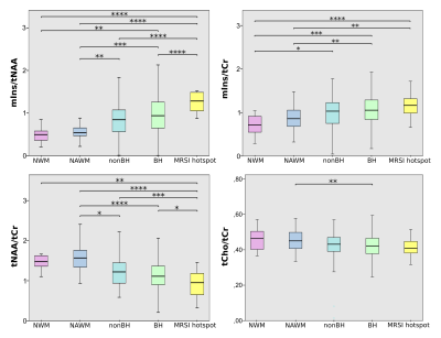 |
Ultra-high field MR spectroscopic imaging at 7 Tesla in Multiple Sclerosis: myo-Inositol as early biomarker for MS pathologies
Alexandra Lipka1,2, Eva Heckova1, Assunta Dal-Bianco3, Gilbert Hangel1, Bernhard Strasser1, Stanislav Motyka1, Lukas Hingerl1, Paulus Rommer3, Fritz Leutmezer3, Petra Hnilicová4, Ema Kantorová4, Stephan Gruber1, Siegfried Trattnig1,2,
and Wolfgang Bogner1,2
1High Field MR Centre, Department of Biomedical Imaging and Image-guided Therapy, Medical University of Vienna, Vienna, Austria, 2Christian Doppler Laboratory for Clinical Molecular MR Imaging, Vienna, Austria, 3Department of Neurology, Medical University of Vienna, Vienna, Austria, 4Jessenius Faculty of Medicine in Martin, Comenius University, Bratislava, Slovakia
Routine T1/T2-weighted magnetic resonance imaging (MRI) is the method of choice for diagnosis and treatment monitoring of Multiple Sclerosis (MS), but is not being able to map the underlying pathological processes. In contrast to T1/T2-lesions which represent the general macroscopic tissue damage, MR Spectroscopic Imaging (MRSI) can detect pathologies on a biochemical level. In 54 relapsing-remitting (RRMS) patients and 16 healthy age/sex-matched controls, we show - enabled through ultra-high resolution Free Induction Decay(FID)-MRSI at 7T - the metabolic distribution within lesions and their close proximity as well as the importance of myo-Inositol as an imaging biomarker in early lesion development.
|
|||
2237.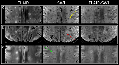 |
Ultra-high field MR spectroscopic imaging at 7 Tesla in Multiple Sclerosis: metabolic fingerprinting of iron rim lesions
Alexandra Lipka1,2, Wolfgang Bogner1,2, Assunta Dal-Bianco3, Gilbert Hangel1, Bernhard Strasser1, Stanislav Motyka1, Lukas Hingerl1, Paulus Rommer3, Fritz Leutmezer3, Stephan Gruber1, Siegfried Trattnig1,2, and Eva Heckova1
1High Field MR Centre, Department of Biomedical Imaging and Image-guided Therapy, Medical University of Vienna, Vienna, Austria, 2Christian Doppler Laboratory for Clinical Molecular MR Imaging, Vienna, Austria, 3Department of Neurology, Medical University of Vienna, Vienna, Austria
Conventional T1/T2-weighted magnetic resonance imaging (MRI) is the method of choice for diagnosis and treatment monitoring of Multiple Sclerosis (MS). Susceptibility weighted imaging (SWI) provides additional information about iron deposition. In addition to these imaging modalities, MR Spectroscopic Imaging (MRSI) can detect pathologies on a biochemical level. In 32 relapsing remitting (RRMS) patients, we showed - with ultra-high spatial resolution Free Induction Decay(FID)-MRSI at 7T - the metabolic changes associated with different types of iron accumulation and the metabolic gradient spanning from within the lesion to its close proximity.
|
|||
2238.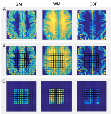 |
Compression of Multivoxel Spectroscopic Data via Visualization of Grey and White Matter Contributions to Metabolite Concentrations
Wufan Zhao1, Eduardo Coello1, Marcia Sahaya Louis1, Katherine Breedlove1, Assaf Tal2, and Alexander Lin1
1Radiology, Brigham and Women's Hospital, Boston, MA, United States, 2Chemical and Biological Physics, Weizmann Institute of Science, Rehovot, Israel
This work introduces a novel way to analyze MR spectroscopic imaging scans by characterizing grey matter and white matter tissue types based on their contributions to metabolite concentrations. This method extracts grey matter and white matter components from anatomical images after tissue segmentation and correlates them with relative metabolite concentrations. The value of the method is shown in visualizing different metabolic profiles for grey and white matter and establishing ranges of metabolite contribution.
|
The International Society for Magnetic Resonance in Medicine is accredited by the Accreditation Council for Continuing Medical Education to provide continuing medical education for physicians.