Digital Posters
Contrast Agents & Preclinical Studies I
ISMRM & SMRT Annual Meeting • 15-20 May 2021

Digital Posters
Contrast Agents & Preclinical Studies I
| Concurrent 2 | 15:00 - 16:00 |
1192.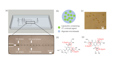 |
Microfluidic preparation of liposomal hydrogel microbeads containing CT agents (CT-lipobeads) to longitudinally monitor pH using CEST MRI
Peng Xiao1, Jianpan Huang1, Xiongqi Han1, Jacinth Wing-Sum Cheu2, Joseph Lai1, Lok Hin Law1, Carmen Chak-Lui Wong2,3, and Kannie Wai Yan Chan1,4,5
1Department of Biomedical Engineering, City University of Hong Kong, Hong Kong, Hong Kong, 2Department of Pathology, Li Ka Shing Faculty of Medicine, The University of Hong Kong, Hong Kong, Hong Kong, 3State Key Laboratory of Liver Research, The University of Hong Kong, Hong Kong, Hong Kong, 4City University of Hong Kong Shenzhen Research Institute, Shen Zhen, China, 5Russell H. Morgan Department of Radiology and Radiological Science, The Johns Hopkins University School of Medicine, Baltimore, MD, United States
Iodinated CT contrast agents(CA) had been investigated as CEST contrast agent while high dose of CA and quick clearance could limit their potential for monitoring pH longitudinally. Here, we aim to introduce CT-lipobeads to address this issue. The CT-lipobeads showed 10% CEST contrast and good pH dependency at pH 6.0-7.0, which is sensitive to distinguish hypoxic medium from normoxic medium. Moreover, CT-lipobeads generated CEST contrast of 9% in the subcutaneous compartment, and contrast remained constant over a week. These findings demonstrated a robust approach for fabrication of CT-lipobeads with decent CEST contrast for longitudinal pH imaging.
|
|||
1193.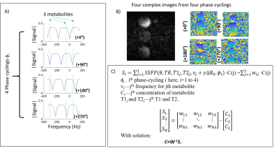 |
Linear combination SSFP for multi-site chemical shift imaging: Applications to Deuterium Metabolic Imaging
Dana C. Peters1, Stefan Markovic2, Qingjia Bao2, Dina Preise2, Keren Sasson2, Lilach Agemy2, Avigdor Scherz2, and Lucio Frydman2
1Radiology and Biomed Eng., Yale University, New Haven, CT, United States, 2Weizmann Institute of Science, Rehovot, Israel
Deuterium metabolic imaging (DMI) is a new method to map abnormal metabolism of tumors. bSSFP might provide high SNR for DMI, due to the high T2/T1 ratios of the injected deuterated glucose, and the lactate, which arises from abnormal metabolism. We developed a linear-combination balanced SSFP method for spectrally resolving glucose, lactate and water, using predicted signal evolutions for different bSSFP phase-cyclings. Our method was able to generate images of the individual compounds in phantoms and in vivo.
|
|||
1194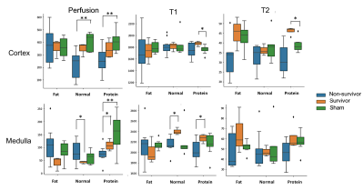 |
Multiparametric MRI of Renal Tissue Shows Dietary Differences Between Survivors and Non-survivors in Sepsis-associated Acute Kidney Injury Video Permission Withheld
Wan-Ting Zhao1,2,3, Karl-Heinz Herrmann1, Martin Krämer1, Renat Sibgatulin1, Ali Nahardani1,2, Jürgen R. Reichenbach1, and Verena Hoerr1,2,4
1Medical Physics Group,Institute of Diagnostic and Interventional Radiology, University Hospital Jena, Jena, Germany, 2Center for Sepsis Control and Care, University Hospital Jena, Jena, Germany, 3Institute of Medical Microbiology, University Hospital Jena, Jena, Germany, 4Clinic for Radiology, University Hospital Muenster, Muenster, Germany
Acute kidney injury (AKI) is a highly lethal complication of sepsis that is frequently asymptomatic with versatile physiological alterations. As diet has gain recognition in the regulation of immune response, we aim to investigate the dietary impact on the course and outcome of sepsis by multiparametric MRI using perfusion, as well as T1 and T2 relaxation time. Cortical T1 and T2 relaxation time was found to differ significantly in animals having received protein-rich diet compared to healthy controls. By combining the cortical T2 relaxation time with perfusion, additional information could be gained, which allowed to distinguish between non-survivors and survivors.
|
|||
1195.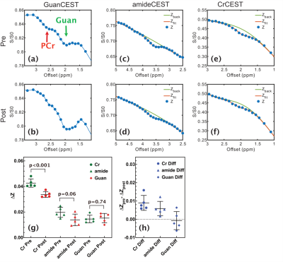 |
The sensitivity of amide, amine, creatine and guanidinium CEST in detecting pH at high MRI field
Lin Chen1,2 and Jiadi Xu1,2
1Department of Radiology and Radiological Science, Johns Hopkins University, Baltimore, MD, United States, 2F.M. Kirby Research Center for Functional Brain Imaging, Kennedy Krieger Research Institute, Baltimore, MD, United States
We compared the sensitivity of several chemical exchange saturation transfer (CEST) contrasts in detecting altered pH at 11.7 T MRI. Studies with egg white phantom revealed that amide and guanidium CEST contrasts in protein are suitable for pH mapping at the physiological relevant range (6.5-7.5), while amineCEST works well for pH lower than 6.5. Hypercapnia (20%) study in mouse brain indicated that creatineCEST showed both higher pH sensitivity and signal intensity compared with amide and guanidium CEST at the physiological pH range and is more suitable for in vivo pH studies at high field.
|
|||
1196.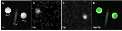 |
Novel phosphorous-based polymer for 31P-magnetic resonance imaging
Natalia Ziółkowska1,2, Ladislav Androvič3, Lucie Woldřichová3, Martin Vít1,4, David Červený1,5, Olga Šebestová Janoušková3, Richard Laga3, and Daniel Jirák1,2
1Site of Computed Tomography, Magnetic Resonance Imaging, and Clinical and Experimental Spectroscopy, Institute for Clinical and Experimental Medicine, Prague, Czech Republic, 2Institute of Biophysics and Informatics, First Faculty of Medicine, Charles University, Prague, Czech Republic, 3Institute of Macromolecular Chemistry, Czech Academy of Sciences, Prague, Czech Republic, 4Faculty of Mechatronics Informatics and Interdisciplinary Studies, Technical University of Liberec, Liberec, Czech Republic, 5Faculty of Health Studies, Technical University of Liberec, Prague, Czech Republic
Our work is focused on the development and investigation of novel polymer-based contrast agent for 31P magnetic resonance (31P MR). The proposed diagnostics is a methacrylate-type polymer containing a phosphorothioate group (p(TMPC)), which causes a significant chemical shift of phosphorus, making it easily distinguishable from other biological phosphorus components, and is thus easily traceable by 31P MRI. Results from 31P MR imaging, spectroscopy and relaxometry obtained at 4.7T show very good MR properties, thus p(TMPC) represent a very promising diagnostic tool for in vivo use.
|
|||
1197.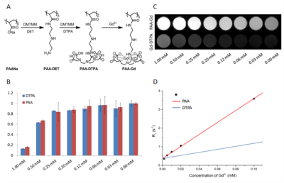 |
PAA-g-(DTPA-Gadolinium): A Versatile Agent to Visualize the Effiencient Interstitial Stream at 7 Tesla
Xiaohan Zhou1,2, Yi Hou3, Wentao Liu1, and Dong Han1,2
1National Centre for Nanoscience and Technology, Beijing, China, 2University of Chinese Academy of Sciences, Beijing, China, 3Beijing University Of Chemical Technology, Beijing, China
Interstitium space and interstitial stream compartmentalize organs distributed in different regions and compose human body among parenchymal tissues. The increase in interests to investigate the behavior and function of interstitial stream, combined with new methodologies propose the demand in new agent to visualize the path itself. A macromolecule gadolinium-based agent PAA-Gd was introduced to outline the path while mass transportation was driven in Balb/c via interstitial stream and histology study was combined to perform in microscopic view as tracer. The novel agent can display its high efficiency to visualize the stream in radiology and pathology study as a versatile enhancer.
|
|||
1198.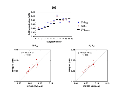 |
Assessment of Gd concentration estimated in DCE-MRI with multiple flip angles
Ayesha Bharadwaj Das1, James Andrew Tranos2, Jin Zhang1, Youssef Zaim Wadghiri2, and Gene Kim1
1Radiology, Weill Cornell Medicine, New York, NY, United States, 2Radiology, New York University School of Medicine, New York, NY, United States
The purpose of this study is to assess the accuracy of Gadolinium-based contrast agent (GBCA) concentration measured using the novel DCE-MRI method with two flip angles, without using the pre-contrast T1 measurement. The accuracy of the proposed method was assessed using the conventional approach with pre-contrast T1 measurement as well as the Gd concentration measurement using Inductively coupled plasma-mass spectrometry using the blood samples collected at the end of each scan. The results exhibited a strong correlation and agreement between the Gd concentration from the proposed two flip angle method and those from the conventional MRI method and ICP-MS analysis.
|
|||
1199.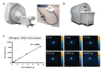 |
Evaluation of cellular sensitivity for magnetic particle imaging and fluorine-19 magnetic resonance imaging
Olivia C Sehl1,2 and Paula J Foster1,2
1Imaging Research Laboratories, Robarts Research Institute, London, ON, Canada, 2Medical Biophysics, University of Western Ontario, London, ON, Canada
Fluorine-19 (19F) MRI and magnetic particle imaging (MPI) are both quantitative modalities used for cell tracking. We evaluated the cellular sensitivity of MPI using ferucarbotran on a preclinical MPI system and 19F MRI using perfluorocarbons on a 3 Tesla clinical system. For both modalities, significant linear increases in 19F and MPI signal were detected with increasing number of mesenchymal stem cells (MSC). As few as 4000 MSC were reliably detected with MPI and 256 x 103 MSC with 19F MRI using the same scan time (1.5 minutes/cell pellet). Both of these detection limits were improved with longer imaging times.
|
|||
1200.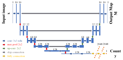 |
Detecting Extreme Small Lesions in MR images with Point Annotations via Multi-task Learning
Xiaoyang Han1, Yuting Zhai1, Botao Zhao1, and Xiao-Yong Zhang1
1Institute of Science and Technology for Brain-Inspired Intelligence, Fudan University, Shanghai, China
Detection of small lesions in magnetic resonance (MR) images is currently one of the most challenging tasks. Compared with detection in natural images, existing methods cannot accurately detect small lesions with a limited amount of information in MR images. To solve the problems, we propose a novel multi-task convolutional neural network, which simultaneously regresses the lesion number and detects the lesion location. We train and evaluate the end-to-end network to count and locate extreme small lesions within 3-5 voxels on a mouse brain MR image dataset with point annotations. Our network outperforms other methods on sensitivity and precision.
|
|||
1201.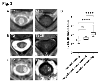 |
Diagnostic value of Gd-contrast administration for spinal cord MRI in MS patients and T2 signal ratio as a predictive marker of lesion activity
Kianush Karimian-Jazi1, Ulf Neuberger1, Katharina Schregel1, Gianluca Brugnara1, Daniel Schwarz1, Laura Jäger2, Wolfgang Wick2, Martin Bendszus1, and Michael Oliver Breckwoldt1
1Neuroradiology, University Clinic of Heidelberg, Heidelberg, Germany, 2Neurology, University Clinic of Heidelberg, Heidelberg, Germany
Our study shows that patients with a stable, spinal T2 lesion load do not benefit from the administration of GBCAs. In contrast to our previous cerebral study, there was no indication of spinal lesion re-activation. The T2 signal ratio has a high sensitivity and specificity to “predict” contrast enhancement of a given lesion and could be used as a predictive marker for “lesion activity”. In combination with previously published studies on the limited value of Gd-administration in cerebral MS follow-up investigations, we propose a “single-shot” MS protocol to combine cerebral and spinal MRI investigations in one standardized single MR protocol.
|
|||
1202.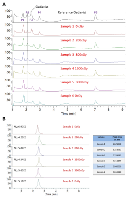 |
Is MRI Contrast Stable in High-Energy Radiation of MR-Linac?
Travis Salzillo1, Yongying Jiang2, Yuri Mackeyev3, Clifton David Fuller1, Caroline Chung1, Seungtaek Choi1, Neil Hughes1, Yao Ding4, Jinzhong Yang4, Sastry Vedam5, Sunil Krishnan3, and Jihong Wang4
1Radiation Oncology, University of Texas MD Anderson Cancer Center, Houston, TX, United States, 2Institute for Applied Cancer Science, University of Texas MD Anderson Cancer Center, Houston, TX, United States, 3Radiation Oncology, Mayo Clinic, Jacksonville, FL, United States, 4Radiation Physics, University of Texas MD Anderson Cancer Center, Houston, TX, United States, 5Radiation Oncology, University of Maryland, Baltimore, MD, United States
Gadolinium-based MRI contrast agents are deemed safe to use in diagnostic exams. Their use would bring value to radiation oncology applications such as daily replanning on the MR-Linac. However, their stability and safe use in the presence of high-energy radiation remains unknown. We analyzed the chemical composition stability of two common MRI contrast agents that were irradiated to various doses using mass spectrometry. We found no detectable amounts of degradation compounds or conformational alterations as a result of irradiation. This is the first step towards demonstrating the safe use of gadolinium-based MRI contrast agents in radiation oncology applications.
|
|||
1203.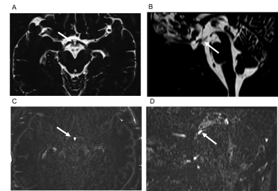 |
Contrast enhancement of the normal infundibular recess using 3D FLAIR
Iichiro Osawa1, Eito Kozawa1, Yuya Yamamoto1, Sayuri Tanaka1, Taira Shiratori1, Akane Kaizu1, Kaiji Inoue1, and Mamoru Niitsu1
1Saitama Medical University Hospital, Saitama, Japan
The infundibular recess (IR) is a cerebrospinal fluid (CSF) space in the third ventricle floor, and its function remains unclear. We retrospectively evaluated contrast enhancement of the normal IR using heavily T2-weighted 3D FLAIR. The enhancement of IR was the strongest on post-contrast images, followed by 4-h delayed post-contrast images. It was also stronger than that of other CSF spaces. This is the first study to report the enhancement of IR after an intravenous gadolinium injection. Evaluations of the enhancement of IR may clarify substance transport, such as gadolinium or hormones into the CSF.
|
|||
1204.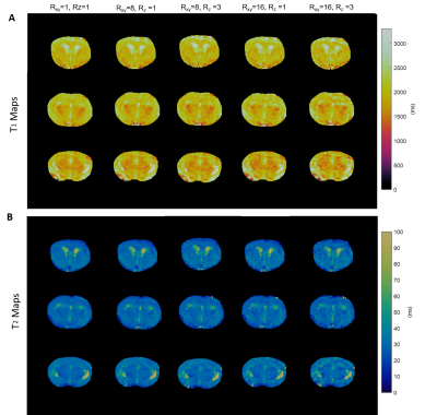 |
Whole-Brain T1 and T2 Mapping in Mouse by 3D Magnetic Resonance Fingerprinting
Yuran Zhu1, Yuning Gu1, Kihwan Kim1, Charlie Androjna2, Chris A. Flask1,3,4,5, Yong Chen3,6, and Xin Yu1,3,7
1Department of Biomedical Engineering, Case Western Reserve University, Cleveland, OH, United States, 2Lerner Research Institute, Cleveland Clinic Foundation, Cleveland, OH, United States, 3Department of Radiology, Case Western Reserve University, Cleveland, OH, United States, 4Department of Pediatrics, Case Western Reserve University, Cleveland, OH, United States, 5Cancer Imaging Program, Case Western Reserve University, Cleveland, OH, United States, 6Biomedical Research Imaging Center (BRIC), University of North Carolina at Chapel Hill, Chapel Hill, NC, United States, 7Department of Physiology and Biophysics, Case Western Reserve University, Cleveland, OH, United States
We present a 3D MR fingerprinting (MRF) sequence for simultaneous T1 and T2 mapping in mouse. Data acquisition and reconstruction covered a field of view (FOV) of 30 × 30 × 9 mm3 with a matrix size of 128 x 128 x 9, yielding a spatial resolution of 0.23 × 0.23 × 1 mm3. Retrospective undersampling analysis was performed on fully-sampled phantom and mouse data to examine the undersampling capacity of the proposed sequence, demonstrating that the method supports up to 16-fold in-plane undersampling with a 3-fold through-plane undersampling.
|
|||
1205.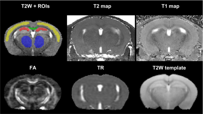 |
In vivo characterization of effect of microglia regeneration after high altitude exposure by quantitative MRI in mice
Alexandru V Korotcov1,2, Caroline A Browne3, Andrew K Knutsen1,2, Dara L Dickstein2,4, Juan Wang4, Xiufen Xu2,5, Kathleen Whiting2,5, Shalini Jaiswal1,2, Allison Nathanael1, Daniel P Perl4, Zygmunt Galdzicki2,5, and Bernard J Dardzinski1,2
1Radiology and Radiological Sciences, Uniformed Services University, Bethesda, MD, United States, 2Center for Neuroscience and Regenerative Medicine, Henry M. Jackson Foundation, Bethesda, MD, United States, 3Department of Pharmacology & Molecular Therapeutics, Uniformed Services University, Bethesda, MD, United States, 4Department of Pathology, Uniformed Services University, Bethesda, MD, United States, 5Department of Anatomy, Physiology and Genetics, Neuroscience Program, Uniformed Services University, Bethesda, MD, United States
Prolonged stays in high-altitude (HA) environments produce pro- and maladaptive physiological and pathological changes. Microglia depletion has been utilized in recovery from a spectrum of neurodegenerative diseases abnormalities. Advanced neuroimaging techniques demonstrate great promise to detect subtle changes in brain activity and morphology related to impairments induced by HA. In this work, we applied advanced MRI and modern behavioral tests to investigate neuropathological consequences of hypobaric hypoxia and the use of microglial regeneration as a novel approach to treat the maladaptive effect of HA.
|
|||
1206.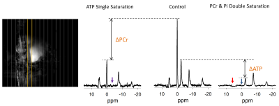 |
Measurement of ATP Hydrolysis Rates in the In Vivo Heart at 7T
Adil Bashir1, Jianyi Zhang2, and Thomas S Denney3
1Department of Electrical and Computer Engineering, Auburn University, Auburn, AL, United States, 2Biomedical Engineering, The University of Alabama at Birmingham, Birmingham, AL, United States, 3Electrical and Computer Engineering, Auburn University, Auburn, AL, United States
Direct measurement of ATP hydrolysis (ATP-->Pi) were elusive in the in vivo myocardium because the level of Pi in the heart is low and the peak attributed to Pi overlaps with the much larger peak for 2,3-disphosphoglycerate (2,3 DPG). We have demonstrated an approach to measure ATP utilization rate indirectly by measuring total ATP utilization and subtracting out the measurable component that can be attributed to ATP flux via CK. The technique was applied in swine and human hearts at 7T. This method can facilitate important insights into biological mechanisms of impaired bioenergetics in myocardium.
|
|||
1207. |
Radial tiny golden angle MRI combined with Cardiac and Respiratory Self-Gating in the Small Animal model – one acquisition, many possibilities
Patrick Metze1, Hao Li2, and Volker Rasche1,2
1Internal Medicine II, Ulm University Medical Center, Ulm, Germany, 2Core Facility Small Animal Imaging (CF-SANI), Ulm University, Ulm, Germany
Radial tiny golden angle MRI is a versatile tool for different applications in the small animal model. Changes on short time-scales can be visualized by sliding-window (and Compressed Sensing) reconstructions, while respiratory and cardiac self-gating enable high-resolution images for cyclic motion patterns – all from the same acquisition.
|
|||
1208.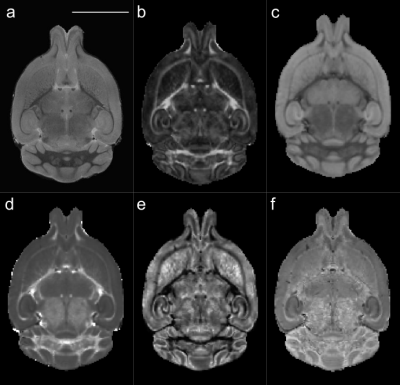 |
Anatomical and microstructural brain alterations in the TDP-M323K mouse model of amyotrophic lateral sclerosis
Aurea B Martins-Bach1, Mohamed Tachrount1, Cristiana Tisca1, Lily Qiu1, Shoshana Spring2, Jacob Ellegood2, Brian J Nieman2, John G. Sled2, Remya Raghavan-Nair 3, Elizabeth Fisher4, Thomas Cunningham3, Jason Lerch1, and Karla Miller1
1Wellcome Centre for Integrative Neuroimaging, University of Oxford, Oxford, United Kingdom, 2Mouse Imaging Centre, The Hospital for Sick Children, Toronto, ON, Canada, 3Mammalian Genetics Unit, MRC Harwell Institute, Oxford, United Kingdom, 4Department of Neuromuscular Diseases, Institute of Neurology, University College London, London, United Kingdom
Amyotrophic lateral sclerosis is a devastating neurodegenerative disease, characterized by aggregates of TDP-43 protein in the brain of most patients. The TDP-M323K mouse has a mutation in the gene encoding the Tdp-43 protein, and presents progressive motor and neurological phenotypes. In this study, we assessed structural and microstructural alterations in the brain of TDP-M323K mice with preclinical MRI (7 tesla). High resolution images showed brain atrophy, but relative volume changes included hypertrophy in the cortex and hippocampus. Diffusion MRI revealed alterations compatible with neurodegeneration in the white matter and striatum. TDP-M323K mice recapitulate brain imaging phenotypes observed in ALS patients.
|
|||
1209.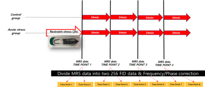 |
Changes in Correlation Between Brain Metabolites Due to Acute Stress in Mouse Hippocampus using Proton Magnetic Resonance Spectroscopy
Chang-Soo Yun1, Yoon Ho Hwang2, Min-Hee Lee3, Wooseung Kim2, Jehyeong Yeon1, Hyeon-Man Baek4, Dong Youn Kim2, and Bong Soo Han1
1Radiation Convergence Engineering, Yonsei University, Wonju-si, Gangwon-do, Korea, Republic of, 2Biomedical Engineering, Yonsei University, Wonju-si, Gangwon-do, Korea, Republic of, 3Institute of Human Genomic Study, Korea University, Ansan-si, Gyeonggi-do, Korea, Republic of, 4Gachon Advanced Institute for Health Sciences and Technology, Gachon University, Incheon-si, Korea, Republic of
We investigated the changes in correlation between mouse brain metabolites due to acute restraint stress using magnetic resonance spectroscopy (MRS). Acquired time series MRS data of the control and acute stress groups were quantified using LCModel. Correlation coefficients between metabolites were calculated from time series metabolite concentration and metabolite pairs with statistically significant correlations were selected using one-sample Sign Test ( p < 0.05 ). We found that Gln-GSH, NAA-Glu and NAA-GSH showed new significant correlations in the acute stress group. Our findings suggest that correlation can be a useful measurement in assessing the effects of stress.
|
|||
1210.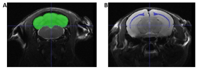 |
Phenotyping a Mouse Model of TRAPPC9-Associated Intellectual Disability using Magnetic Resonance Imaging
Mark David Platt1, Harish Poptani1, Antonius Plagge2, and Mahon Maguire1
1Molecular and Clinical Cancer Medicine, University of Liverpool, Liverpool, United Kingdom, 2Cellular and Molecular Physiology, University of Liverpool, Liverpool, United Kingdom
Loss of function mutations in the TRAPPC9 gene causes autosomal recessive intellectual disability and microcephaly. A mouse model was generated to investigate the effects of TRAPPC9 knockout in mice. In vivo and ex vivo MRI were used alongside behavioural tests to probe differences in brain volume and learning ability. In vivo MRI demonstrated significant differences between the whole brain, cerebellar and corpus callosum volumes of adult TRAPPC9 knockout mice and wildtype controls. Behavioural assays also revealed significant differences in learning ability. These results align with the human condition and suggest the model is appropriate for further study of TRAPPC9 knockout.
|
|||
1211.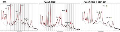 |
Cerebral metabolic derangements as translatable biomarkers in pantothenate kinase-associated neurodegeneration mouse model via 1H MRS
Puneet Bagga1, Jeffrey Steinberg2, Walter Akers2, Zoltan Patay1, Beth McCarville1, Chitra Subramanian3, Charles O Rock3, and Suzanne Jackowski3
1Department of Diagnostic Imaging, St Jude Children's Research Hospital, Memphis, TN, United States, 2Center for In Vivo Imaging and Therapeutics (CIVIT), St Jude Children's Research Hospital, Memphis, TN, United States, 3Department of Infectious Diseases, St Jude Children's Research Hospital, Memphis, TN, United States
Pantothenate kinase (PanK) is a metabolic enzyme that is the first and rate-controlling step in the only pathway for cellular coenzyme A (CoA) biosynthesis. A rare, life-threatening neurological disorder known as pantothenate kinase-associated neurodegeneration (PKAN), formerly known as Hallervorden-Spatz disease, arises from mutations in the human PANK2 gene. Here, we studied the neurochemical effects of a drug capable of allosterically activating PanK protein isoforms as a potential PKAN therapeutic. We applied 1HMRS to quantify changes in cerebral metabolites including Glx, GABA, lactate, and NAA in a mouse model of CoA deficiency employing the neuron-specific deletion of Pank1 and Pank2 genes.
|
The International Society for Magnetic Resonance in Medicine is accredited by the Accreditation Council for Continuing Medical Education to provide continuing medical education for physicians.