Digital Posters
Contrast Agents & Preclinical Studies II
ISMRM & SMRT Annual Meeting • 15-20 May 2021

Digital Posters
Contrast Agents & Preclinical Studies II
| Concurrent 2 | 15:00 - 16:00 |
1212.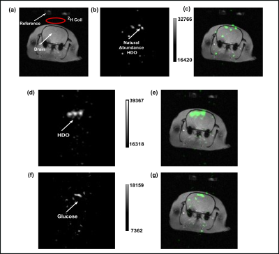 |
Deuterium MRI for HDO Imaging of the Rat Brain Following Metabolism of [2H7]glucose
Rohit Mahar1, Huadong Zeng2, Anthony Giacalone3, Mukundan Ragavan3, Thomas H. Mareci3, and Matthew E. Merritt3
1Biochemistry and Molecular Biology, University of Florida, Gainesville, FL, United States, 2Advanced Magnetic Resonance Imaging and Spectroscopy (AMRIS) Facility, University of Florida, Gainesville, FL, United States, 3Department of Biochemistry and Molecular Biology, University of Florida, Gainesville, FL, United States
In this study, we demonstrate the application of deuterium MRS(I) for in vivo rat brain imaging. Administration of [2H7]glucose via the tail vein produced deuterated lactate and glx analogous to earlier reports, but here we show for the first time that HDO generated from metabolism can be imaged in the rodent brain. By using HDO evolution as a metric of glucose utilization, we introduce a biomarker of cerebral glycolysis and oxidative flux that is imaged with superior sensitivity and resolution.
|
|||
 |
1213. |
Tracking cancer cells in the mouse brain with magnetic resonance imaging (MRI) and magnetic particle imaging (MPI)
Natasha N Knier1,2 and Paula J Foster1,2
1University of Western Ontario, London, ON, Canada, 2Robarts Research Institute, London, ON, Canada
Magnetic particle imaging (MPI) is an emerging modality that may overcome challenges associated with quantification of iron-based MRI cell tracking. The magnetic properties of an SPION tracer heavily influences the sensitivity and resolution in MPI. Synomag-D™ has been explored as a sensitive tracer for MPI. Currently, no studies exist using this tracer to track cancer cells. Here, we utilize Synomag-D with MPI to detect and quantify iron-labeled breast cancer cells in the mouse brain and compare to MRI. Employing MPI for experimental cancer cell tracking will allow for quantification of the arrest, clearance, and retention of cancer cells in vivo.
|
||
1214. |
Consolidated simulation of 23Na Dynamics in Biological Tissue
Chengchuan Wu1, Yasmin Blunck1, and Leigh A. Johnston1
1Department of Biomedical Engineering, The University of Melbourne, Parkville, Australia
This work presents a mathematical framework that enables the simulation for the dynamics of the spin-3/2 23Na density operator. The simulator is featured with easy implementation for 23Na in biological environment. This work demonstrates the simulator in two 23Na pulse sequences.
|
|||
1215. |
α-ATP suppression in 31P MR spectroscopy by homonuclear BIRD filter: An approach to quantify NAD+ and NADH at 3T in vivo
Julian Mevenkamp1, Yvonne M.H. Bruls1,2, Robin A. de Graaf3, Joachim E. Wildberger1, Matthijs K.C. Hesselink2, Lucas Lindeboom1,2, and Vera B. Schrauwen-Hinderling1,2
1Department of Radiology & Nuclear Medicine, Maastricht University Medical Center, Maastricht, Netherlands, 2Department of Nutrition & Movement Sciences, Maastricht University, Maastricht, Netherlands, 3Department of Radiology & Biomedical Imaging, Yale School of Medicine, New Haven, CT, United States
NAD+ and NADH play important roles in metabolism and metabolic health. Therefore, a non-invasive 31P MRS for the quantification of those metabolites on clinical 3T scanners was developed. This sequence suppresses α-ATP resonances based on homonuclear Bilinear Rotation Decoupling (BIRD). With reduced α-ATP intensity, neighbouring NAD+/H resonances appear as a separate peak. Results in eight young, lean and healthy volunteers show that the sequence succeeded in considerable α-ATP suppression. However, a clear separation of NADH and NAD+ resonances remains challenging due to remaining α-ATP signal.
|
|||
1216.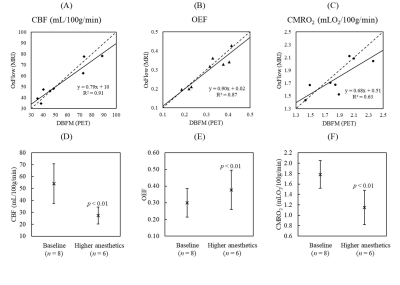 |
Validation of OxFlow Measurements of Whole-Brain CBF, OEF and CMRO2 by Simultaneous PET/MRI
Lucas Narciso1,2, Tracy Ssali1,2, Linshan Liu1, Heather Biernaski1, John Butler1, Laura Morrison1, Jennifer Hadway1, Jeffrey Corsaut1, Justin W. Hicks1, Michael C. Langham3, Felix W. Wehrli3, Hidehiro Iida4, and Keith St Lawrence1,2
1Lawson Health Research Institute, London, ON, Canada, 2Department of Medical Biophysics, Western University, London, ON, Canada, 3University of Pennsylvania Perelman School of Medicine, Philadelphia, PA, United States, 4University of Turku and Turku PET Centre, Turku, Finland
OxFlow is a non-invasive MRI method for measuring whole-brain oxidative metabolism. This study presents its validation in an animal model by comparing to a PET-only method, as well as its sensitivity to alterations in cerebral metabolism. Strong correlations were observed between measurements from the MRI and PET techniques, with no significant differences. Furthermore, OxFlow proved sensitive to changes in metabolism induced by an increase in anesthetics.
|
|||
1217.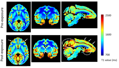 |
CNS Involvement in a Non-human Primate Model Infected with Aerosolized Ebola Virus
Byeong-Yeul Lee1, Jeffrey M. Solomon2, Marcelo Castro1, Dong-Yun Kim3, Joseph Laux1, Matthew G. Lackemeyer1, Jordan K. Bohannon4, Anna N. Hanko5, Dima Hammoud6,7, and Ji Hyun Lee1
1Integrated Research Facility at Fort Detrick, National Institute of Allergy and Infectious Diseases, National Institutes of Health, Frederick, MD, United States, 2Clinical Monitoring Research Program Directorate, Frederick National Laboratory for Cancer Research sponsored by the National Cancer Institute, Frederick, MD, United States, 3Office of Biostatistics Research, National Heart, Lung and Blood Institute, Bethesda, MD, United States, 4National Biodefense Analysis and Countermeasures Center, Frederick, MD, United States, 5Microbiology, Boston University School of Medicine, Boston, MA, United States, 6Radiology and Imaging Sciences, Clinical Center, National Institutes of Health, Bethesda, MD, United States, 7Center for Infectious Disease Imaging, National Institutes of Health, Bethesda, MD, United States
We performed a quantitative neuroimaging study to determine central nervous system involvement following exposure with Ebola virus (EBOV) variant Makona via the aerosolized route in a rhesus monkey model. Using MR relaxometry, we found increases in T1 and R2* values in multiple brain regions. Notably, R2* changes corresponding to the deep cerebral venous system highly correlated with viral loads in CSF. These results provide in vivo evidence of brain involvement with EBOV and emphasize the potential of advanced imaging techniques to better understand the pathophysiology of organ involvement in various infectious diseases, including the brain.
|
|||
1218.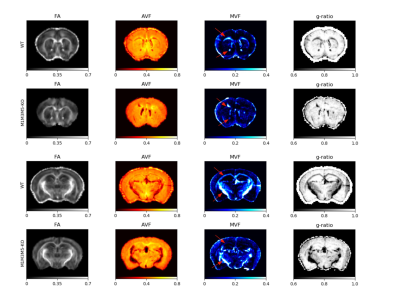 |
Characterization of a Novel Hypomyelination Mouse Model Using Microstructural Imaging of Myelin Volume Fraction and Axon g-ratio
Vladimir Grouza1,2, Zhe Wu1,3, Marius Tuznik1,2, Hooman Bagheri4, Dan Wu5, Alan C Peterson2,4,6, and David Rudko1,2,7
1McConnell Brain Imaging Centre, Montreal Neurological Institute and Hospital, Montreal, QC, Canada, 2Department of Neurology and Neurosurgery, McGill University, Montreal, QC, Canada, 3Techna Institute, University Health Network, Toronto, ON, Canada, 4Department of Human Genetics, McGill University, Montreal, QC, Canada, 5Russell H. Morgan Department of Radiology & Radiological Science, Johns Hopkins University, Baltimore, MD, United States, 6Gerald Bronfman Department of Oncology, McGill University, Montreal, QC, Canada, 7Department of Biomedical Engineering, McGill University, Montreal, QC, Canada
We investigated a novel mouse model of CNS hypomyelination using T2* weighted mGRE and diffusion weighted imaging. By integrating a recently developed method for MWF estimation from mGRE data (BSS-rPCA), we observed strong correlations between Mbp/Golli mRNA expression levels and MVF in select WM tracts in the brain. In addition, WM tract g-ratio values computed from BSS-rPCA MVF combined with NODDI-derived parameters were consistent with those found in the literature computed using differing methods. These results support integrating BSS-rPCA MWF estimates into quantitative microstructure evaluation workflows.
|
|||
1219.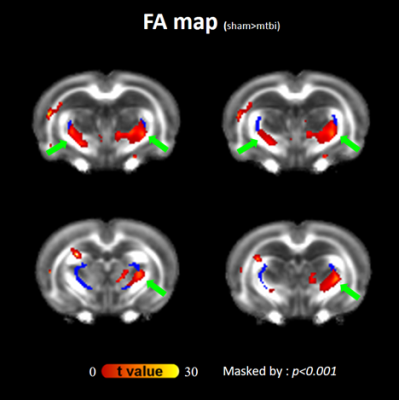 |
Thalamic reticular nucleus injury as the cause of thalamocortical dysrhythmia in mild traumatic brain injury: a rodent model DTI study
Duen-Pang Kuo1,2, Yi-Tien Li1,3, Chen-Yin Ou1, Yung-Chieh Chen1,2, and Cheng-Yu Chen1,2
1Translational Imaging Research Center, Taipei Medical University Hospital, Taipei, Taiwan, 2Department of Medical Imaging, Taipei Medical University Hospital, Taipei, Taiwan, 3Neuroscience Research Center, Taipei Medical University, Taipei, Taiwan Thalamocortical dysrhythmia (TCD) has been implicated in the neuropsychiatric disorders in patients following mild traumatic brain injury (mTBI). We investigated the time-course diffusion tensor changes in the rodent brain up to 35 days using a controlled closed head injury model. Significant and persistent elevation of fractional anisotropy (FA) and reduced radial and mean diffusivities were found at the boundary of bilateral thalami where high shear stress was expected, suggesting that disinhibition of inhibitory circuits from the thalamic reticular nucleus (TRN) may play a role in TCD. |
|||
1220.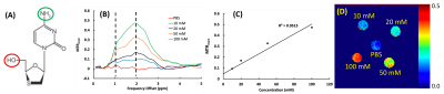 |
HIV Theranostics Based on Intrinsic CEST Contrasts of Antiretroviral Drugs
Aditya Bade1, Howard Gendelman1, and Yutong Liu2
1Pharmacology and Exp Neuroscience, University of Nebraska Medical Center, Omaha, NE, United States, 2Radiology, University of Nebraska Medical Center, Omaha, NE, United States
HIV theranostics that enables the in vivo imaging of antiretroviral drugs is a powerful tool for the development of antiretrovirals (ARVs) targeting HIV reservoirs, and the development of long-acting ARVs that affect drug adherence and as such reduce viral transmission, prevent new infections, and limit the emergence of viral drug resistance. We tested the possibility to develop HIV theranostics based on the intrinsic CEST contrasts of ARVs. Herein, we measured the CEST effects of first-line ARVs including 3TC (lamivudine) and FTC (emtricitabine) and developed CEST MRI methods for in vivo detection of ARVs in the central nervous system (CNS).
|
|||
1221.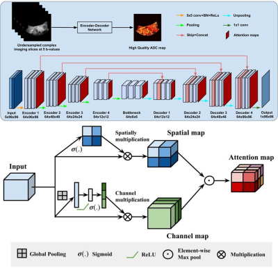 |
Deep Learning of ADC Maps from Under-sampled Diffusion-Weighted Radially Sampled MRI
Yuemeng Li1, Hee Kwon Song1, Miguel Romanello Giroud Joaquim1, Stephen Pickup1, Rong Zhou1, and Yong Fan1
1Radiology, University of Pennsylvania, Philadelphia, PA, United States
Respiratory motion and high magnetic fields pose challenges for quantitative diffusion weighted MRI (DWI) of mouse abdomen on preclinical MRI systems. EPI-based DWI method yields inadequate suppression of motion and magnetic susceptibility artifacts. Diffusion-weighted radial spin-echo (Rad-SE-DW) produces artifact-free images but require substantially longer acquisition times. Here, we demonstrate a new deep learning concept for accelerating acquisition of RAD-SE-DW. Fully sampled Rad-SE-DW images are used to train a convolution neural network for directly extracting apparent diffusion coefficient (ADC) maps from highly under-sampled Rad-SE-DW data. Comparisons with standard ADC extraction and acceleration methods are made to support this concept.
|
|||
1222.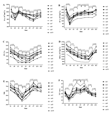 |
Multiparametric functional magnetic resonance imaging for longitudinal evaluation of liver regeneration after 70% hepatectomy in a rat model
shuangshuang xie1, caixin qiu1, yajie sun1, jinxia zhu2, Robert Grimm3, and wen shen1
1Tianjin First Central Hospital, Tianjin, China, 2MR Collaboration, Siemens Healthcare Ltd., Beijing, China, 3MR Application Development, Siemens Healthcare GmbH, Erlangen, Germany
This study investigated the feasibility of multiparametric magnetic resonance imaging (MRI) (IVIM, DKI, BOLD) to evaluate the liver regeneration process after 70% hepatectomy in rats. MRI examination and pathologic samples were performed at multiple time-points. The results showed that D, D*, FP, MD, MK, and T2* can be used to monitor the microstructural changes after surgery, and D* and MK were significantly correlated with proliferative indexes of Ki - 67 and hepatocyte size. This suggests that multiparametric MRI can aid in noninvasive radiologic monitoring of liver regeneration and intrinsic microstructure evaluation
|
|||
1223.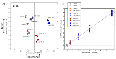 |
Investigating differences in lung cancer cells with induced and de-induced cisplatin resistance by 1H HR-MAS NMR metabolomics
Martina Vermathen1, Hendrik von Tengg-Kobligk2,3, Martin Nils Hungerbühler2,3, Christoph Kempf2,3, Peter Vermathen2,3, and Nico Ruprecht2,3
1Department of Chemistry and Biochemistry, University Bern, Bern, Switzerland, 2University Institute of Diagnostic, Interventional and Pediatric Radiology, University Bern, Bern, Switzerland, 3Department of BioMedical Research, University Bern, Bern, Switzerland
Cisplatin (cisPt) is an important drug that is used against various cancers, including advanced lung cancer. However, drug resistance is a major problem. Here, a high resolution magic angle spinning (HR-MAS) NMR study is presented determining the metabolic profile of lung cancer cells (A24) and metabolic adaptations in different levels of induced cisPt-resistance as well as in their de-induced counterparts. More than 40 metabolites were identified. Metabolic adaptations were determined, which increased with higher cisPt-resistance. Importantly, de-induced cell lines demonstrated similar metabolic adaptations. Metabolites predominantly changed in cisPt resistant cells and their de-induced counterparts include glutathione or taurine.
|
|||
1224.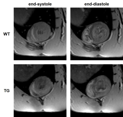 |
Characterization of cardiac function in early-stage pulmonary arterial hypertension in asymptomatic BMPR2 mutant rats by MRI and 31P MRS
Dounia El Hamrani1,2, Julie Magat1,2, Jérôme Naulin1,2, Frédéric Perros3, Marilyne Campagnac2, David Benoist1,2, Christelle Guibert2, and Bruno Quesson1,2
1IHU LIRYC, Bordeaux, France, 2INSERM U1045, CRCTB, Bordeaux, France, 3INSERM UMR_S 999, Université Paris–Saclay, Le Kremlin Bicêtre, France
The transgenic rat line studied has a monoallelic mutation of Bmpr2 which is a model of heritable pulmonary arterial hypertension. The purpose of this study is to evaluate cardiac differences between wild type and asymptomatic mutated rats by cardiac MRI and 31P MRS. Transgenic rats showed a decrease in end-diastolic and end-systolic volumes of ventricles leading to a reduced stroke volume. These results indicate a significant decrease in cardiac output but without ventricular hypertrophy (constant wall thickness) and no significant variation of ejection fraction. The cardiac energy balance (phosphocreatine/ATPβ) remained unaltered in Bmpr2 rats at rest and post-injection of dobutamine.
|
|||
1225.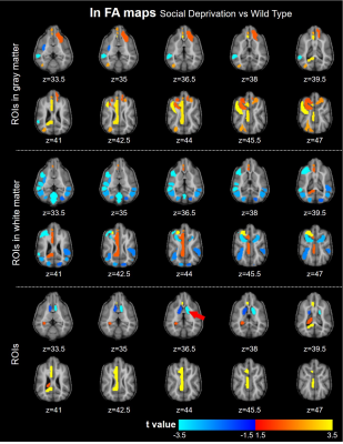 |
A pilot study of socially deprivated brain alterations using fMRI and dMRI in domestic dogs
Xueru Liu1,2, Huilin Hong3, Zhentao Zuo1,2,4, Hui Zhao3, Rui Tian3, Yongqing Zhang2,3,4, and Yan Zhuo1,2,4
1State Key Laboratory of Brain and Cognitive Science, Institute of Biophysics, Chinese Academy of Sciences, BeiJing, China, 2University of Chinese Academy of Sciences, Beijing, China, 3State Key Laboratory of Molecular Developmental Biology, Institute of Genetics and Developmental Biology, Chinese Academy of Sciences, Beijing, China, 4CAS Center for Excellence in Brain Science and Intelligence Technology, Chinese Academy of Sciences, Beijing, China
Social interactions, like sleep, is the basic needs for humans and animals, its neural mechanism is not fully understood. Domestic dog has similar emotional and social processing with humans, it would be a promising animal model for social interaction. In this study, 3 beagles were social deprived (SD) 4-week, and performed resting-state fMRI and diffusion MRI. The functional connectivity and diffusion metrics were compared between SD and wild type dogs. We found the functional connectivity strengthened within prefrontal cortex and weakened between prefrontal cortex with visual and auditory cortex in SD. Demyelination observed in frontal, temporal, and insula regions.
|
|||
1226.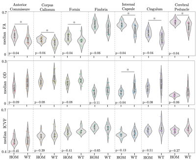 |
Vcan mutation leads to sex-specific changes in white matter microstructure in mice
Cristiana Tisca1, Mohamed Tachrount 1, Frederik Lange1, Chaoyue Wang1, Lily Qiu1, Javier Barallobre-Barreiro2, Marika Fava2, Manuel Mayr2, Jason Lerch1,3,4, Aurea Martins-Bach1, and Karla Miller1
1Wellcome Centre for Integrative Neuroimaging, FMRIB, Nuffield Department of Clinical Neurosciences, University of Oxford, Oxford, United Kingdom, 2British Heart Foundation Centre of Research Excellence, King's College London, London, United Kingdom, 3Mouse Imaging Centre, Hospital for Sick Children, Toronto, ON, Canada, 4Department of Medical Biophysics, University of Toronto, Toronto, ON, Canada
Recent genome-wide association studies using UK Biobank datasets have shown associations between diffusion-weighted MRI (dMRI) phenotypes and genetic loci associated with the human VCAN gene, spanning most white matter tracts. To understand the biological cause of this association, we acquired ex vivo structural and dMRI data in a Vcan mouse model and evaluated whether it displays similar changes in white matter microstructure. We show that male Vcan mice display related dMRI phenotypes to those associated with mutations in VCAN in humans. This gives us the ability to investigate the biological interpretations of human GWAS results using histology.
|
|||
1227.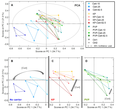 |
Metabolic Responses of HeLa Cells upon addition of the Photosensitizer Chlorin e4 and Carriers in the Dark by 1H HR-MAS NMR
Martina Vermathen1, Tobias Emmanuel Kämpfer1,2, and Peter Vermathen2
1Department of Chemistry and Biochemistry, University Bern, Bern, Switzerland, 2Departments of BioMedical Research and Radiology, University Bern, Bern, Switzerland
Porphyrinic photosensitizers (PSs) like chlorin e4 (Ce4) are applied with polymer-based carriers for photodynamic therapy of cancer. However, the impact of PSs and their carriers on the metabolic state of cells in the dark-interval before light treatment are not clear. Here, HR-MAS NMR metabolomics was applied to cancer cells treated with Ce4 alone or combined with polyvinylpyrrolidone (PVP) or block-copolymer micelles (BCMs). A Ce4-concentration dependent metabolic response was observed that was clearly attenuated by BCMs and more pronounced by PVP indicating the beneficial role of the carriers. Direct interaction of Ce4 with glycero-phosphocholine was observed suggesting intracellular membrane localization.
|
|||
1228. |
Absolute quantification of cardiac 31P metabolites using surface loop and dipole array coils at 7T
Jabrane Karkouri1, Stanislav Frištyk2, Lucian AB Purvis3, Christopher T. Rodgers*1, and Ladislav Valkovic*3
1Wolfson Brain Imaging Center, University of Cambridge, Cambridge, United Kingdom, 2Department of Electromagnetic and Biomedical Engineering, University of Žilina, Zilina, Slovakia, 3Oxford Centre for Clinical Magnetic Resonance Research, University of Oxford, Oxford, United Kingdom
Phosphorus magnetic resonance spectroscopy (31P-MRS) delivers unique information to aid our understanding of cardiac metabolism. To quantify “absolute” concentrations of phosphorus metabolites (i.e. in recognised units such as mmol / kg wet weight), sensitivity calibration and transmit field correction are required. Ultrahigh field (>7 T) magnets and the use of multichannel RF coils make this even more challenging. In this study, we investigate the feasibility of absolute concentration of phosphorus metabolites in the human heart at 7T, compare our findings to literature values and evaluate reproducibility with different coils and different MR systems.
|
|||
1229. |
Deuterium MRI brain mapping of absence seizure medication: ethosuximide illustration
Andrey N. Pravdivtsev1, Arne Brahms2, Jens Gröbner3, Rosa Rojas4, Eva Peschke1, Anna-Sophia Buschhoff4, Frowin Ellermann1, Rainer Herges2, Jan-Bernd Hövener1, and Peer Wulff4
1Section Biomedical Imaging, MOIN CC, Kiel University, Kiel, Germany, 2Otto Diels Institute for Organic Chemistry, Kiel University, Kiel, Germany, 3Fachhochschule Südwestfalen, Fachbereich Elektrotechnik und Informationstechnik, Lüdenscheid, Germany, 4Institute of Physiology, Kiel University, Kiel, Germany
The mechanisms of action of many common antiepileptics are currently not fully understood. These drugs have a narrow therapeutic window with some severe side effects. Here, we are setting up a pipeline to study the distribution of CNS-targeting drugs non-invasively with MRI, using deuterium (2H) labelling and deuterium magnetic resonance spectroscopy.
|
|||
1230.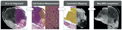 |
Correlating patterns in tumor cytoarchitecture with multiparametric MR signal in preclinical models of sarcoma
Stephanie J Blocker1, James Cook1, Yvonne M Mowery2, Jeffrey I Everitt3, Yi Qi1, Kathryn Hornburg1, Gary P Cofer1, Fernando Zapata1, Alex M Bassil2, Cristian T Badea1, David G Kirsch2, and G. Allan Johnson1
1Radiology, Duke University, Durham, NC, United States, 2Radiation Oncology, Duke University, Durham, NC, United States, 3Pathology, Duke University, Durham, NC, United States
Identification of MR imaging biomarkers for solid tumors requires a reliable means of correlating multiparametric MR and pathological data. We have constructed a platform for registration of multi-scale preclinical imaging libraries that utilizes MR histology and cytometric feature mapping for correlative studies. With this platform we have identified a selection of cytometric features in murine sarcomas which demonstrate correlative trends with ex vivo and in vivo MR, including ADC and T2*.
|
|||
1231. |
PET-MR imaging of reactive macrophages in an orthotopic model of ovarian cancer
Catherine Foss1, Desmond Jacob1, Flonné Wildes1, and Marie-France Penet1
1The Russell H. Morgan Department of Radiology and Radiological Science, The Johns Hopkins University School of Medicine, Baltimore, MD, United States
Major limitations of radiological evaluation of ovarian cancer arise from the inability to consistently detect small lesions. Here, we combine the anatomic imaging capabilities of MRI with the highly sensitive molecular imaging capabilities of PET in an orthotopic syngeneic model of ovarian cancer. [124I]iodo-DPA-713, a macrophage-specific imaging agent, is used to detect and stage ovarian cancer. Specific uptake of the radiotracer was observed within and proximal to the primary tumor at early stages, and in the peritoneal ascitic fluid and within metastases at later stage. These studies will provide insights into ovarian cancer progression that will complement emerging host-directed therapeutics.
|
The International Society for Magnetic Resonance in Medicine is accredited by the Accreditation Council for Continuing Medical Education to provide continuing medical education for physicians.