Digital Posters
Diffusion: Phantoms & Simulations
ISMRM & SMRT Annual Meeting • 15-20 May 2021

Digital Posters
Diffusion: Phantoms & Simulations
| Concurrent 4 | 19:00 - 20:00 |
 |
3401.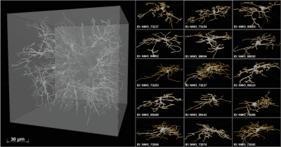 |
Realistic simulations of diffusion MR spectroscopy: The effect of glial cell swelling on non-Gaussian and anomalous diffusion
André Döring1, Maryam Afzali1, Elena Kleban1, Roland Kreis2, and Derek K Jones1
1Cardiff University Brain Research Imaging Centre (CUBRIC), Cardiff University, Cardiff, United Kingdom, 2Departments of Radiology and Biomedical Research, University of Bern, Bern, Switzerland
An existing Monte-Carlo simulation was refined to allow realistic modeling of 150x150x150μm³ tissue samples. The approach was validated on a reference sample, where ground truth is known. The non-Gaussian and anomalous diffusion behavior in diffusion MR spectroscopy experiments was modeled in realistic tissue samples composed of 30 glial cells addressing a hypothesized cell swelling evoked by glial activation. The simulated results agree well with in vivo literature values and may help to improve the linkage between diffusion and histology.
|
||
3402.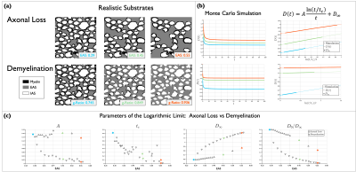 |
Characterizing time-dependent diffusion in the extra-axonal space of white matter for axon loss and demyelination
Ricardo Coronado-Leija1, Hong-Hsi Lee1, Els Fieremans1, and Dmitry S. Novikov1
1Radiology, New York University School of Medicine, New York, NY, United States
In this work, we use Monte Carlo simulations to show how time-dependent diffusion, D(t), in the extra-axonal space provides information about the packing geometry of the axons. In particular we relate the correlation-length obtained from simulations to the correlation-length computed from the power spectrum of the axon packings. We also show through Monte Carlo simulations, in geometries of cylinders and realistic substrates, that D(t) can provide information that differentiates between axon loss and demyelination.
|
|||
3403.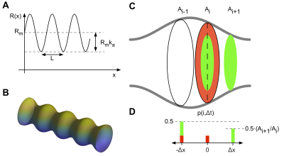 |
A minimal geometrical model for Monte Carlo simulations of time dependent diffusion in axons
Henrik Lundell1 and Samo Lasič1,2
1Danish Research Centre for Magnetic Resonance, Centre for Functional and Diagnostic Imaging and Research, Copenhagen University Hospital Hvidovre, Hvidovre, Denmark, 2Random Walk Imaging, Lund, Sweden
Understanding the link between axonal geometry and diffusion is of large relevance for diffusion weighted MRI (DW-MRI) of white matter. Identifying the minimal geometrical features needed to describe time dependent diffusion allows for faster simulations adequate for the experimentally feasible level of abstraction. We propose an augmented 1D random walk model that within relevant limits mimics the time dependent diffusion in a 3D model of an axon with varying radius.
|
|||
3404.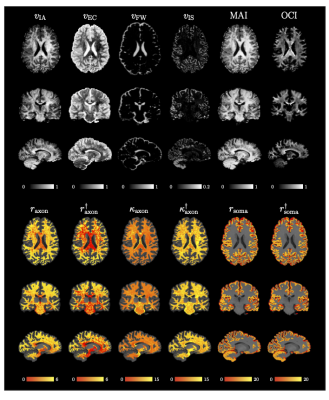 |
Quantifying Cell Size and Membrane Permeability with Microstructure Fingerprinting
Khoi Minh Huynh1,2, Ye Wu2, and Pew-Thian Yap1,2
1Biomedical Engineering, UNC Chapel Hill, Chapel Hill, NC, United States, 2Department of Radiology and Biomedical Research Imaging Center (BRIC), UNC Chapel Hill, Chapel Hill, NC, United States
In diffusion MRI, biophysical models offer a non-invasive means of probing the tissue micro-architecture of the human brain. However, most models rely on closed-form formulas derived with simplifying assumptions such as short gradi- ent pulse, Gaussian phase distribution, and the absence of compartmental ex- change. We present a fast microstructure fingerprinting framework for accurate estimation of axon/soma radii and membrane permeability without relying on these assumptions.
|
|||
3405.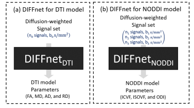 |
DIFFnet: Diffusion parameter mapping network generalized for input diffusion gradient directions and b-values
Juhyung Park1, Woojin Jung1, Eun-jung Choi1, Se-Hong Oh2, Dongmyung Shin1, Hongjun An1, and Jongho Lee1
1Seoul National University, Seoul, Korea, Republic of, 2Hankuk University of Foreign Studies, Gyeonggi-do, Korea, Republic of
A deep neural network, referred to as DIFFnet, was developed to reconstruct the diffusion parameters from data with reasonable b-value and gradient scheme (gradient direction and the number of gradients). For the generalization, Qmatrix was proposed via the projection and quantization of q-space. DIFFnet was trained by simulated datasets with various b-values and gradient schemes. Two DIFFnets, one for DTI and the other for NODDI were developed. DIFFnet successfully reconstructs the diffusion parameter maps of two in-vivo datasets with different b-values and gradient schemes.
|
|||
3406.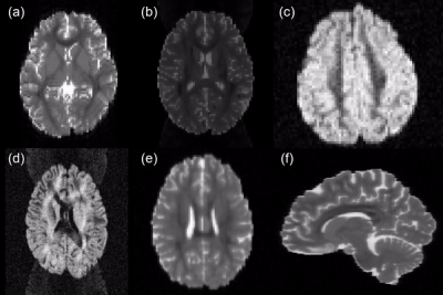 |
Providing realistic ground truth and AI-ready data for fiber tractography: The 99 simulated brains dataset
Peter Neher1 and Klaus Maier-Hein1,2,3
1Medical Image Computing, German Cancer Research Center (DKFZ), Heidelberg, Germany, 2Medical Faculty, University of Heidelberg, Heidelberg, Germany, 3Pattern Analysis and Learning Group, Heidelberg University Hospital, Heidelberg, Germany
We present our new dataset of 99 simulated brains, suitable for fiber tractography training, validation and beyond. With the proposed approach it was possible to create a large dataset of 792 simulated MR images based on 99 subjects. A large variety of acquisition settings and artifacts could be realized. This dataset is the first large collective of diversely simulated brain-like MRI datasets. We believe that this unique dataset is an important contribution to the ongoing efforts of the tractography community to enable proper validation with a real ground truth and to further enable the training of new machine-learning based approaches.
|
|||
3407.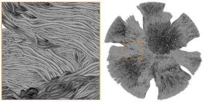 |
A novel in silico phantom for microstructure, tractography and quantitative connectivity estimation
Gabriel Girard1,2,3, Jonathan Rafael-Patino3, Raphael Truffet4, Marco Pizzolato3,5, Emmanuel Caruyer4, and Jean-Philippe Thiran1,2,3
1University Hospital Center (CHUV) and University of Lausanne (UNIL), Lausanne, Switzerland, 2CIBM Center for BioMedical Imaging, Lausanne, Switzerland, 3Swiss Federal Institute of Technology Lausanne (EPFL), Lausanne, Switzerland, 4Univ Rennes, Inria, CNRS, Inserm, IRISA UMR 6074, Empenn ERL U-1228, Rennes, France, 5Technical University of Denmark, Kongens Lyngby, Denmark
In this work, we propose a novel phantom obtained from Monte-Carlo simulations of spins dynamics to improve testing and validation of DW-MRI quantitative structural connectivity. The DiSCo (Diffusion-Simulated Connectivity) phantom is composed of 16 regions of interest placed on a sphere of 1 millimeter in diameter, interconnected by 12,196 axon-like tubular fibers ranging from 1.4um to 4.2um in diameter. Its associated connectivity matrix is weighted by their cross-sectional areas. This in silico phantom, with both microscopic and macroscopic complexity, aims at improving the development and the validation of white matter connectivity estimation methods.
|
|||
3408.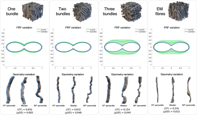 |
Impact of within-voxel heterogeneity in fibre geometry on spherical deconvolution
Ross Callaghan1, Daniel C Alexander1, Marco Palombo1, and Hui Zhang1
1Department of Computer Science and Centre for Medical Image Computing, University College London, London, United Kingdom
Axons in white matter have been shown to have varying geometries within a bundle, but what does this mean for spherical deconvolution, which assumes a single diffusion MRI (dMRI) fibre response function (FRF) for all axons within a voxel? We demonstrate, using advanced dMRI simulations, that variable fibre geometry leads to a variable FRF across axons and that a different choice of FRF can lead to large differences in the recovered fibre orientation distribution function (fODF). This finding suggests that assuming a single FRF can lead to misestimation of the fODF, causing further downstream errors in techniques such as tractography.
|
|||
3409.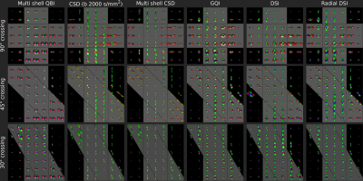 |
A closer look at diffusion and fiber ODFs in a ground truth crossing fiber phantom
Steven H. Baete1,2, Patryk Filipiak1,2, Lee Basler3, Anthony Zuccolotto3, Ying-Chia Lin1,2, Dimitris G. Placantonakis4, Timothy Shepherd1,2, Walter Schneider3, and Fernando E. Boada1,2
1Center for Advanced Imaging Innovation and Research (CAI2R), NYU School of Medicine, New York, NY, United States, 2Center for Biomedical Imaging, Dept. of Radiology, NYU School of Medicine, New York, NY, United States, 3Psychology Software Tools, Inc., Pittsburgh, PA, United States, 4Department of Neurosurgery, Perlmutter Cancer Center, Neuroscience Institute, Kimmel Center for Stem Cell Biology, NYU School of Medicine, New York, NY, United States
High quality diffusion acquisitions are routinely used to study brain connectivity. In each voxel complex intra-voxel fiber crossings may be captured in Orientation Distribution Functions (ODFs). Direct comparison of ODFs calculated with different methods challenging due to a lack of ground truth. Here, we compare different q-space sampling schemes and ODF-reconstructions on a clinical 3T scanner for a known ground truth of crossing Taxons (textile water filled tubes). This comparison illustrates difficulties separating fibers crossing at less than 45° and estimating relative fiber bundle densities using conventional fiber peak identification. Use of more advanced methods is thus recommended.
|
|||
3410.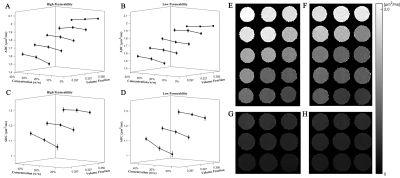 |
Assessment of the effects of cellular properties in tissue on ADC measurements by an experimental study
Xiaodong Li1, Yafei Bai1, Yupeng Liao1, and Sherman Xuegang Xin1
1South China University of Technology, Guangzhou, China
The apparent diffusion coefficient (ADC) provided by diffusion-weighted magnetic resonance imaging (DW-MRI) has been used clinically for nearly three decades. Some hypotheses have been proposed to explain the change in ADC, including the change in membrane permeability, intracellular volume fraction (IVF), tortuosity of extracellular spaces, and intracellular diffusivity. However, no experimental study has been conducted to quantitatively assess the effects of these parameters on the ADC measurements. Experimental study is helpful to understand the biophysical mechanisms underlying the change in the ADC. Here, we designed a series of multi-parameter phantoms to conduct a quantitative experimental study.
|
|||
3411.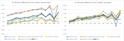 |
Normalization of Temperature Effects for Improved Quantitative Prostate Apparent Diffusion Coefficient (ADC) Imaging Across Multiple Sites
Ken-Pin Hwang1, R. Jason Stafford2, Joshua Yung2, and Aradhana M. Venkatesan3
1The University of Texas M.D. Anderson Cancer Center, Houston, TX, United States, 2Department of Imaging Physics, The University of Texas M.D. Anderson Cancer Center, Houston, TX, United States, 3Department of Abdominal Radiology, The University of Texas M.D. Anderson Cancer Center, Houston, TX, United States
Performing a standardized measurement of ADC requires maintaining a phantom temperature of 0oC. To expand quality assurance of ADC measurement on a network of scanners at room temperatures, a model for temperature dependence was applied as a correction factor. A diffusion phantom was scanned on 1.5T and 3T scanners at four locations using sequence parameters from a prostate protocol. With temperature of the phantom measured at each acquisition, ADC measurements on identically configured scanners exhibited reduced variance when normalized to modeled values. The use of the temperature model is potentially useful in developing a QA program for ADC measurements.
|
|||
3412.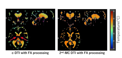 |
Measurement of intraventricular temperature in the whole brain using second order motion compensation DTI
Shuhei Shibukawa1, Tetsu Niwa2, Tosiaki Miyati3, Misaki Saito4, Tetsuo Ogino5, Daisuke Yoshimaru6, and Kagayaki Kuroda7
1Tokai university hospital, Kanagawa, Japan, 2Tokai University School of Medicine, Isehara, Japan, 3Kanazawa University, Kanazawa, Japan, 4Tokai university hospital, Isehara, Japan, 5Philips Japan, Tokyo, Japan, 6RIKEN Center for Brain Science, saitama, Japan, 7Course of Electrical and Electronic Engineering, Graduate School of Engineering, Tokai University, Isehara, Japan
The intraventricular cerebrospinal fluid (CSF) temperature calculated from the diffusion coefficient is affected by the CSF pulsation. Therefore, we investigated the second-order motion compensation DTI (2nd-MC DTI) in consideration of fractional anisotropy (FA) for the CSF to the determination of the intraventricular temperature to improve that accuracy. The measurement of the intraventricular temperature with 2nd-MC DTI showed the least SD and can be more accurately estimated than conventional DTI.
|
|||
3413.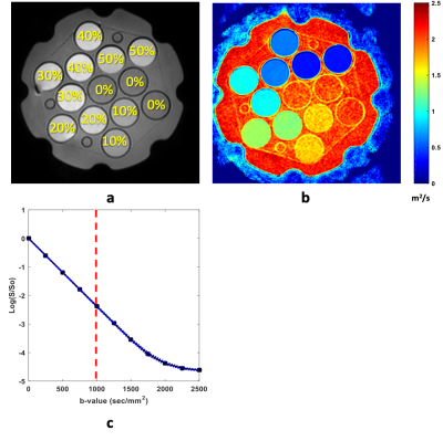 |
Temperature and Concentration Dependence of PVP Phantom Diffusion
Ghoncheh Amouzandeh1, Dariya I Malyarenko1, Yuxi Pang1, Brian D Ross1, and Thomas L Chenevert1
1Radiology, University of Michigan, Ann arbor, MI, United States
This study investigates temperature and concentration dependence of apparent diffusion coefficient (ADC) for aqueous solutions of polyvinylpyrrolidone (PVP). Our objective is to report accurate diffusion values for PVP in the scanner room temperature range. We performed ADC measurements of PVP (0-50% concentration) with K40 polymer moiety at multiple room temperatures while simultaneously measuring internal phantom temperature using an MRI-compatible optical thermometer. To reduce measurement bias, we also performed “gradient calibration” of each physical gradient channel using water ADC at 0oC. Our results are consistent with Arrhenius model for water ADC dependence on temperature and PVP concentration.
|
|||
3414.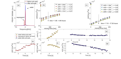 |
Detection of alterations in water transport across the cell membrane by filter-exchange spectroscopy
Athanasia Kaika1, Geoffrey J. Topping1, Mathias Schillmaier1, and Franz Schilling1
1Department of Nuclear Medicine, Technical University of Munich, School of Medicine, Klinikum rechts der Isar, Munich, Germany
Filter-Exchange Spectroscopy (FEXSY) is performed using a double-diffusion magnetic resonance pulse sequence, which encodes information dependent on transmembrane water exchange. Permeabilized baker’s yeast cells were examined with a FEXSY sequence and trypan blue staining. Upon permeabilization with isopropanol, TritonX-100 and sonication, the AXR value of yeast cells increased and trypan blue staining verified the membrane permeabilization. In presence of low isopropanol concentration, a progressive increase of the AXR value was observed, which stopped after isopropanol removal. No differences were detected in the trypan blue staining over time, in presence and in absence of isopropanol.
|
|||
3415.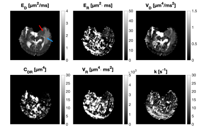 |
Cumulant expansions for measuring restricted diffusion and water exchange
Arthur Chakwizira1, Filip Szczepankiewicz2, Linda Knutsson1,3, Pia Sundgren2,4,5,6, and Markus Nilsson2
1Department of Medical Radiation Physics, Lund University, Lund, Sweden, 2Department of Diagnostic Radiology, Lund University, Lund, Sweden, 3Russell H. Morgan Department of Radiology and Radiological Science, Johns Hopkins University School of Medicine, Baltimore, MD, United States, 4Lund University Bioimaging Center, Lund University, Lund, Sweden, 5Department for Medical Imaging and Physiology, Skåne University Hospital, Lund, Sweden, 6Department of Radiology, University of Michigan, Ann Arbor, MI, United States
Cell sizes and membrane permeability can be inferred by probing time–dependent diffusion due to restricted diffusion and water exchange. However, restriction and exchange have opposing effects on the diffusion weighted signal when varying the diffusion time, making it a challenge to disentangle the two phenomena. We explore the applicability of a unified theoretical framework that includes size and exchange in living cells by applying it to healthy and diseased human brain in vivo as well as a yeast phantom. Results highlight the potential of the framework for differentiating between healthy and abnormal tissue.
|
|||
3416.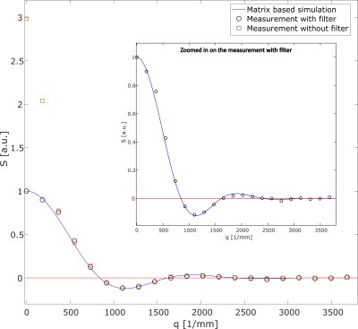 |
Filtered water diffusion pore imaging on a 14.1T spectrometer using strong gradients and capillary phantoms in the presence of extraporal fluid
Dominik Ludwig1,2, Frederik B. Laun3, Karel D. Klika4, Mark E. Ladd1,2,5, Peter Bachert1,2, and Tristan A. Kuder1
1Medical Physics in Radiology, German Cancer Research Center (DKFZ), Heidelberg, Germany, 2Faculty of Physics and Astronomy, Heidelberg University, Heidelberg, Germany, 3Institute of Radiology, University Hospital Erlangen, Friedrich-Alexander-Universität Erlangen-Nürnberg (FAU), Erlangen, Germany, 4Molecular Structure Analysis, German Cancer Research Center (DKFZ), Heidelberg, Germany, 5Faculty of Medicine, Heidelberg University, Heidelberg, Germany
Diffusion pore imaging (DPI) can be used to retrieve the pore space function of arbitrary closed pores. In this study, we show that DPI of glass capillaries is possible even under difficult experimental conditions. By separating the long gradient into a CPMG-like gradient echo train, matching the magnetic susceptibility and adding an additional filter diffusion weighting, it was possible to acquire diffraction patterns of glass capillaries that were placed orthogonally to the main magnetic field. Furthermore, the feasibility of doing DPI in the presence extraporal water using our filtered approach was demonstrated.
|
|||
| 3417. | Lamellar liquid crystal phantom for validating MRI methods to distinguish oblate and prolate diffusion tensors on whole-body scanners
Hong Jiang1, João Pedro de Almeida Martins1, Dan Lundberg2, Chantal M. W. Tax3, and Daniel Topgaard1
1Physical Chemistry, Lund University, Lund, Sweden, 2CR Competence AB, Lund, Sweden, 3Cardiff University Brain Research Imaging Centre (CUBRIC), Cardiff University, Cardiff, United Kingdom
Conventional diffusion MRI can yield planar tensors in tissues such as epidermoid cysts, comprising tightly packed planar cells, and brain tissue "sheet" structures with populations of axonal fibers crossing at nearly right angles. The two cases may be resolved by multidimensional diffusion encoding as previously demonstrated with various liquid crystal phantoms on preclinical equipment. For method validation also on whole-body scanners, we here develop a lamellar liquid crystal phantom giving microscopically planar diffusion tensors and having sufficiently long T2 for use with single-shot EPI signal read-out.
|
|||
3418.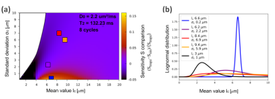 |
Microstructure size-distribution estimations with smooth and sharp non-uniform oscillating gradient spin-echo modulations
Melisa Lucía Giménez1,2, Pablo Jiménez1,2, Leandro Andrés Pedraza Pérez1,2, Diana Betancourth2, Analía Zwick2,3,4, and Gonzalo Agustín Álvarez1,2,3,4
1Departamento de Física Médica, Instituto Balseiro, Universidad Nacional de Cuyo, CNEA, San Carlos de Bariloche, Argentina, 2Centro Atómico Bariloche, CNEA, San Carlos de Bariloche, Argentina, 3Consejo Nacional de Investigaciones Científicas y Técnicas de Argentina (CONICET), San Carlos de Bariloche, Argentina, 4Instituto de Nanociencia y Nanotecnología, CNEA,CONICET, San Carlos de Bariloche, Argentina
Morphological changes related to neurological diseases occur at micrometer scales. Obtaining such information non-invasively opens new paradigms for clinical diagnosis. We use the Non-uniform Oscillating Gradient Spin-Echo sequence to estimate microstructure size-distributions with high sensitivity based on probing a signal “decay-shift” rather than a signal decay-rate. The “decay-shift” arises with sharp gradient modulations. As fast ramps are prohibitive in clinical diagnosis, we evaluate the method using sharp and smooth gradient modulations. We show using simulations and proof-of-principle experiments with phantoms that mimic axon-bundles, that optimal estimation of the underlying microstructure-size distribution is obtained either using sharp or smooth modulations.
|
|||
3419.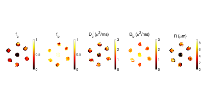 |
Estimating the pore size in a biomimetic phantom using free gradient waveforms
Maryam Afzali1, Tomasz Pieciak2,3, Lars Mueller1, Andre Doring1, Dan Ma4, Marco Pizzolato5,6, and Derek K Jones1
1Cardiff University Brain Research Imaging Centre (CUBRIC), School of Psychology, Cardiff University, Cardiff, United Kingdom, 2AGH University of Science and Technology, Kraków, Poland, 3LPI, ETSI Telecomunicación, Universidad de Valladolid,, Valladolid, Spain, 4Biomedical Engineering, Case Western Reserve University, Cleveland, OH, United States, 5Department of Applied Mathematics and Computer Science, Technical University of Denmark,, Kongens Lyngby, Denmark, 6Signal Processing Lab (LTS5), École polytechnique fédérale de Lausanne (EPFL), Lausanne, Swaziland
Diffusion magnetic resonance imaging is a non-invasive tool to probe the microstructural features of a sample. One of these properties is the restriction size that can be measured by changing the diffusion time or alternatively changing the frequency content of the gradient waveform. B-tensor encoding was proposed recently to disentangle microstructural features of the tissue. Here we use the combination of linear, planar, and spherical tensor encoding to estimate the pore size in a biomimetic phantom, for which ground truth size estimates were available. The results show a good agreement between the estimated sizes and ground truth values.
|
|||
3420.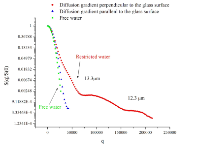 |
A New Phantom to Study a Restricted Diffusion Introduction
Sergey Magnitsky1
1CHOP, Philadelphia, PA, United States
Diffusion-weighted imaging is instrumental in the evaluation of bone quality. However, an interpretation of the data obtained from porous material is complex due to the effects of restricted diffusion. In this study, we are presenting a new restriction diffusion phantom, which was developed for an optimization of acquisition protocols for bone studies. The phantom consists of microscopic-slides separated by glass spheres (~10 μm). The space between slides was filled with water. NMR data were collected and diffusion-properties of the phantom were documented. The proposed phantom can be easily replicated in any laboratory and will assist in investigations of restriction diffusion.
|
The International Society for Magnetic Resonance in Medicine is accredited by the Accreditation Council for Continuing Medical Education to provide continuing medical education for physicians.