Digital Posters
Brain Microstructure: Gray Matter, Pathology & Preclinical Validation
ISMRM & SMRT Annual Meeting • 15-20 May 2021

Digital Posters
Brain Microstructure: Gray Matter, Pathology & Preclinical Validation
| Concurrent 3 | 15:00 - 16:00 |
2023.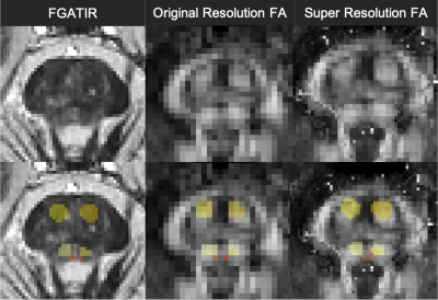 |
Super-resolution and CNN denoising to improve the accuracy of small brainstem structure characterization with in vivo diffusion MRI
Benjamin Ades-Aron1, Hong-Hsi Lee1, Heidi Schambra2, Dmitry S. Novikov1, Els Fieremans1, and Timothy Shepherd1
1Radiology, NYU School of Medicine, New York, NY, United States, 2Neurology, NYU Langone, New York, NY, United States
Diffusion MRI should be sensitive to early pathology or functional re-organization changes for small internal brainstem structures associated with ischemia, multiple sclerosis or neurodegeneration. Application of diffusion MRI to brainstem studies is challenged by limited spatial resolution, image distortion from skull base artifacts and bias introduced if diffusion contrast is also used for structure segmentation. We describe and evaluate a novel combination of Fast Gray Matter Acquisition T1 Inversion Recovery (FGATIR) denoising with deep learning, multi-modal nonlinear image co-registration and super-resolution techniques to improve the accuracy of small internal brainstem structure segmentation on advanced diffusion MRI data.
|
|||
2024.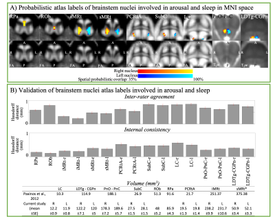 |
Probabilistic structural atlas and connectome of brainstem nuclei involved in arousal and sleep by 7 Tesla MRI in living humans
Maria Guadalupe Garcia Gomar1, Kavita Singh1, and Marta Bianciardi1
1Radiology, Athinoula A. Martinos Center for Biomedical Imaging, Massachusetts General Hospital and Harvard Medical School, Boston, MA, United States, Charlestown, MA, United States
Brainstem nuclei such as the medullary reticular formation, raphe nuclei, pontine nuclei, locus coeruleus and subcoeruleus, among others, play a crucial role in arousal and sleep. Despite their involvement in critical functions, their study in living humans is challenging due to limited resolution and contrast of conventional MRI. First, we precisely delineated nine arousal/sleep brainstem nuclei in 7 Tesla MRI of healthy humans and generated their validated in-vivo stereotaxic probabilistic atlas. Second, we evaluated their structural connectivity using 7 Tesla High-Angular-Resolution-Diffusion-Imaging and probabilistic tractography. This atlas and connectome might help evaluating disorders of consciousness, sleep disorders and neurological disorders.
|
|||
2025.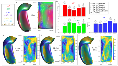 |
Investigating the Relationship Between Morphology and Microstructure in the Hippocampus
Bradley Karat1, Jordan DeKraker1, Uzair Hussain2, and Ali Khan1,2
1Department of Neuroscience, Schulich School of Medicine & Dentistry, Western University, London, ON, Canada, 2Centre for Functional and Metabolic Mapping, Robarts Research Institute, Western University, London, ON, Canada
Aberrations of hippocampal subfields and microstructure are well studied, however, current in-vivo measures of microstructure are non-specific towards intra-hippocampal pathways and neurites within subfields. This is partly due to its complex and folded structure. Although highly folded, the hippocampus maintains an intrinsic coordinate system with well defined axes along the anterior-posterior, proximal-distal, and inner-outer dimensions. Importantly, hippocampal microstructure tends to align with these axes. Specific macrostructural measures such as gradient vector fields can be derived from these axes. Leveraging these measures, we were able to find systematic correlations between macrostructure and local microstructure in the hippocampus.
|
|||
2026.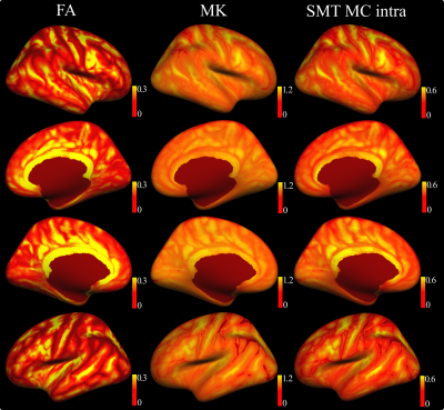 |
Probing human cortical microstructure using diffusion MRI: Insights from N=17,646 individuals of the UK Biobank.
Ivan Maximov1,2, Dennis van der Meer2, Ann-Marie de Lange2, Tobias Kaufmann2, and Lars T. Westlye2
1Department of Health and Functioning, Western Norway University of Applied Sciences, Bergen, Norway, 2NORMENT, University of Oslo, Oslo, Norway
While diffusion MRI (dMRI) has been established as a powerful tool for studying human brain white matter, its potential for characterising structural grey matter properties waits for to be fully realised. dMRI is capable of providing unique complementary information about cortical microstructure, function and connectivity. Here, we describe the spatial distribution and individual differences in cortical grey matter microstructure across 17,646 UK Biobank (UKB) participants using three diffusion MRI approaches, namely, diffusion tensor imaging, kurtosis tensor imaging and spherical mean technique, and demonstrate its predictive value for brain age prediction using machine learning.
|
|||
2027.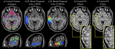 |
Using Sub-Millimeter Isotropic DTI to Observe the Cortical Depth Dependence of Diffusion Anisotropy and Diffusivity in-vivo
Iain P Bruce1, Yixin Ma1, and Allen W Song1
1Brain Imaging and Analysis Center, Duke University, Durham, NC, United States
The cerebral cortex is comprised of short column structures that span across the cortical layers between white matter and the pial surface. Subtle abnormalities in this microstructure, which may not be visible on anatomical scans, can have a large effect on brain function and development. In an effort to delineate such abnormalities, this study presents a technique to accurately and effectively observe the cortical depth dependence of diffusion anisotropy and diffusivity in-vivo using sub-millimeter isotropic resolution diffusion tensor imaging.
|
|||
2028.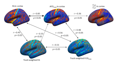 |
Surface based analysis of cortical diffusion metrics: associations with cortical myeloarchitecture and underlying white matter anisotropy
Tonima Sumya Ali1 and Fernando Calamante1,2
1School of Biomedical Engineering, The University of Sydney, Sydney, Australia, 2Sydney Imaging, The University of Sydney, Sydney, Australia
We employ structural and diffusion MRI metrics to evaluate their effectiveness for cortical segregation based on myelination, neurite density and microstructure in cortex. Three cortical measures (T1w/T2w, FA and total apparent fibre density, AFDtotal, demonstrated significant inter-correlations. Two track-weighted parameters (FOD and FA, measured in white matter adjacent to cortex), also showed significant correlations to cortical FA and AFDtotal. The spatial cortical patterns corresponding to these parameters suggest they provide complementary information on cortical features, and may help improve our understanding of cortical organisation in vivo and eventually lead to a further multi-parametric model for cortical parcellation.
|
|||
2029.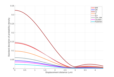 |
Generalized anisotropy profiles distinguish cortical and subcortical structures in ex vivo diffusion MRI
Robert Jones1, Qiyuan Tian1, Chiara Maffei1, Jean Augustinack1, Aapo Nummenmaa1, Susie Huang1, and Anastasia Yendiki1
1Athinoula A. Martinos Center for Biomedical Imaging, Massachusetts General Hospital and Harvard Medical School, Charlestown, MA, United States
We propose an approach to extracting a generalized anisotropy profile from the ensemble average propagator, which can be obtained either from diffusion spectrum imaging (DSI) data or from a generalized DSI analysis of diffusion MRI data acquired on q-shells. We validate this approach on data from ex vivo human tissue, acquired at 9.4T with 0.25 mm spatial resolution. We show that the proposed anisotropy profile can be used to distinguish between various white- and gray-matter structures, revealing fundamental differences in their microstructure.
|
|||
2030.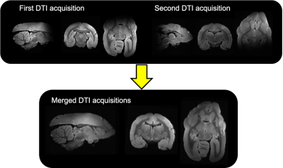 |
Comparison of high-resolution DTI in ex vivo newborn and adult marmoset brain
Emmanuelle Weber1, Erpeng Dai1, Christoph Leuze1, Nikola T. Markov2, Nicholas Tran1, Mariko Bennett1, Samuel Baker1, Kerriann Casey1, Kalanit Grill-Spector1, Keren Haroush1, and Jennifer A. McNab1
1Stanford, Stanford, CA, United States, 2Buck Institute, Novato, CA, United States
High-resolution (0.15 mm isotropic) diffusion MRI of newborn and an adult marmoset brains were acquired. For the adult marmoset brain the diffusion MRI data of anterior and posterior parts of the brain were separately acquired and subsequently co-registered. Across the whole brain, fractional anisotropy (FA) was lower and mean diffusivity (MD) was higher (Fig. 4) in the newborn compared to the adult. The biggest differences were observed in white matter. Cingulate cortex region A25 showed the smallest difference, which aligns with previous studies showing its early stage development
|
|||
2031.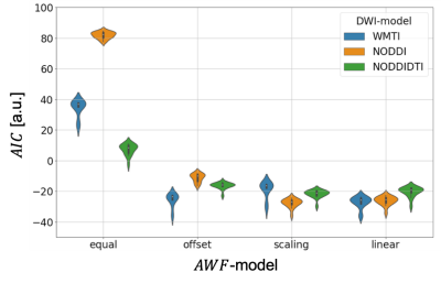 |
Is calibration necessary to relate NODDI, NODDIDTI, WMTI to axonal volume fraction? - A joint ex vivo MRI and histology study.
Sebastian Papazoglou1, Mohammad Ashtarayeh1, Martina F. Callaghan2, Mark D. Does3,4,5,6, and Siawoosh Mohammadi1,7
1Department of Systems Neurosciences, University Medical Center Hamburg-Eppendorf, Hamburg, Germany, 2Wellcome Centre for Human Neuroimaging, UCL Queen Square Institute of Neurology, University College London, London, United Kingdom, 3Department of Biomedical Engineering, Vanderbilt University, Nashville, TN, United States, 4Institute of Imaging Science, Vanderbilt University Medical Center, Nashville, TN, United States, 5Department of Radiology and Radiological Sciences, Vanderbilt University Medical Center, Nashville, TN, United States, 6Department of Electrical Engineering, Vanderbilt University, Nashville, TN, United States, 7Department of Neurophysics, Max Planck Institute for Human Cognitive and Brain Sciences, Leipzig, Germany
It is unknown whether the accuracy of DWI-model based estimates of the axonal water fraction could be enhanced by additional calibrations. In this study we compared three DWI-models (WMTI, NODDI, NODDIDTI) using histology data as gold standard in ex vivo mouse models with a broad dynamic range of axonal metrics. We found that all DWI-models improved with additional calibration. Models with fixed diffusivities benefited efficiently from an additional scaling, while the WMTI model was stronger affected by an additional offset. Furthermore, without any additional calibration, the axonal compartment biomarker from NODDIDTI could explain the data better than WMTI or NODDI.
|
|||
2032. |
b-Value Dependency of Diffusion Parameters Derived from the DTI and DKI Models in Postmortem Human Brain Hemispheres
Junye Yao1, Zihan Zhou1, Benjamin C. Tendler2, Karla L. Miller2, Lei Zhang3, Keqing Zhu3, Aimin Bao3, Hongjian He1, and Jianhui Zhong1,4
1Center for Brain Imaging Science and Technology, College of Biomedical Engineering and Instrumental, Zhejiang University, Hangzhou, China, 2Wellcome Centre for Integrative Neuroimaging, FMRIB, Nuffield Department of Clinical Neurosciences, University of Oxford, London, United Kingdom, 3National Human Brain Bank for Health and Disease, School of Brain Science and Brain Medicine, Zhejiang University, Hangzhou, China, 4Department of Imaging Sciences, University of Rochester, Rochester, NY, United States
Diffusion weighted images of formalin-fixed human brain hemispheres were used to study b-value dependence of diffusion parameters with the diffusion tensor and diffusion kurtosis model. Consistent decreases of FA with increasing b-values were observed in all hemispheres. Meanwhile, the mean kurtosis was found to increase with the discrepancy of FA between high and low b-value datasets. These results indicate that the b-value dependence of FA could be explained by non-gaussian diffusion effects consistent with tissue microstructure.
|
|||
2033.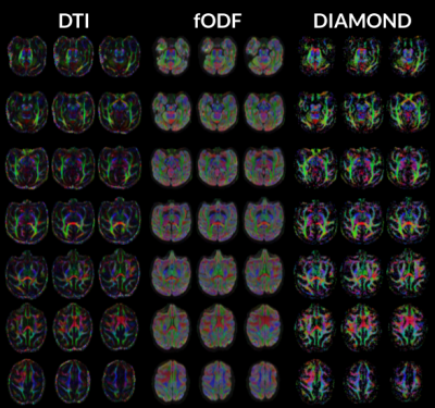 |
Analysis of high-resolution 3T diffusion MRI data obtained with minimal CUSP acquisition scheme from a Non-Human Primate
Alex Colin Valcourt Caron1, Amir Shmuel2, Ilana R. Leppert2, and Maxime Descoteaux1
1Université de Sherbrooke, Sherbrooke, QC, Canada, 2Montreal Neurological Institute, McGill University, Montreal, QC, Canada
The validation of advanced diffusion MRI methods requires acquisition at high-resolution and high SNR, which is difficult to obtain in humans. The use of anesthetized Non-Human Primates (NHPs) is key to unlocking valid microstructural maps. However, data acquisition and analysis tools must be adapted and configured for successful application to NHPs imaging. Here we propose a processing pipeline allowing for the analysis of diffusion data using DTI, CSD, and DIAMOND reconstruction, tailored for the analysis of data from the macaque brain. We present results obtained by applying the pipeline to a dataset acquired with a minimal CUSP 90 directions scheme.
|
|||
2034. |
Scanning post mortem fixed whole human brain for advanced higher order diffusion modelling using a 300 mT/m whole-body MRI scanner
Luke Joel Edwards1, Evgeniya Kirilina1,2, Carsten Jäger1,3, Kirsten Garus4, Markus Cremer4, Katrin Amunts4,5, and Nikolaus Weiskopf1,6
1Department of Neurophysics, Max Planck Institute for Human Cognitive and Brain Sciences, Leipzig, Germany, 2Center for Cognitive Neuroscience Berlin, Freie Universität Berlin, Berlin, Germany, 3Paul Flechsig Institute of Brain Research, Leipzig University, Leipzig, Germany, 4Institute of Neuroscience and Medicine, Research Centre Jülich, Jülich, Germany, 5C. and O. Vogt Institute for Brain Research, Heinrich Heine University Düsseldorf, Düsseldorf, Germany, 6Felix Bloch Institute for Solid State Physics, Leipzig University, Leipzig, Germany
Higher order diffusion modelling (diffusional kurtosis and multi-tissue CSD) of a whole post mortem human brain with excellent tissue quality was enabled by ultra-high b values from a whole-body 3T scanner with ultra-strong gradients. This was complemented by a novel gradient nonlinearity correction scheme to interpolate signals onto diffusion shells before estimation of fibre distributions. The brain is part of the BigBrain initiative, and will undergo an extensive atlasing procedure. This dataset will thus extend the studies possible on the future BigBrain atlas, allowing investigation into diffusion microstructure imaging and tractography.
|
|||
2035.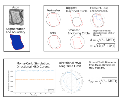 |
Adapted Microscopy Estimation of Axon Diameters for Diffusion MRI Comparison
Michael Paquette1, Cornelius Eichner1, Guillermo Gallardo1, and Alfred Anwander1
1Neuropsychology, Max Planck Institute for Human Cognitive and Brain Sciences, Leipzig, Germany
The quantification of axon diameter from electron microscopy (EM) images requires a choice of heuristics to approximate the dimensions. This choice impacts the resulting size distributions and should be adapted to the task. In diffusion MRI, complex shapes are encoded based on their directional mean squared displacement. We compute diffusion-specific ground truth diameters using Monte-Carlo simulations and evaluate typical choices of EM image analysis heuristics against them. Further, we propose a new and simple heuristic based on the mean square displacement inside an ellipse to compute better axon diameter quantifications from electron microscopy.
|
|||
2036.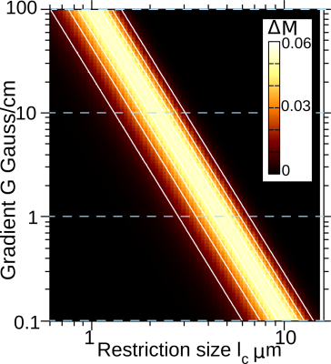 |
Selective microstructure-size filters for non-invasive quantitative MRI
Milena Capiglioni1,2,3, Analía Zwick1, Pablo Jimenez1, and Gonzalo Álvarez1,2
1Laboratorio de Espectroscopía e Imágenes por RMN, Departamento de Física Médica, Centro Atómico Bariloche - CNEA - CONICET, Bariloche, Argentina, 2Instituto Balseiro, CNEA, Universidad Nacional de Cuyo, Bariloche, Argentina, 3Support Center for Advanced Neuroimaging (SCAN), Institute for Diagnostic and Interventional Neuroradiology, University Hospital of Bern, University of Bern, Bern, Switzerland
Extracting quantitative microstructure information of living-tissue by non-invasive imaging is an outstanding challenge for understanding disease mechanisms. We introduce a method to make selectively images of microstructure-sizes by probing molecule diffusion with MRI. It relies on designing dynamical control of nuclear spins to sense magnetization “decay-shifts” rather than the commonly used spin-echo decay-rates. The feasibility and performance of the method are illustrated with proof-of-principle experiments and simulations on typical size-distributions of white-matter in the mouse brain. These results position spin-echo decay-shifts as a promising MRI tool to perform non-invasive histology without assuming a microstructure distribution model.
|
|||
2037.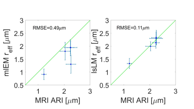 |
Validation of MRI-based axon radius index estimation using large-scale light microscopy and deep learning
Mohammad Ashtarayeh1, Laurin Mordhorst1, Maria Morozova2,3, Tobias Streubel1,2, Jan Malte Oeschger1, Joao Periquito4, Andreas Pohlmann4, Henriette Rusch3, Carsten Jäger2, Thoralf Niendorf4, Nikolaus Weiskopf2,5, Markus Morawski2,3, and Siawoosh Mohammadi1,2
1Department of Systems Neurosciences, University Medical Center Hamburg-Eppendorf, Hamburg, Germany, 2Department of Neurophysics, Max Planck Institute for Human Cognitive and Brain Sciences, Leipzig, Germany, 3Paul Flechsig Institute of Brain Research, University of Leipzig, Leipzig, Germany, 4Berlin Ultrahigh Field Facility (B.U.F.F.), Max-Delbrueck-Center for Molecular Medicine in the Helmholtz Association, Berlin, Germany, 5Felix Bloch Institute for Solid State Physics, Faculty of Physics and Earth Sciences, Leipzig University, Leipzig, Germany
We used a new method for validation of MRI-based axon radius index (ARI) mapping using large-scale light microscopy (lsLM) that provides a good representation of the fraction of large axons - the main contributors to the MRI-based ARI. The proposed method captures 100-1000 times more axons than current standard small field of view microscopy. We showed that the one-to-one correspondence between MRI-based ARI and lsLM-based effective axon radius is superior to the current standard method.
|
|||
2038.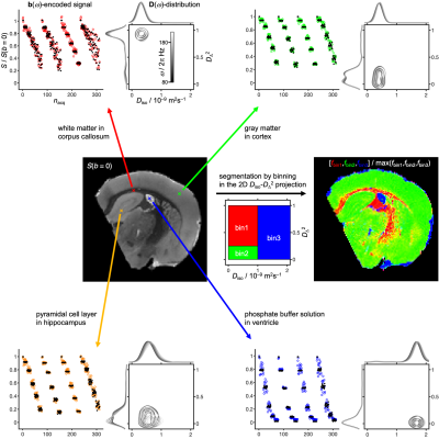 |
Nonparametric D(ω)-distributions for model-free analysis of b(ω)-encoded multidimensional diffusion MRI on ex vivo rat brain
Omar Narvaez1, Maxime Yon2, Alejandra Sierra1, and Daniel Topgaard3
1A.I. Virtanen Institute for Molecular Sciences, University of Eastern Finland, Kuopio, Finland, 2CEMHTI, French National Centre for Scientific Research, Paris, France, 3Department of Chemistry, Lund University, Lund, Sweden
Nonparametric distributions of cell sizes or diffusion tensors have recently been applied to analyze clinically relevant data acquired with advanced diffusion encoding schemes building on oscillating gradients, targeting the frequency-dependence and cell size, or more general q-vector trajectories focusing on the tensorial aspects. We introduce nonparametric D(ω)-distributions as a joint analysis framework taking both frequency-dependence and tensorial properties into account, and demonstrate the approach with ex vivo rat brain data acquired with gradient waveforms exploring the relevant dimensions of the tensor-valued encoding spectrum b(ω).
|
|||
2039.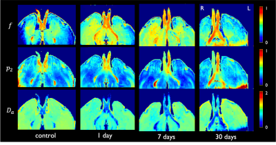 |
Biophysical modeling of ex vivo diffusion MRI for the longitudinal characterization of axonal degeneration in the optic nerve
Ricardo Coronado-Leija1, Santiago Coelho1, Omar Narvaez2, Jorge Larriva-Sahd3, Alonso Ramirez-Manzanares4, Luis Concha3, Dmitry S. Novikov1, and Els Fieremans1
1Radiology, New York University School of Medicine, New York, NY, United States, 2University of Eastern Finland, Kuopio, Finland, 3Instituto de Neurobiologia, Universidad Nacional Autonoma de Mexico, Queretaro, Mexico, 4Centro de Investigacion en Matematicas, Guanajuato, Mexico In this work, we evaluated the parameters of the Standard Model (SM), augmented with a free water compartment, on ex vivo diffusion MRI, to detect longitudinal changes caused by axonal degeneration. Parameters of SM were estimated from the rotational invariants of the diffusion signal using a supervised machine learning approach. Axonal degeneration was induced by nerve injury using a rat retinal ischemia model. Comparisons with 2D histology derived metrics revealed that SM parameters are sensitive to changes caused by axonal degeneration, particularly axon loss. SM parameters also reveal presence of microglia, and increased orientation dispersion. |
|||
2040.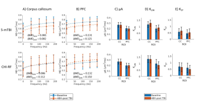 |
Microstructural Diffusion MRI in Mouse Models of Severe and Repetitive Mild Traumatic Brain Injury
Naila Rahman1,2, Kathy Xu2, Nico Arezza1,2, Kevin Borsos1,2, Matthew Budde3, Arthur Brown2,4, and Corey Baron1,2
1Medical Biophysics, Western University, London, ON, Canada, 2Robarts Research Institute, London, ON, Canada, 3Neurosurgery, Medical College of Wisconsin, Milwaukee, WI, United States, 4Anatomy and Cell Biology, Western University, London, ON, Canada
Microstructural diffusion MRI (dMRI) improves the specificity required to detect microstructure changes related to pathophysiology. Oscillating gradient spin-echo (OGSE) dMRI is sensitive to structural disorder and microscopic anisotropy (µA) dMRI is sensitive to water diffusion anisotropy independent of neuron fiber orientation. In this work, OGSE and µA protocols were implemented to enable in vivo longitudinal scanning at 9.4T. Preliminary data in a rodent model of traumatic brain injury (TBI) revealed changes in mean diffusivity dependence on OGSE frequency post-TBI and changes in spherical tensor kurtosis (sensitive to cell size heterogeneity), compared to healthy mice.
|
|||
 |
2041.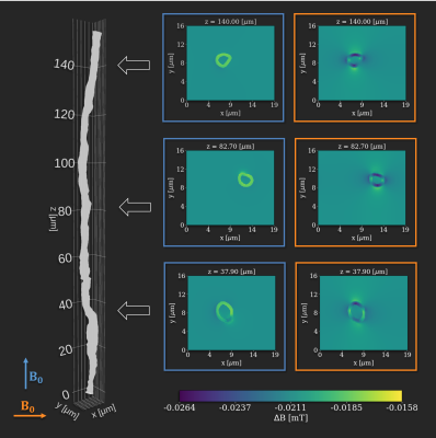 |
Susceptibility-induced fiber orientation dependency of the DWI signal in white matter measured in ex vivo rat brain at 7 T
Sidsel Winther1,2, Mariam Andersson1,2, Henrik Lundell2, and Tim Dyrby1,2
1DTU Compute, Technical University of Denmark, Kongens Lyngby, Denmark, 2Danish Research Center for Magnetic Resonance, Functional and Diagnostic Imaging and Research, Copenhagen University Hospital Hvidovre, Hvidovre, Denmark
Magnetic susceptibility of myelin induces morphology- and orientation-dependent perturbations of the B_0-field. At the microstructural scale of brain white matter, the main contribution to these perturbations comes from myelin. Here, we show that a consequence of this is a fiber-orientation-dependent bias of the DWI-signal across white matter regions in an ex vivo rat brain. This implies that biophysical modeling must regard susceptibility at the microstructural scale in order not to become biased as well. Higher field strength will increase the bias and vice versa. Hence, especially pre-clinical scans are affected.
|
||
2042.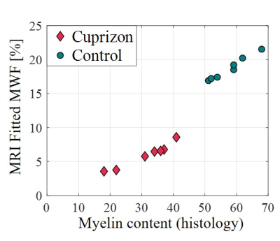 |
In vivo validation of a data driven algorithm for multicomponent T2 mapping on a mice model of demyelination
Noam Omer1, Ella Wilczynski1, Neta Stern1, Tamar Blumenfeld-Katzir1, Meirav Galun2, and Noam Ben-Eliezer1,3,4,5,6
1Department of Biomedical Engineering, Tel-Aviv University, Tel-Aviv, Israel, 2Department of Computer Science and Applied Mathematics, Weitzman institute of science, Rehovot, Israel, 3Department of Orthopedics, Shamir Medical Center, Zerifin, Israel, 4Sackler Faculty of Medicine, Tel-Aviv University, Tel-Aviv, Israel, 5Sagol School of Neuroscience, Tel-Aviv University, Tel-Aviv, Israel, 6Center for Advanced Imaging Innovation and Research (CAI2R), New-York University Langone Medical Center, New York, NY, United States
Multicomponent T2 analysis (mcT2) can provide a clinically-useful myelin biomarker in vivo. Its clinical applicability, however, is hindered by lack of gold standard technique that can overcome the ambiguity of fitting several T2 components to a single experimental signal. In this study we aimed to validate the utility of a novel data-driven mcT2 mapping algorithm for quantifying myelin content. To that end, we applied the mcT2 algorithm to 14 mice divided into two groups of mice: cuprizone-induced demyelination model, and controls. Results show excellent agreement between the mcT2 based myelin biomarker and ground truth quantification of myelin from immunohistochemical staining.
|
The International Society for Magnetic Resonance in Medicine is accredited by the Accreditation Council for Continuing Medical Education to provide continuing medical education for physicians.