Digital Posters
Microstructure: Models, Sampling & Analysis
ISMRM & SMRT Annual Meeting • 15-20 May 2021

Digital Posters
Microstructure: Models, Sampling & Analysis
| Concurrent 4 | 13:00 - 14:00 |
3637.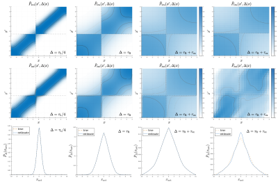 |
Recovering almost everything that diffusion could reveal
Evren Özarslan1,2
1Department of Biomedical Engineering, Linköping University, Linköping, Sweden, 2Center for Image Science and Visualization, Linköping University, Linköping, Sweden
Diffusion magnetic resonance has been employed for determining the distribution of net displacements (ensemble average propagator), moments and correlations of net displacements, and the steady-state distribution of magnetized particles. All such quantities are accessible via the diffusion propagator, which characterizes the diffusion process fully. Here, a novel diffusion encoding and data analysis framework is introduced with which the diffusion propagator can be recovered.
|
|||
3638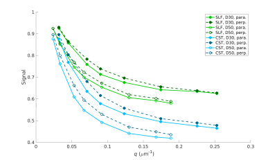 |
Validation of between-bundle differences and within-bundle continuity of microstructural indices in ex vivo human brain tissue Video Permission Withheld
Robert Jones1, Chiara Maffei1, Qiuyun Fan1, Jean Augustinack1, Barbara Wichtmann2, Aapo Nummenmaa1, Susie Huang1, and Anastasia Yendiki1
1Radiology, Athinoula A. Martinos Center for Biomedical Imaging, Massachusetts General Hospital and Harvard Medical School, Charlestown, MA, United States, 2Department of Radiology, University Hospital Bonn, Bonn, Germany, Bonn, Germany
We take advantage of the high spatial resolution that is feasible ex vivo on a preclinical 9.4T system to investigate between-bundle differences and within-bundle continuity of microstructural indices. In a human brain sample that has also undergone optical imaging to obtain direct measurements of axonal orientations, we identify regions occupied by motor fibers or association fibers. We collect diffusion-weighted images with two diffusion times and eight q-shells and use them to estimate the parallel and perpendicular diffusion coefficient in each fiber bundle. We show that the diffusion coefficients vary smoothly along each bundle, and have different profiles between bundles.
|
|||
3639.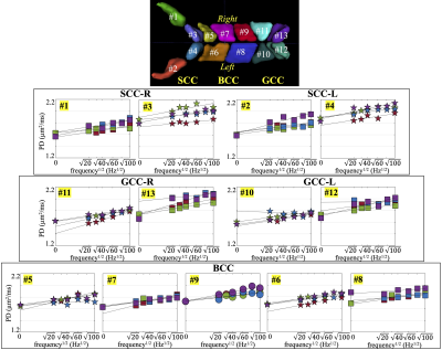 |
Oscillating Gradient Spin Echo-Based Time-dependent Diffusivity Reflects Regional Microstructure Differences in Human White Matter
Ante Zhu1, J. Kevin DeMarco2,3, Robert Y. Shih2,3, Radhika Madhavan1, Tim Sprenger4, Chitresh Bhushan1, Maureen Hood2,3, Luca Marinelli1, Vincent B. Ho2,3, and Thomas K.F. Foo1
1GE Global Research, Niskayuna, NY, United States, 2Uniformed Services University of the Health Sciences, Bethesda, MD, United States, 3Walter Reed National Military Medical Center, Bethesda, MD, United States, 4GE Healthcare, Stockholm, Sweden
Diffusivity measurements of the human brain have been shown to increase at short diffusion time by using oscillating gradient spin echo (OGSE). The time-dependent diffusivity averaged in large white matter parcels has been measured to assess microstructure characteristics. In this study, we assessed time-dependent diffusivity in finer white matter parcels to study the regional microstructure differences. In four healthy volunteers, we consistently observed higher parallel diffusivity values and higher increasing rate over OGSE frequencies in two sub-parcels of the genu and two of the splenium in the corpus callosum, compared to other regions, indicating regional microstructure differences of the brain.
|
|||
3640. |
A tale of two frequencies: optimizing oscillating gradients for frequency dependent differential kurtosis mapping
Kevin B Borsos1,2, Desmond HY Tse2, Paul I Dubovan1,2, and Corey A Baron1,2,3
1Department of Medical Biophysics, Western University, London, ON, Canada, 2Centre for Functional and Metabolic Mapping, Western University, London, ON, Canada, 3Robarts Research Institute, Western University, London, ON, Canada
Frequency dependent diffusion kurtosis has historically been difficult to measure with oscillating gradient spin echo (OGSE) sequences and even more so without the use of gradient insert coils. Here we present a new OGSE gradient waveform and determine the optimal frequency to observe kurtosis differences between PGSE and OGSE acquisitions using a conventional gradient system. Using this method we present in vivo differential kurtosis maps based on the frequency dependence.
|
|||
3641.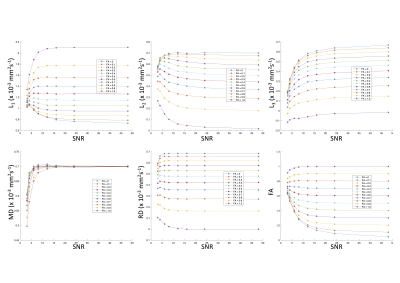 |
When Averaging Only Gets You So Far: Repulsion and Peanut Squashing in Diffusion Tensor MRI
Samuel Bryce Jones1, Emre Kopanoglu2, Chantal Tax2, and Derek Jones2
1Radyr Comprehensive School, Cardiff, United Kingdom, 2CUBRIC, School of Psychology, Cardiff, United Kingdom
This work asks a very simple but important question: To what extent can we recover lost SNR by signal averaging in a standard diffusion tensor MRI experiment? Based on the theory that if SNR of a signal is reduced by a factor (1/m), then m2 signal averages will recover that SNR, the diffusion MRI literature often assumes that averaging is the 'magic bullet'. Here, using Monte Carlo simulations, we show that this only true to a certain extent and, despite averaging, low SNR data results in biased DTI estimates.
|
|||
3642.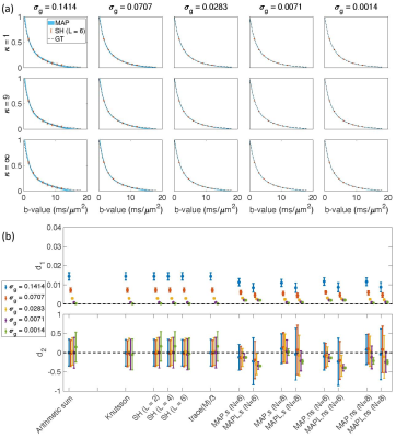 |
Computing the Orientational-Average of Diffusion-Weighted MRI Signals: A Comparison of Different Techniques
Maryam Afzali1, Hans Knutsson2,3, Evren Özarslan2,3,4, and Derek K Jones1,5
1Cardiff University Brain Research Imaging Centre (CUBRIC), School of Psychology, Cardiff University, Cardiff, United Kingdom, 2Department of Biomedical Engineering, Linköping University, Linköping, Sweden, 3Center for Medical Image Science and Visualization, Linköping University, Linköping, Sweden, 4These authors share last authorship, Linköping, Sweden, 5These authors share last authorship, Cardiff, United Kingdom
Numerous applications in diffusion MRI involve computing the orientationally-averaged diffusion-weighted signal. Most approaches assume that the gradient vectors are uniformly distributed on a sphere, computing the orientationally-averaged signal through arithmetic averaging. One challenge is that not all acquisition schemes have gradient vectors distributed over perfect spheres. Alternative averaging methods include: weighted signal averaging; spherical harmonic; and Mean Apparent Propagator MRI (MAP-MRI). Here, these methods are compared under different signal-to-noise (SNR) realizations. With dense and isotropically-distributed sampling, all methods give comparable results. As the SNR and number of data points are reduced, MAP-MRI-based approaches give pronounced improvements over the other methods.
|
|||
3643.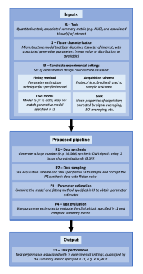 |
Towards a computational framework for task-driven experimental design
Sean C Epstein1, Timothy J.P. Bray2, Margaret A. Hall-Craggs2, and Hui Zhang1
1Department of Computer Science & Centre for Medical Image Computing, University College London, London, United Kingdom, 2Centre for Medical Imaging, University College London, London, United Kingdom
We present a novel computational method for quantitative assessment of experimental design choices for diffusion-weighted imaging (DWI). This approach is motivated by the observation that real-world tasks (e.g. clinical classification) are assessed by metrics (e.g. AUC) that depend non-trivially on the accuracy and precision of DWI-derived parameters. The proposed method enables, for the first time, the assessment of such metrics in the course of computational experimental design. Evaluation with clinical datasets demonstrates its ability to accurately predict real-world task performance for a range of experimental designs. Illustrative use cases are presented to demonstrate its advantages over existing computational approaches.
|
|||
3644.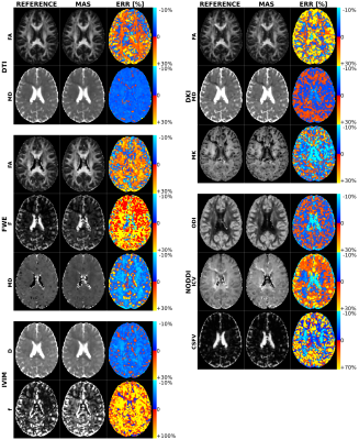 |
Multi-component diffusion technique acquisition protocol optimization for different microstructure models
Tommaso Ciceri1, Alberto De Luca2,3, Filippo Arrigoni1,4, and Denis Peruzzo1
1NeuroImaging Unit, Scientific Institute IRCCS Eugenio Medea, Bosisio Parini, Italy, 2Neurology Department, UMC Utrecht, Utrecht, Netherlands, 3PROVIDI Lab, Image Sciences Institute UMC Utrecht, Utrecht, Netherlands, 4Radiology Unit, Fatebenefratelli Hospital, Milan, Italy
Complementary aspects of the tissue microstructure can be addressed using different models to quantify diffusion MRI. However, there is no consensus on a common acquisition scheme within a clinical feasible time to support the quantification of multiple models. We acquired a large dataset with multiple b-values and directions, and recursively subsampled it to identify the minimum acquisition scheme (MAS) for each model. Finally, we investigated the impact of the MAS on the parameter estimates in the main fiber bundles of the brain. This work supports the transition of advanced analyses to the clinical practice.
|
|||
3645.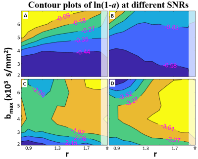 |
Optimizing DWI b-value Sampling for Accurate Metabolic and Cytometric Parameter Extraction: Activity MRI [aMRI]
Xin Li1, Eric M. Baker1, Brendan Moloney1, Cory Wyatt1, Eric Baetscher1, Erin W. Gilbert2, Charles S. Springer1, Alexander R. Guimaraes1,3, and William D. Rooney1
1Advanced Imaging Research Center, Oregon Health & Science University, Portland, OR, United States, 2Surgery, Oregon Health & Science University, Portland, OR, United States, 3Diagnostic Radiology, Oregon Health & Science University, Portland, OR, United States
The optimal data acquisition strategy for accurate metabolic and cytometric parameter extraction using a digital DWI library is investigated using simulations under the constant data-acquisition time (same number of b-values) constraint. Base on DWI data from pancreatic tail tissue and using a seven b-value approach, the optimal maximum b-value is found to be data signal-to-noise ratio dependent, and generally should be below 3,000 s/mm2. In addition, evenly-spaced b-values are generally within the optimal b-spacing range.
|
|||
3646.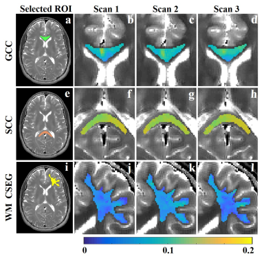 |
Data driven algorithm for multicomponent T2 analysis based on identification of spatially global sub-voxel features
Noam Omer1, Neta Stern1, Tamar Blumenfeld-Katzir1, Meirav Galun2, and Noam Ben-Eliezer1,3,4,5,6
1Department of Biomedical Engineering, Tel-Aviv University, Tel-Aviv, Israel, 2Department of Computer Science and Applied Mathematics, Weitzman institute of science, Rehovot, Israel, 3Department of Orthopedics, Shamir Medical Center, Zerifin, Israel, 4Sackler Faculty of Medicine, Tel-Aviv University, Tel-Aviv, Israel, 5Sagol School of Neuroscience, Tel-Aviv University, Tel-Aviv, Israel, 6Center for Advanced Imaging Innovation and Research (CAI2R), New-York University Langone Medical Center, New York, NY, United States
Multicomponent T2 analysis (mcT2) yields a voxel-wise distribution of T2 values, which can be used to estimate sub-voxel information such as myelin content. Producing such data, however, remains challenging due to the large ambiguity in the T2 space. We present a data-driven approach for mcT2 analysis, which learns the anatomy in question and identifies microscopic tissue-specific features as a preprocessing step. It then utilizes them for analyzing each voxel locally using a designated optimization scheme. Experiments in human brain data show reproducible myelin content estimations at clinical settings without any prior assumptions.
|
|||
3647.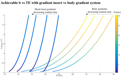 |
Ultra-strong gradient diffusion MRI at 7T with a head insert
Chantal Tax1,2, Edwin Versteeg1, Dennis J.W. Klomp1, Martijn F. Froeling1, Alberto de Luca1, and Jeroen C.W. Siero1,3
1University Medical Center Utrecht, Utrecht, Netherlands, 2CUBRIC, Cardiff University, Cardiff, United Kingdom, 3Spinoza Centre for Neuroimaging Amsterdam, Amsterdam, Netherlands
Diffusion weighting is achieved by the application of external field gradients typically for tens of milliseconds, during which the signal substantially decays due to inherent T2 relaxation. This work focuses on the benefits of strong gradients - here provided by a gradient head insert - for high SNR and short TE diffusion imaging at 7T. Proof-of-principle images show that a short TE (21 ms) at a b-value of 1000 s/mm2 is achievable at 7T using an EPI-readout.
|
|||
3648.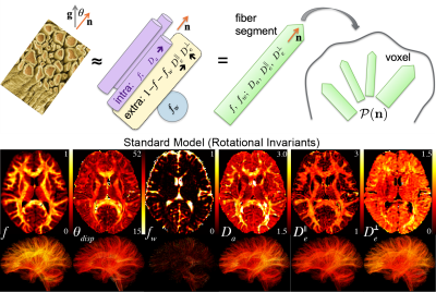 |
Brain microstructure at 1.5mm resolution via RMT reconstruction on a high-slew rate MAGNUS system
Gregory Lemberskiy1, Santiago Coelho1, Thomas K.F. Foo2, Radhika Madhavan2, Luca Marinelli2, Jaemin Shin3, Els Fieremans1, and Dmitry S Novikov1
1Radiology, NYU School of Medicine, New York, NY, United States, 2GE Research, Niskayuna, NY, United States, 3GE Healthcare, New York, NY, United States
A multishell diffusion neuro protocol at 1.5 mm isotropic resolution was acquired in 10 minutes with a high performance MAGNUS head gradient coil (200 mT/m with 500 mT/m/ms slew) at 3.0 T, and reconstructed using random matrix theory (RMT) denoising at the coil level. The SNR enhancement due to RMT reconstruction, and the nearly distortion-free images due to high slew rate, yield precise diffusion and kurtosis maps, fiber-dispersion, as well as microstructure parameters estimated using 3-compartment Standard Model of diffusion. We draw compartment fractions and diffusivities on fiber-tracts reconstructed based on fiber ODFs estimated by deconvolving model-based local fiber response.
|
|||
3649.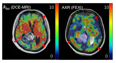 |
Comparison of DCE-MRI and FEXI in the measurement of vascular water exchange in high-grade glioma
Zejun Wang1, Bao Wang2, Yingchao Liu3, and Ruiliang Bai1,4
1Key Laboratory of Biomedical Engineering of Ministry of Education, College of Biomedical Engineering and Instrument Science, Zhejiang University, Hangzhou, China, 2Department of Radiology, Qilu Hospital of Shandong University, Jinan, China, 3Department of Neurosurgery, Shandong Provincial Hospital Affiliated to Shandong First Medical University, Jinan, China, 4Department of Physical Medicine and Rehabilitation, Interdisciplinary Institute of Neuroscience and Technology, The Affiliated Sir Run Run Shaw Hospital, School of Medicine, Zhejiang University, Hangzhou, China
Vascular water exchange is a highly sensitive marker of BBB dysfunction and a potential biomarker of metabolic activity. In this study, we compared two different MRI methods for vascular water exchange measurement, including shutter speed (SS) DCE-MRI and filter-exchange imaging (FEXI) in high-grade glioma patents. Our results demonstrated consistent vascular water exchange assessments by SS DCE-MRI and FEXI in both normal-appearing white matter and tumor.
|
|||
3650.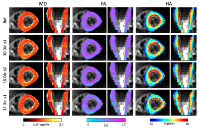 |
Directions or Averages? An Ablation Study for in vivo Cardiac DTI
Jaume Coll-Font1,2,3, Shi Chen1, Robert A Eder1, and Christopher T. Nguyen1,2,3
1Cardiovascular Research Center, Cardiology Division, Massachusetts General Hospital, Charlestown, MA, United States, 2Athinoula A. Martinos Center for Biomedical Imaging, Charlestown, MA, United States, 3Harvard Medical School, Boston, MA, United States
Recent advances in in vivo cardiac DTI have enabled rapid acquisition of the required diffusion weighted (DW) images. With the possibility of acquiring more DW images, it is not clear what is the best sampling strategy. Here we evaluate whether there are any differences between acquiring twice as many directions versus duplicating the number of repetitions. Our results indicate that it is marginally better to acquire multiple directions and that the loss of accuracy is produced by the reduction in the total number of DW images.
|
|||
3651.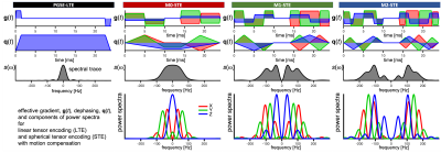 |
Time-dependent anisotropic diffusion in the mouse heart: feasibility of motion compensated tensor-valued encoding on a 7T preclinical scanner
Samo Lasic1,2, Henrik Lundell1, Beata Wereszczyńska3, Matthew Budde4, Nadira Yuldasheva3, Filip Szczepankiewicz5, Erica Dall’Armellina3, Jürgen E. Schneider3, and Irvin Teh3
1Danish Research Centre for Magnetic Resonance, Centre for Functional and Diagnostic Imaging and Research, Copenhagen University Hospital Hvidovre, Copenhagen, Denmark, 2Random Walk Imaging, Lund, Sweden, 3Leeds Institute of Cardiovascular and Metabolic Medicine, University of Leeds, Leeds, United Kingdom, 4Department of Neurosurgery, Medical College of Wisconsin, Milwaukee, WI, United States, 5Clinical Sciences, Lund University, Lund, Sweden
Tensor-valued diffusion encoding with simultaneous nulling of velocity, acceleration and concomitant gradients can be applied with high b-values on a preclinical 7T scanner. Results for ex-vivo mouse hearts confirm that time-dependent diffusion can significantly affect estimation of mean diffusivity. The estimated restriction sizes are consistent with results from pig hearts. Signal attenuations at high b-values suggest relatively low microscopic anisotropy and a strong influence of time-dependent diffusion on microstructure characterization.
|
|||
3652.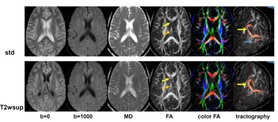 |
Evaluation of Synthetic-DWI with T2-based Water Suppression for DTI
Tokunori Kimura1, Kousuke Yamashita1, and Kouta Fukatsu1
1Department of Radiological Science, Shizuoka College of Medicalcare Science, Hamamatsu, Japan
We evaluated our proposed T2wsup-DWI method of reducing CSF-partial volume effects (PVE) on DTI. We assessed the errors in ADC and FA and the comparison of the ADC SNRs with several methods with simulation, and brain study. We clarified that our proposed T2wsup-DWI technique was superior to already proposed water suppression DWI methods of FLAIR and non-b-zero (NZE) methods in both of the ADC-SNR and the reduction effects of CSF-PVE in DTI parameters of ADC, FA, and tractograpy, typically at the portion of the fornix crus or genu of the corpus callosum which are close to the ventricle.
|
|||
3653.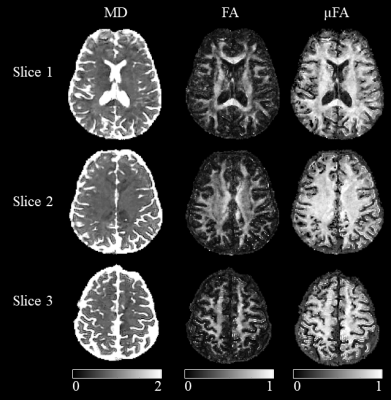 |
Toward high-resolution mapping of microscopic anisotropy in the cortex using b-tensor diffusion imaging with a spiral readout at 7 Tesla
Sajjad Feizollah1,2 and Christine L. Tardif1,2,3
1Department of Neurology and Neurosurgery, McGill University, Montreal, QC, Canada, 2McConnell Brain Imaging Center, Montreal Neurological Institute, McGill University, Montreal, QC, Canada, 3Department of Biomedical Engineering, McGill University, Montreal, QC, Canada
High-resolution diffusion-weighted imaging has been used to investigate the microstructure of the cortex in vivo. However, conventional linear diffusion encoding provides limited insight into the underlying microstructural differences between cortical areas. b-Tensor encoding disentangles macroscopic from microscopic anisotropy in voxels with complex fiber geometries. The SNR and thus resolution of these scans are limited by the longer diffusion encoding times. A DWI sequence with b-tensor encoding was implemented at 7T with a spiral readout trajectory and dynamic field monitoring to image cortical microstructure. Microscopic anisotropy maps of the brain are presented at 1.4 mm isotropic, within minimal distortions and blurring.
|
|||
3654.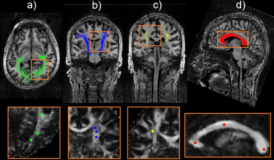 |
Reducing Rician noise bias in axial-symmetric Diffusion Kurtosis Imaging and biophysical tissue models
Jan Malte Oeschger1, Karsten Tabelow2, and Siawoosh Mohammadi1,3
1Institute of Systems Neuroscience, University Medical Center Hamburg-Eppendorf, Hamburg, Germany, 2Weierstrass Institute for Applied Analysis and Stochastics, Berlin, Germany, 3Department of Neurophysics, Max Planck Institute for Human Cognitive and Brain Sciences, Leipzig, Germany
Five out of eight axial-symmetric Diffusion Kurtosis Imaging (AxDKI) parameters are directly related to biophysical microstructure parameters including intra- and extra-axonal diffusivities, fiber dispersion and axonal-water fraction. Their estimation, however, is biased at small signal-to-noise ratios (SNR). Here, based on simulations, we investigated the Rician noise bias’s effect and its correction (RBC) on estimated AxDKI and biophysical parameters at varying SNRs for the standard and AxDKI model. Our study suggests AxDKI to be better than standard DKI, here least biased AxDKI estimators were produced with RBC while for biophysical parameters results were branch-dependent (SNR≥23 with RBC and SNR≥33 without RBC).
|
|||
3655.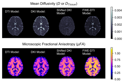 |
Cerebrospinal Fluid Partial Volume Effects in Microscopic Fractional Anisotropy Imaging
Nico J. J. Arezza1,2 and Corey A. Baron1,2
1Medical Biophysics, Western University, London, ON, Canada, 2Centre for Functional and Metabolic Mapping, Robarts Research Institute, London, ON, Canada
Microscopic fractional anisotropy (μFA) quantifies diffusion anisotropy independent of fiber orientation, giving it high specificity to microstructure. However, it is underestimated in brain regions containing cerebrospinal fluid (CSF) partial volumes. Here, we investigated two methods to reduce CSF partial volume effects: the free-water elimination method and a shifted kurtosis method. Compared to diffusion tensor imaging and diffusion kurtosis imaging, these techniques produced more accurate diffusivity estimates in simulations and higher μFA estimates in brain tissue. This preliminary work demonstrates the potential for CSF-suppressing techniques to improve μFA estimation in regions where CSF partial volume effects are prevalent.
|
|||
3656.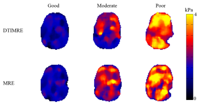 |
Development of in vivo human brain DTI-MRE: Optimization of experimental parameters
Shujun Lin1, Bradley Sutton2, Richard Magin1, Aaron Anderson2, and Dieter Klatt1
1Richard and Loan Hill Department of Bioengineering, University of Illinois at Chicago, Chicago, IL, United States, 2Beckman Institute, University of Illinois at Urbana-Champaign, Urbana-Champaign, IL, United States
Simultaneous acquisition of diffusion tensor imaging (DTI) and magnetic resonance elastography (MRE) has been proven feasible in a preliminary study of in vivo human brain. However, the experimental parameters have to be optimized in order to prevent mutual interferences of DTI and MRE acquisitions. We identified in simulations three experimental parameter sets for in vivo human brain DTI-MRE that we classify as good, moderate and poor with regard to optimization and present a pilot study using these parameter sets. The experimental results verify the simulations as we found the best performance of DTI-MRE for the good parameters set.
|
The International Society for Magnetic Resonance in Medicine is accredited by the Accreditation Council for Continuing Medical Education to provide continuing medical education for physicians.