Digital Posters
Diffusion Acquisition
ISMRM & SMRT Annual Meeting • 15-20 May 2021

| Concurrent 6 | 15:00 - 16:00 |
1312.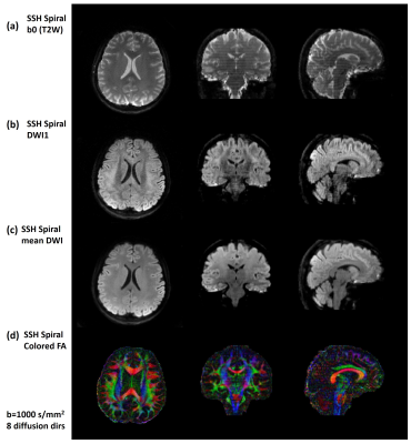 |
Whole-brain Diffusion Tensor Imaging Using Single-Shot Spiral Sampling
Guangqi Li1, Xin Shao1, Xinyu Ye1, Xiaodong Ma2, and Hua Guo1
1Center for Biomedical Imaging Research, Department of Biomedical Engineering, School of Medicine, Tsinghua University, Beijing, China, 2Center for Magnetic Resonance Research, Radiology, Medical School, University of Minnesota, Minneapolis, MN, United States
Single-shot acquisition techniques are commonly used to acquire diffusion weighted images. Single-shot spiral sampling allows shorter TE acquisition, thus provides higher SNR compared to EPI acquisition. However, spiral acquisition is sensitive to field inhomogeneity. Blurring effects degrade the quality of spiral images. In this study, compared to previous studies, a relatively large acceleration factor was used to reduce the spiral readout duration, and off-resonance correction was implemented for deblurring. The results show that the proposed single-shot spiral sampling can achieve whole-brain diffusion tensor imaging.
|
|||
1313.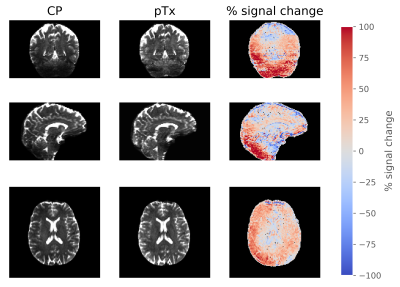 |
Dynamic parallel transmission for diffusion MRI at 7T
Belinda Ding1, Iulius Dragonu2, Patrick Liebig3, Robin M Heidemann3, and Christopher T Rodgers1
1Wolfson Brain Imaging Centre, University of Cambrige, Cambridge, United Kingdom, 2Siemens Healthcare Limited, Firmley, United Kingdom, 3Siemens Healthineers, Erlangen, Germany
In this study, we showed the novel application of dynamic pTx pulses for diffusion MRI on a Siemens 7T Terra scanner, with an 8Tx32Rx Nova head coil. We compared the performance of subject-specific spokes pulses against traditional circularly polarised pulses in a healthy volunteer. We observed that pTx pulses improves the signal across the whole brain, especially in lower brain regions like the cerebellum. This leads to improved definition of diffusion tracts and higher FA values in the regions of interest. In conclusion, this work demonstrated the feasibility and benefits of using pTx pulses for diffusion MRI at 7T.
|
|||
1314.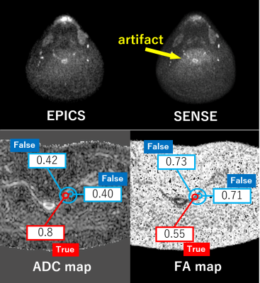 |
Noise reduction in diffusion tensor imaging of the brachial plexus using single-shot DW-EPI with Compressed SENSE
Takayuki Sada1, Hajime Yokota2, Takafumi Yoda1, Ryuna Kurosawa1, Koji Matsumoto1, Takashi Namiki3, Masami Yoneyama3, Yoshitada Masuda1, and Takashi Uno2
1Department of Radiology, Chiba University Hospital, Chiba, Japan, 2Diagnostic Radiology and Radiation Oncology, Graduate School of Medicine, Chiba University, Chiba, Japan, 3Philips Japan, Tokyo, Japan
The brachial plexus is a difficult region to obtain high-quality DTI. In DTI with SENSE, noise-like artifacts appear in the center of the image, resulting in poor image quality. It has been reported that C-SENSE (EPICS), which combines parallel imaging and compressed sensing, can reduce parallel imaging-derived artifacts and may be a more robust method for quantitative imaging than SENSE. This study compared EPICS with SENSE and SENSE×2 (same conditions as SENSE with increased NEX) in the brachial plexus region, and EPICS was a robust imaging method producing better reproducibility of ADC and FA values than SENSE and SENSE×2.
|
|||
1315.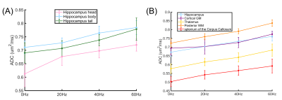 |
Inversion-recovery prepared 3D oscillating gradient sequence (IR-OGprep-GRASE) improves time-dependency measurements in the human brain
Haotian Li1, Yi-Cheng Hsu2, Tao Zu1, Zhiyong Zhao1, Ruibin Liu1, Yi Sun2, Yi Zhang1, and Dan Wu1
1Key Laboratory for Biomedical Engineering of Ministry of Education, Department of Biomedical Engineering, College of Biomedical Engineering & Instrument Science, Zhejiang University, Hangzhou, China, 2MR Collaboration, Siemens Healthcare China, Shanghai, China
Oscillating gradient diffusion MRI enables diffusion measurement at short diffusion-time (td), but it is challenging on the clinical system. Here we proposed an inversion-recovery prepared 3D oscillating gradient (IR-OGprep-GRASE) sequence to improve td–dependency measurements in the human brain. The result indicated that in brain regions that are possibly contaminated by CSF signals, such as the hippocampus, td-dependent ADC changes were not evident with OGprep-GRASE but can be recovered by adding the IR module. With IR-OGprep-GRASE, we identified different td-dependent patterns between the gray and white matter, as well as between the head, body, and tail of hippocampus.
|
|||
1316.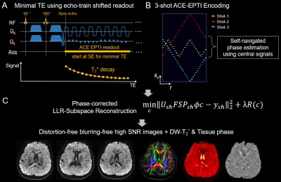 |
ACcelerated Echo-train shifted EPTI (ACE-EPTI) for fast distortion-blurring-free high-resolution diffusion imaging with minimal echo time
Zijing Dong1,2, Fuyixue Wang1,3, Lawrence L. Wald1,3, and Kawin Setsompop4,5
1Athinoula A. Martinos Center for Biomedical Imaging, Massachusetts General Hospital, Charlestown, MA, United States, 2Department of Electrical Engineering and Computer Science, MIT, Cambridge, MA, United States, 3Harvard-MIT Health Sciences and Technology, MIT, Cambridge, MA, United States, 4Department of Radiology, Stanford University, Stanford, CA, United States, 5Department of Electrical Engineering, Stanford University, Stanford, CA, United States
ACcelerated Echo-train shifted EPTI (ACE-EPTI) is proposed for fast and high-fidelity dMRI and diffusion-relaxometry. Distortion- and blurring-free imaging at high resolution is achieved using the time-resolved imaging approach, which recovers sharp multi-contrast images at each echotime across a continuous readout window. A new spatiotemporal encoding is proposed for rapid acquisition, requiring only 3 shots for submillimeter imaging with the capability of self-navigated physiological phase correction. An echo-training shifting acquisition is also incorporated into ACE-EPTI to enable shortest possible TE, which provides 30-40% higher SNR-efficiency over single-shot EPI. High-quality diffusion images and diffusion-weighted quantitative maps acquired by ACE-EPTI are demonstrated in-vivo.
|
|||
1317.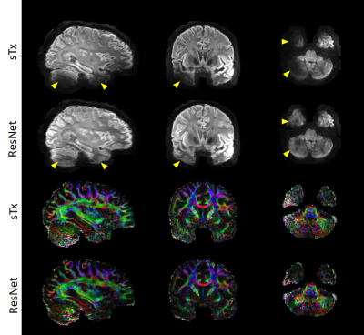 |
Creating parallel-transmission-style MRI with deep learning (deepPTx): a feasibility study using high-resolution whole-brain diffusion at 7T
Xiaodong Ma1, Kamil Uğurbil1, and Xiaoping Wu1
1Center for Magnetic Resonance Research, Radiology, Medical School, University of Minnesota, Minneapolis, MN, United States
Parallel transmission (pTx) has proven capable of addressing two RF-related challenges at ultrahigh fields (≥7 Tesla): RF non-uniformity and power deposition in tissues. However, the conventional pTx workflow is tedious and requires special expertise. Here we propose a novel deep-learning framework, dubbed deepPTx, which aims to train a deep neural network to directly predict pTx-style images from images obtained with single transmission (sTx). The feasibility of deepPTx is demonstrated using 7 Tesla high-resolution, whole-brain diffusion MRI. Our preliminary results show that deepPTx can substantially enhance the image quality and improve the downstream diffusion analysis.
|
|||
1318.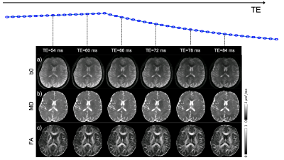 |
Distortion-Free Diffusion-Relaxometry Imaging with Self-navigated Cartesian-based Echo-Planar Time Resolved Acquisition (cEPTI)
Erpeng Dai1, Philip K Lee1,2, Zijing Dong3,4, Fanrui Fu1, Kawin Setsompop1,2, and Jennifer A McNab1
1Department of Radiology, Stanford University, Stanford, CA, United States, 2Department of Electrical Engineering, Stanford University, Stanford, CA, United States, 3Athinoula A. Martinos Center for Biomedical Imaging, Massachusetts General Hospital, Charlestown, MA, United States, 4Department of Electrical Engineering and Computer Science, MIT, Cambridge, MA, United States
Recently, there has been a growing interest in developing distortion-free EPI acquisitions for high-fidelity diffusion, relaxometry and/or functional MRI. Point spread function (PSF)-based techniques have been proposed for distortion-free diffusion imaging. Another technique, echo-planar time resolved imaging (EPTI), has been demonstrated for distortion-free relaxometry with an EPI readout. Additionally, a combination of PROPELLER and EPTI has been reported for motion-robust simultaneous diffusion and relaxometry imaging. In this study, we develop a self-navigated Cartesian-based EPTI (cEPTI) acquisition for distortion-free diffusion-relaxometry imaging. In vivo human brain data demonstrate that high-quality distortion-free diffusion and relaxometry images can be acquired with the proposed cEPTI.
|
|||
1319.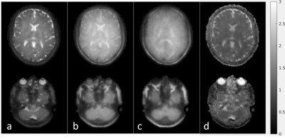 |
Distortionfree multishot 3D diffusion weighted turbo spin echo imaging using cartesian spiral acquisition and data rejection
Tim Schakel1, Tom Bruijnen1,2, and Marielle Philippens1
1Department of Radiotherapy, University Medical Center Utrecht, Utrecht, Netherlands, 2Computational Imaging Group for MRI diagnostics and therapy, Centre for Image Science, Utrecht, Netherlands In this work a multishot 3D turbo spin echo (TSE) sequence is combined with a diffusion preparation module and a modified implementation of the cartesian spiral (CASPR) k-space sampling pattern. CASPR samples the center of k-space in each shot, providing redundancy and enabling self-navigated identification of corrupt data. Using low resolution reconstructions per shot, weights are assigned for each shot enabling a soft-weighted compressed sense reconstruction. Results are obtained in a phantom and healthy volunteers, demonstrating the feasibility in acquiring undistorted 3D diffusion images.
|
|||
1320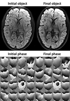 |
Highly Segmented Multishot Diffusion Imaging With Spiral Readouts Video Permission Withheld
Yoojin Lee1, Franz Patzig1, and Klaas P. Pruessmann1
1Institute for Biomedical Engineering, ETH Zurich and University of Zurich, Zürich, Switzerland
Multi-shot acquisition for diffusion MRI is challenging due to shot-to-shot phase variations caused by motion. Multiplexed sensitivity-encoding (MUSE) tackles this problem by extracting phase estimates from reconstruction of individual shot images. In this approach, the feasible number of shots is limited by increasing g-factor noise penalty. Against this background, the present work studies the feasibility of highly segmented MUSE with spiral acquisition, which offers particularly benign g-factor behavior. To stabilize reconstruction, we explore the utility of moving from separate estimation of phase offsets to joint optimization of phase biases and image content.
|
|||
1321.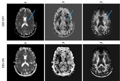 |
Simple improvement of Multi-Dimensional diffusion MRI(MD-dMRI) image quality by double-sampled EPI
Nicolas Geades1, Oscar Jalnefjord2,3, Guillaume Gilbert4, and Maria Ljungberg2,3
1MR Clinical Science, Philips, Gothenburg, Sweden, 2Department of Medical Physics and Biomedical Engineering, Sahlgrenska University Hospital, Gothenburg, Sweden, 3Department of Radiation Physics, University of Gothenburg, Gothenburg, Sweden, 4MR Clinical Science, Philips, Markham, ON, Canada
This study presents a novel implementation of established methods to eliminate ghosting artifacts in Multi Dimensional diffusion MRI (MD-dMRI) images. By replacing scan time used for acquiring images purely used for averaging (when applicable), with EPI acquisitions at opposing readout directions, ghost artifacts can be eliminated. The implementation provides ghost-free images with a small increase in SNR and a minimal (7%) increase in scan time.
|
|||
1322.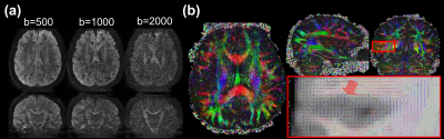 |
Navigator-Free Submillimeter Diffusion MRI using Multishot-encoded Simultaneous Multi-slice (MUSIUM) Imaging
Wei-Tang chapel Chang1, Khoi Minh Huynh2, Pew-Thian Yap1, and Weili Lin2
1Radiology, UNC at Chapel Hill, Chapel Hill, NC, United States, 2BRIC, UNC at Chapel Hill, Chapel Hill, NC, United States
One major challenge in submillimeter dMRI is the inherently low signal-to-noise ratio (SNR). to To address this issue, the simultaneous multislab (SMSlab) approaches were proposed but susceptible to slab boundary artifacts and require additional navigators for phase estimation. The gSlider sequences require relatively high RF power and peak amplitude, increasing SAR and complicating RF excitation. Here, we introduce a navigator-free approach called multishot-encoded simultaneous multi-slice (MUSIUM) imaging for enhanced SNR, low RF power and peak amplitude, and freedom from slab boundary artifacts.
|
|||
1323.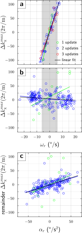 |
Motion-Insensitive Brain Diffusion MRI using Intra-Sequence Motion Updates: Interaction between TE and Tracking Frame Rate
Artan Kaso1 and Thomas Ernst1
1Diagnostic Radiology and Nuclear Medicine, University of Maryland School of Medicine, Baltimore, MD, United States
Uncorrected head movement during DWI acquisitions can cause signal dropouts, due to rotation-induced imbalances in gradient moments. Using a fast optical tracking system, signal losses can be reversed by the realignment of the scanner’s gradient axes with the moving head. However, while experimentally the lost signals were recovered to a great extent, the residual gradient moments were larger than anticipated by simulations. We demonstrated that this was due to the discretized nature and asynchronous application of motion updates in relation to the pulse sequence, which can effectively cause bias in the correction of gradient moments.
|
|||
1324.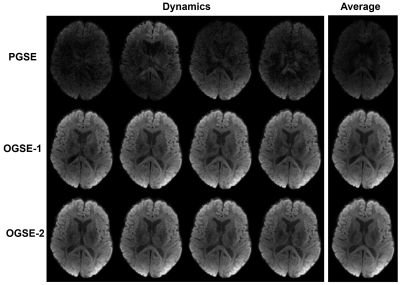 |
Multi-shot diffusion MRI of the human brain with motion-compensated oscillating gradients
Eric Seth Michael1, Franciszek Hennel1, and Klaas Paul Pruessmann1
1Institute for Biomedical Engineering, ETH Zurich and University of Zurich, Zurich, Switzerland
High-resolution multi-shot acquisitions are commonly avoided in diffusion MRI because motion-related phase instability, which can result from diffusion encoding schemes with a nonzero first moment, often hampers image reconstruction. This issue can be circumvented through the use of motion-compensated diffusion gradient shapes derived from oscillating gradient spin-echo (OGSE) methodologies. The utility of this solution is demonstrated here for interleaved spiral scans performed using a high-performance gradient system. The robustness against motion of OGSE sequences provided a notable advantage compared to a standard diffusion sensitization sequence for the phase stability and subsequent quality of multi-shot acquisitions.
|
|||
1325.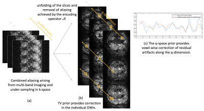 |
Highly Accelerated Multi-shot EPI based Diffusion MRI Using SMS and Joint k-q Under-sampling Enabled Using Deep Learned Manifold Priors
Merry Mani1, Vincent Magnotta1, and Mathews Jacob1
1University of Iowa, Iowa City, IA, United States
We propose a new acceleration and reconstruction method for highly accelerated multi-shot dMRI. The acceleration makes use of multi-band excitation in combination with joint k-q undersampling. We develop new iterative reconstruction with deep learned q-space manifold priors to enable the recovery for the DWIs from 12-fold under-sampled data. The reconstruction error is shown to be less than 3%. The proposed method enables utilization of multi-shot EPI trajectories for diffusion microstructure and connectivity studies requiring high q-space coverage, without prolonging scan time.
|
|||
1326.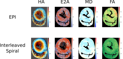 |
Optimising spiral diffusion tensor cardiovascular magnetic resonance for high resolution ex-vivo STEAM imaging on a clinical scanner
Malte Roehl1,2, Peter D Gatehouse1,2, Pedro F Ferreira1,2, Sonya V Babu-Narayan1,2, David N Firmin1,2, Dudley J Pennell1,2, Sonia Nielles-Vallespin1,2, and Andrew D Scott1,2
1National Heart and Lung Institute, Imperial College London, London, United Kingdom, 2Cardiovascular Magnetic Resonance Unit, Royal Brompton Hospital, London, United Kingdom
We demonstrate STEAM spiral diffusion tensor cardiovascular magnetic resonance (DT-CMR) for high resolution ex-vivo imaging (1x1x2mm2) on a clinical 3T scanner. We optimized the coil combination method, diffusion weighting, number of spiral interleaves and averages. A comparison in ex-vivo porcine myocardium shows substantial improvements in image quality when using the spiral method over a single-shot STEAM EPI protocol acquired with matched duration, spatial resolution and diffusion encoding.
|
|||
1327.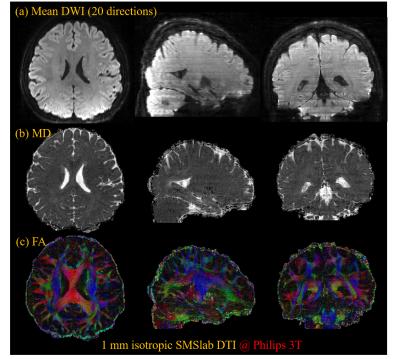 |
The Influence of Navigator Acquisition on 3D Multi-slab DWI Reconstruction: A Comparison between 2D, 3D Acquired and Synthesized Navigator
Simin Liu1, Erpeng Dai2, and Hua Guo1
1Center for Biomedical Imaging Research, Department of Biomedical Engineering, School of Medicine, Tsinghua University, Beijing, China, 2Department of Radiology, Stanford University, Stanford, CA, United States
3D multi-slab is SNR-efficient for isotropic high-resolution diffusion imaging. It can be combined with simultaneous multi-slice, namely SMSlab, for optimal SNR efficiency. In multi-slab, either 2D or 3D navigators can be acquired for inter-shot phase correction, while in SMSlab, only 2D navigators can be acquired. One study has proposed to synthesize a 3D navigator from a 2D navigator in SMSlab, yet lacking a comparison. This study compares the performance of 2D, 3D acquired and synthesized navigators. The synthesized 3D navigator shows similar performance with the acquired 3D navigator in multi-slab and outperforms the 2D navigator in both multi-slab and SMSlab.
|
|||
1328.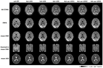 |
Four-shot Navigator-free Spiral Acquisition Strategy for High-resolution Diffusion Imaging
Guangqi Li1, Xinyu Ye1, Xin Shao1, Xiaodong Ma2, and Hua Guo1
1Center for Biomedical Imaging Research, Department of Biomedical Engineering, School of Medicine, Tsinghua University, Beijing, China, 2Center for Magnetic Resonance Research, Radiology, Medical School, University of Minnesota, Minneapolis, MN, United States
Multi-shot acquisition is widely used for high-resolution diffusion weighted imaging (DWI). For multi-shot spiral DWI, one of the challenges is correcting shot-to-shot phase variations. Uniform density spiral (UDS) is a navigator-free acquisition scheme with high efficiency of spatial encoding. Previous studies indicated that POCS-ICE algorithm can iteratively solve the phase errors for navigator-free acquisitions in multi-shot diffusion imaging. Another challenge is off-resonance correction. In this study, we proposed two acquisition strategies to reduce spiral readout duration for alleviating blurring effects. High-resolution diffusion-weighted images can be acquired using a 4-shot navigator-free spiral acquisition.
|
|||
1329.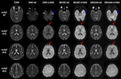 |
Super-resolution and distortion-corrected diffusion-weighted imaging using 2D super-resolution generative adversarial network
Pu-Yeh Wu1, Weiqiang Dou1, Hongyuan Ding2, Jiulou Zhang3, Yong Shen1, Guangnan Quan1, Zhangxuan Hu1, and Bing Wu1
1GE Healthcare, Beijing, China, 2Radiology Department, The First Affiliated Hospital of Nanjing Medical University, Nanjing, China, 3Artificial Intelligence Imaging Laboratory, School of Medical Imaging, Nanjing Medical University, Nanjing, China
We proposed a deep learning-based method for super-resolution and distortion-corrected DWI reconstruction with a visual perception-sensitive super-resolution network SRGAN and multi-shot DWI as target. Our preliminary results demonstrated that the proposed model could produce satisfactory reconstruction of super-resolution diffusion images at b = 0 and 1000 s/mm2, and the geometric distortions in prefrontal cortex and temporal pole were well corrected. Furthermore, SRGAN reconstructed images provide comparable texture details to that of multi-shot DWI. With these findings, this developed model may be considered an effective tool for detecting subtle alterations of diffusion properties with only regular T2WI and DWI as inputs.
|
|||
1330.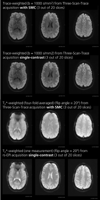 |
Evaluation of Saturation Effects in Simultaneous Multi-Contrast (SMC) Imaging
Nora-Josefin Breutigam1, Daniel Christopher Hoinkiss1, Mareike Alicja Buck 1,2, Klaus Eickel1,2, Matthias Günther1,2, and David Porter3
1Imaging Physics, Fraunhofer MEVIS, Bremen, Germany, 2Faculty 01 (Physics/Electrical Engineering), University Bremen, Bremen, Germany, 3Imaging Centre of Excellence (ICE), University of Glasgow, Glasgow, Scotland
Simultaneous multi-contrast imaging (SMC) can be used to combine acquisition of diffusion-weighted (DW) and T2*-weighted (T2*W) images into a single scan. Compared to conventional single-contrast imaging, SMC reduces the total scan time and improves image registration. However, saturation effects can reduce SNR and alter contrast. In this study, these effects are investigated in simulations, in phantoms, and in vivo. By using the results of this study to control saturation effects in SMC, the method enables rapid acquisition of distortion-matched, high-quality, well-registered DW and T2*W imaging, which could support rapid diagnosis and treatment of acute stroke.
|
|||
1331.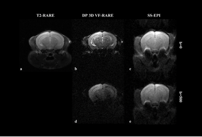 |
Distortion-free high-resolution diffusion weighted imaging of mouse brain using diffusion-prepared 3D VF-RARE
Qiang Liu1,2, Yuanbo Yang1,2, Xinyuan Zhang1,2, Yingjie Mei1,2,3, Qiqi Lu1,2, Guoxi Xie4, and Yanqiu Feng1,2
1School of Biomedical Engineering, Southern Medical University, Guangzhou, China, 2Guangdong Provincial Key Laboratory of Medical Image Processing, Southern Medical University, Guangzhou, China, 3Philips Healthcare, Guangzhou, China, 4Department of Biomedical Engineering, School of Basic Sciences, Guangzhou Medical University, Guangzhou, China
The purpose of the present work was to achieve three-dimensional (3D) high spatial resolution diffusion weighted imaging (DWI) of mouse brain using diffusion-prepared 3D VF-RARE (DP 3D VF-RARE) sequence at 7 Tesla. To employ long echo trains while prospectively controlling signal variation during acquisition, variable flip angles (VF) were implemented in the refocusing pulse train. The results showed improved image quality with less distortion over 2D single-shot EPI and comparable signal-to-noise ratio (SNR). With the application of the presented sequence, undistorted diffusion-weighted mouse brain images were obtained with high spatial resolution and potential SNR efficiency.
|
The International Society for Magnetic Resonance in Medicine is accredited by the Accreditation Council for Continuing Medical Education to provide continuing medical education for physicians.