Digital Posters
Lung: Methods
ISMRM & SMRT Annual Meeting • 15-20 May 2021

| Concurrent 5 | 17:00 - 18:00 |
3225.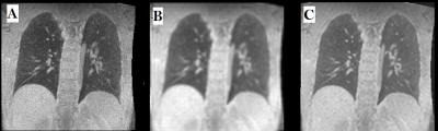 |
Deep learning improves retrospective free-breathing 4D-ZTE thoracic imaging: Initial experience
Dorottya Papp1, Jose M. Castillo T.1, Piotr A. Wielopolski1, Pierluigi Ciet1, Gyula Kotek1, Jifke F Veenland1, and Juan Antonio Hernandez-Tamames2
1Radiology and Nuclear Medicine, Erasmus Medical Center, Rotterdam, Netherlands, 2Erasmus Medical Center, Rotterdam, Netherlands
Although fully convolutional neural networks (FCNNs) have been widely used for MR imaging, they have not been extended for improving free-breathing lung imaging yet. Our aim was to improve the image quality of retrospective respiratory gated version of a Zero Echo Time (ZTE) MRI sequence (4D-ZTE) in free-breathing using a FCNN so enabling free-breathing acquisition in those patients who cannot perform breath-hold imaging. Our model obtained a MSE of 0.08% on the validation set. When tested on unseen data (4D-ZTE) the predicted images from our model had improved visual image quality and artifacts were reduced in free-breathing 4D-ZTE.
|
|||
3226.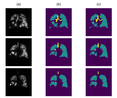 |
Simultaneous segmentation of airways and ventilated lung in hyperpolarised-gas MR images by deep learning
Fabien J Bertin1, Guilhem J Collier2, Paul JC Hughes2, Laurie Smith2, James Aeden2, Helen Marshall2, Jim M Wild2,3, and Alberto M Biancardi2
1Télécom SudParis, Paris, France, 2POLARIS, Department of Infection, Immunity and Cardiovascular Disease, The University of Sheffield, Sheffield, United Kingdom, 3Insigneo Institute for in silico Medicine, The University of Sheffield, Sheffield, United Kingdom
The assessment of pulmonary hyperpolarised(HP)-gas MR images is instrumental in identifying potential pathologies, directing treatment, or monitoring disease progression. HP-gas images quantify the amount of gas concentration, whose distribution within the lungs is analysed. A key role in this process is played the main airways that are identified for quality control and excluded for the analysis. Currently, this task is performed manually and existing deep-learning (DL) applications do not provide an explicit labelling of the airways. A specific tailoring of a well-known DL approach was developed with the aim of replacing the manual editing thanks to its good performance.
|
|||
3227.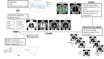 |
A Flexible Physics-Based Digital Phantom for Functional Lung MRI Validation
Sarah H. Needleman1, Jamie R. McClelland2, Björn Eiben2, and Geoff J. M. Parker1,3
1Centre for Medical Image Computing, Quantitative Imaging Group, Department of Medical Physics and Biomedical Engineering, University College London, London, United Kingdom, 2Centre for Medical Image Computing, Radiotherapy Image Computing Group, Department of Medical Physics and Biomedical Engineering, University College London, London, United Kingdom, 3Bioxydyn Limited, Manchester, United Kingdom We describe a flexible framework incorporating an anatomically realistic digital thorax phantom with physics-based simulation, respiratory motion and functional contrasts. We demonstrate the framework in the context of dynamic oxygen-enhanced MRI (OE-MRI).
The framework is designed to provide ground-truth for assessment of novel scan protocols and analysis methods, of utility for OE-MRI as derived lung function measures are limited in accuracy due to motion artefacts, blurring and poor signal-to-noise ratio. The framework was applied to a 2D inversion-prepared spoiled gradient echo dynamic OE-MRI readout. The simulated series displayed respiratory motion; quantitative measures describing hyperoxia-induced contrast enhancement agreed with experimental literature. |
|||
3228.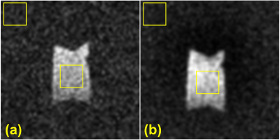 |
A 3D Stack-of Spirals Approach for Inert Fluorinated (19F) Gas MRI of the Lungs
Brandon Zanette1, Yonni Friedlander1,2, Marcus J Couch3,4,5, and Giles Santyr1,2
1Translational Medicine, The Hospital for Sick Children, Toronto, ON, Canada, 2Department of Medical Biophysics, University of Toronto, Toronto, ON, Canada, 3Siemens Healthcare Limited, Montreal, QC, Canada, 4McConnell Brain Imaging Centre, Montreal Neurological Institute, Montreal, QC, Canada, 5Department of Neurology and Neurosurgery, McGill University, Montreal, QC, Canada
Inert fluorinated (19F) gas MRI is an emergent technology that has potential to be a lower cost, reduced infrastructure alternative to hyperpolarized gas MRI for lung imaging. In this work we present an approach for non-Cartesian spiral imaging in a phantom. Compared to typical gradient echo imaging with the same spatial resolution, the proposed approach increased SNR by 44% in approximately a third of the acquisition time. Further simulation and extrapolation to clinical imaging experiments indicate significant potential for this approach to improve SNR and image quality.
|
|||
3229.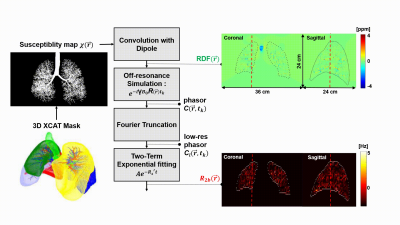 |
Simulation of Lung Parenchyma MRI and Field-Strength Dependence
Bochao Li1, Nam G. Lee1, and Krishna Nayak2
1Biomedical Engineering, University of Southern California, Los Angeles, CA, United States, 2Ming Hsieh Department of Electrical and Computer Engineering, University of Southern California, Los Angeles, CA, United States
MRI of lung parenchyma is limited by low proton density and susceptibility effects at air-tissue interfaces. Recent high-performance low-field MRI systems have provided a new opportunity to mitigate the latter issue. In this work, we demonstrate a framework for simulating lung MRI across B0 field strengths. We estimate apparent transverse relaxation due to proximity to A) sub-voxel alveoli, and B) bronchial tree. Alveoli are modeled using face-centered cubic close packing. The bronchial tree is modeled using a recent XCAT phantom.
|
|||
3230.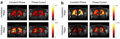 |
Phase-Cycled Balanced Steady-State Free Precession Imaging for Functional Lung Imaging at 1.5 and 3 Tesla
Efe Ilicak1, Jascha Zapp1, Safa Ozdemir1, Lothar R. Schad1, and Frank G. Zöllner1,2
1Computer Assisted Clinical Medicine, Medical Faculty Mannheim, Heidelberg University, Mannheim, Germany, 2Mannheim Institute for Intelligent Systems in Medicine, Medical Faculty Mannheim, Heidelberg University, Mannheim, Germany
Functional lung imaging is of great importance for diagnosis of pulmonary diseases. Previously, a non-contrast-enhanced method called Fourier Decomposition was proposed for assessing pulmonary functions based on balanced steady-stated free precession pulse sequence. However, this pulse sequence is known to be sensitive to magnetic field inhomogeneities. Here, we propose a phase-cycled acquisition for improved robustness against field inhomogeneities. In vivo results from 1.5 T and 3 T scanners are provided to demonstrate the performance of phase-cycled acquisitions for functional lung imaging. Preliminary results indicate that phase cycling can be a viable option for reducing limitations arising from magnetic field inhomogeneities.
|
|||
3231.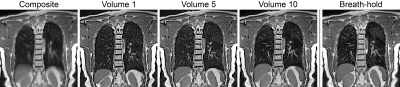 |
Free-breathing thoracic imaging using balanced steady-state free precession with half-radial dual-echo readout (bSTAR)
Grzegorz Bauman1,2 and Oliver Bieri1,2
1Department of Radiology, Division of Radiological Physics, University of Basel Hospital, Basel, Switzerland, 2Department of Biomedical Engineering, University of Basel, Allschwil, Switzerland
In this work, we demonstrate the application of free-breathing respiratory self-gated thoracic MRI with balanced steady-state free precession half-radial dual-echo imaging technique (bSTAR) in human subjects. The technique combines an efficient minimal-TR readout sampling with an interleaved randomly tilted Archimedean spiral trajectory. The methodological improvements result in high-quality visualization of pulmonary parenchyma and vessel structure. The proposed k-space sampling scheme allows reconstruction of multi-volume data sets at different respiratory phases.
|
|||
3232.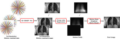 |
Improving iMoCo through Group-wise Registration and Motion State Weighted Reconstruction
Zekang Ding1, Huajun She1, and Yiping Du1
1School of Biomedical Engineering, Shanghai Jiao Tong University, Shanghai, China
In original iMoCo algorithm, a single frame image was reconstructed by solving the iMoCo reconstruction model including the estimated motion fields and TGV sparse constraint. Since motion fields is critical in iMoCo algorithm, errors in motion estimation would deteriorate the final reconstructed image. In this study, we improved the performance of iMoCo through (1) reconstructing the full resolution dynamic images for motion estimation, (2) estimating motion fields through nonrigid group-wise registration, and (3) using a motion state weighted iMoCo reconstruction model. Residual streaking artifacts and certain image blurring were suppressed using the proposed algorithm in comparison with the original iMoCo.
|
|||
3233.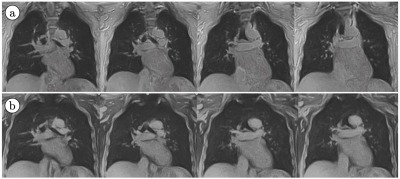 |
Free-Breathing 3D MRI: T2*, Inspiration/Expiration Lung Volume, and Pulmonary Vasculature
Vadim Malis1, Won Bae1, Asako Yamamoto1, Yoshimori Kassai2, Andrew Yen1, Susan Hopkins1, Yoshiharu Ohno3, and Mitsue Miyazaki1
1Radiology, UC San Diego, San Diego, CA, United States, 2Canon Medical, Tochigi, Japan, 3Radiology, Fujita Health University, Toyoake, Japan
We have developed both the acquisition methodology and post-processing algorithms for 3D volume MRI. Using 3D MRI, T2*, lung volume changes between inspiration and expiration as a measure of regional specific ventilation (δV/V0), and pulmonary vasculature can be obtained using 3D ultra-short TE (UTE) imaging during free breathing.
|
|||
3234.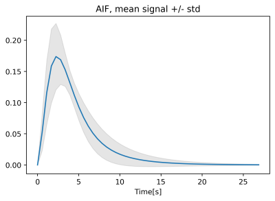 |
Population Arterial Input Function for Lung Perfusion Imaging
Marta Tibiletti1, Jo Naish1,2, John C Waterton1,3, Paul JC Hughes4, James A Eaden4, Jim M Wild4, and Geoff JM Parker1,5
1Bioxydyn Ltd, Manchester, United Kingdom, 2MCMR, Manchester University NHS Foundation Trust, Manchester, United Kingdom, 3Centre for Imaging Sciences,, University of Manchester, Manchester, United Kingdom, 4POLARIS, Imaging Sciences, Department of Infection, Immunity and Cardiovascular Disease, The University of Sheffield, Sheffield, United Kingdom, 5Centre for Medical Image Computing, University College London, London, United Kingdom
In this work, we explore the possibility of extracting a population Arterial Input Function (AIF) to be used in the quantification of lung perfusion using T1-weighted contrast agent-based perfusion imaging. The population AIF averages the shape of first pass of the CA bolus at high temporal resolution in the pulmonary arteries from 90 scans acquired in 50 patients with interstitial lung disease. The population AIF is then scaled by the patient weight. The results of the analysis using a measured AIF and the population AIF are compared.
|
|||
3235.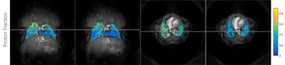 |
Lung Imaging with Tiny Golden Angle UTE in Mice
Anke Balasch1, Hao Li2, Patrick Metze1, Alireza Abaei2, and Volker Rasche1
1Department of Internal Medicine II, Ulm University Medical Center, Ulm, Germany, Ulm, Germany, 2Core Facility Small Animal Imaging (CF-SANI), Ulm University, Ulm, Germany, Ulm, Germany
Lung imaging in small animals is particular difficult due to small anatomy dimensions, high respiratory and heart rate and the very short T2* value in high magnetic fields. A 2D tiny golden angle UTE sequence was tested in free-breathing healthy mice to generate high quality lung images. The functional ventilation and proton density was evaluated.
|
|||
3236.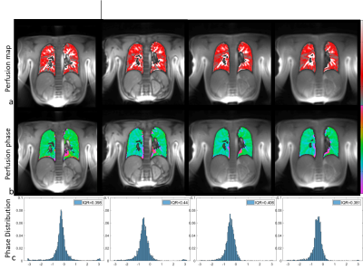 |
Functional lung imaging with SENCEFUL using a 2D radial UTE-sequence at different echo times
Anne Slawig1, Andreas Max Weng1, Bernhard Petritsch1, Simon Veldhoen1, and Herbert Köstler1
1Department of Diagnostic and Interventional Radiology, University Hospital Würzburg, Würzburg, Germany
MR imaging of the lung is a challenge due to the low proton density in lung parenchyma and the very short relaxation times. To overcome low signal and SNR problems UTE-sequences have successfully been applied to lung imaging. Here, the benefit of a 2D UTE-acquisition for the determination of ventilation and perfusion, as determined by SENCEFUL-MRI, is evaluated. It is shown that SENCEFUL-MRI clearly benefits from the use of a UTE-sequence.
|
|||
3237.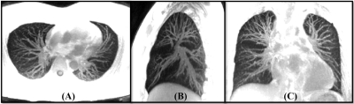 |
Free Breathing Phase-Resolved Lung Imaging Using a 3D UTE Cones Sequence with Randomized Encoding
Ya-Jun Ma1, Michael Carl2, Hyungseok Jang1, Saeed Jerban1, Eric Y Chang1,3, Seth Kligerman1, and Jiang Du1
1UC San Diego, San Diego, CA, United States, 2GE Healthcare, San Diego, CA, United States, 3VA Health system, San Diego, CA, United States
MR imaging of lung is challenging due to its short T2* and low proton density. In this study, we proposed a free breathing phase-resolved lung imaging technique using a 3D UTE sequence with an efficient Cones trajectory. Five different respiration phases were resolved using a self-navigator technique, and the corresponding lung images were reconstructed with a combined parallel imaging and compressed sensing algorithm. Small pulmonary vessels were well-displayed in these images.
|
|||
3238.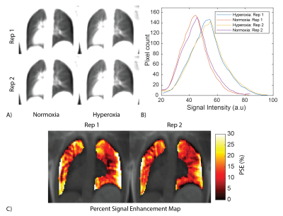 |
Free-breathing oxygen-enhanced pulmonary imaging with self-gated stack-of-spirals ultra-short echo time sequence at 0.55T
Ahsan Javed1, Rajiv Ramasawmy1, Pan Su2, Thomas Benkert3, Waqas Majeed2, and Adrienne E Campbell-Washburn1
1Cardiovascular Branch, Division of Intramural Research, National Heart, Lung, and Blood Institute, National Institutes of Health, Bethesda, MD, United States, 2Siemens Medical Solutions USA Inc., Malvern, PA, United States, 3Siemens Healthcare GmbH, Erlangen, Germany
Recently, low-field MRI has been shown to improve the sensitivity of oxygen-enhanced (OE) MRI due to the increased relaxivity of oxygen at lower field strengths. In this work, we demonstrate that using self-gated 3D stack-of-spiral OE-MRI we can further improve the sensitivity and repeatability by: 1) using longer free-breathing acquisitions to increase the signal to noise ratio (SNR) of the acquired images, and 2) improving the consistency of respiratory position between two oxygen states. We also demonstrate that these two improvements give us higher temporal SNR which is a measure of sensitivity in quantitative MRI.
|
|||
3239.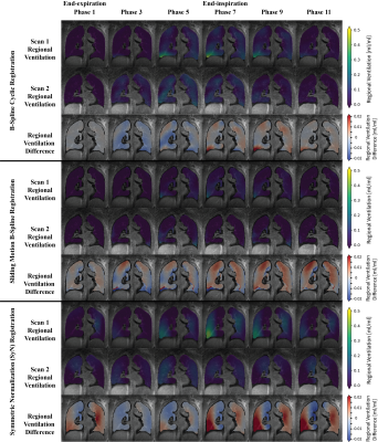 |
Pulmonary Ventilation Analysis Using 1H Ultra-Short Echo Time (UTE) Lung MRI: A Reproducibility Study
Fei Tan1, Xucheng Zhu2, and Peder E.Z. Larson1,3
1Bioengineering, UC Berkeley - UCSF, San Francisco, CA, United States, 2GE Healthcare, Menlo Park, CA, United States, 3Radiology and Biomedical Imaging, University of California, San Francisco, San Francisco, CA, United States
In this abstract, we conducted a reproducibility study on the Jacobian determinant-based regional ventilation quantification of 1H phase-resolved ultrashort-echo (UTE) lung MRI. We evaluated its feasibility on five healthy adult volunteers and the influence of three registration approaches, cyclic registration, multi-b-spline, and symmetric image normalization (SyN). Regional ventilation maps of two scans, split violin plots of each individual’s regional ventilation distribution, within-subject coefficient of variation, and Bland-Altman plot and linear regression of total ventilation are presented. We demonstrated that Jacobian determinant ventilation quantification and the registration methods are reproducible and may be applied to patient studies in the future.
|
|||
3240.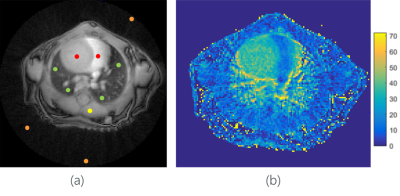 |
Perfusion evaluation in small animal with 2D tiny golden angle UTE
Anke Balasch1, Hao Li2, Patrick Metze1, Alireza Abaei2, and Volker Rasche1
1Department of Internal Medicine II, Ulm University Medical Center, Ulm, Germany, Ulm, Germany, 2Core Facility Small Animal Imaging (CF-SANI), Ulm University, Ulm, Germany, Ulm, Germany
In this study, a two dimensional ultra-short echo-time technique was combined with tiny golden angle angular ordering for investigating its feasibility of qualitative assessment of contrast agent dynamics in the lungs after systemic injection.
|
|||
3241.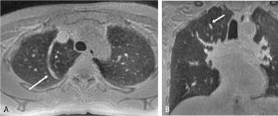 |
Feasibility of free-breathing 3D isotropic whole-lung zero echo time imaging
Yu Deng1, Qiuxi Lin2, Xinchun Li2, Weiyin Vivian Liu3, Lei Zhang4, Qi Wan2, and Chongpeng Sun2
1Radiology, The first affiliated hospital of Guangzhou Medical University, Guangzhou, China, 2The first affiliated hospital of Guangzhou Medical University, Guangzhou, China, 3MR Research, GE Healthcare, Beijing, China, 4GE Healthcare, Beijing, China
The study assessed the feasibility and image quality of free-breathing 3D isotropic whole-lung zero echo time (ZTE) imaging. The overall image quality of the lung in most healthy subjects was good to excellent. The visibility of pulmonary arteries (up to the 7th generation) was better than the bronchi (up to the 5th generation). The demonstration of fissures was poor. In addition, one volunteer with azygos fissure was discovered in our study, which encourage us promote ZTE as a routine lung examination tool. The whole lung ZTE with free-breathing is feasible and can serve as an alternative method of chest imaging.
|
|||
3242.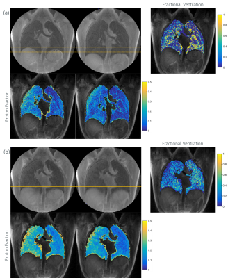 |
Breathhold vs. free-breathing tyGA SoS for Lung Imaging
Anke Balasch1, Patrick Metze1, Kilian Stumpf1, Meinrad Beer2, Wolfgang Rottbauer1, and Volker Rasche1
1Department of Internal Medicine II, Ulm University Medical Center, Ulm, Germany, Ulm, Germany, 2Department of Radiology, Ulm University Medical Centre, Ulm, Germany, Ulm, Germany
Lung MRI is challenging due to the short T2* and respiratory and cardiac motion. In this study an UTE stack-of-stars sequence has been combined with tiny golden angle (tyGA SoS) for imaging of lung morphology (proton fraction, PF) and function (fractional ventilation, FV). Application of the techniques to smoker and non-smokers revealed significant differences in the PF thus indicating its sensitivity.
|
|||
3243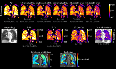 |
Pulmonary functional MR imaging with simultaneous multiple-breath washout tests Video Permission Withheld
Anne-Christianne Kentgens1, Kathryn Ramsey1, Grzegorz Bauman2,3, Francesco Santini2,3, Christoph Corin Willers1, Philipp Latzin1, Oliver Bieri2,3, and Orso Pusterla2,3,4
1Division of Respiratory Medicine, Department of Pediatrics, Inselspital, University of Bern, Bern, Switzerland, 2Division of Radiological Physics, University Hospital Basel, Basel, Switzerland, 3Department of Biomedical Engineering, University Hospital Basel, Basel, Switzerland, 4Institute for Biomedical Engineering, University and ETH Zurich, Zurich, Switzerland
Simultaneous acquisition of oxygen-enhanced MRI and Multiple Breath Washout (MBW) testing is an unexplored and innovative methodology for lung function assessment. Until date, these techniques have been conducted separately. Combined use provides complementary and simultaneous information on pulmonary ventilation, diffusion and perfusion, and has several advantages compared to separate measurements. In this study, we performed oxygen-enhanced relaxometry and non-contrast enhanced ventilation and perfusion mapping using matrix pencil MRI, simultaneously with the MBW.
|
|||
3244.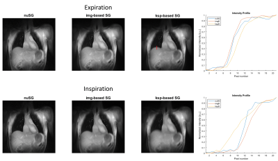 |
Quantitative Evaluation of Self-Gating and nonuniform self-gating for highly irregular respiratory patterns
Patrick Metze1, Tobias Speidel1, Fabian Straubmüller1, and Volker Rasche1,2
1Internal Medicine II, Ulm University Medical Center, Ulm, Germany, 2Core Facility Small Animal Imaging (CF-SANI), Ulm University, Ulm, Germany
In this work we evaluate the proposed nonuniform self-gating (nuSG) method for lung imaging in highly irregular respiratory patterns and compare its performance to image-based and k-space-based gating methods and to a breath-hold reference. Functional parameters (proton fraction, fractional ventilation) and image sharpness are evaluated, with the nuSG algorithm showing a superior performance in case of fractional ventilation and image sharpness. In more uniform motion patterns its performance is similar to image-based SG and both techniques outperform k-space based gating.
|
The International Society for Magnetic Resonance in Medicine is accredited by the Accreditation Council for Continuing Medical Education to provide continuing medical education for physicians.