Digital Posters
Diffusion Acquisition & Post-Processing
ISMRM & SMRT Annual Meeting • 15-20 May 2021

Digital Posters
Diffusion Acquisition & Post-Processing
| Concurrent 6 | 15:00 - 16:00 |
1332.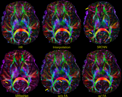 |
Improved Super-Resolution reconstruction for DWI using multi-contrast information
Xinyu Ye1, Pylypenko Dmytro1, Yuan Lian1, Yajing Zhang2, and Hua Guo1
1Center for Biomedical Imaging Research, Department of Biomedical Engineering, School of Medicine, Tsinghua University, Beijing, China, 2MR Clinical Science, Philips Healthcare, Suzhou, China
In clinical scans, the acquired DWI images usually has limited resolution. Super resolution method has the potential to improve the image resolution without adding scan time. Here we propose a deep-learning based multi-contrast super resolution network with gradient-map guidance and a novel FA loss function to reconstruct high-resolution DWI images from low-resolution DWI images and high–resolution anatomical images. In-vivo DWI data are used to test the proposed method. The results show that the image quality can be improved.
|
|||
1333.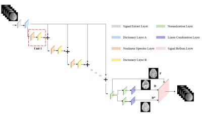 |
A Model-driven Deep Learning Method Based on Sparse Coding to Accelerate IVIM Imaging in Fetal Brain
Tianshu Zheng1, Cong Sun2, Guangbin Wang2, Weihao Zheng1, Wen Shi1, Yi Sun3, Yi Zhang1, Chuyang Ye4, and Dan Wu1
1Key Laboratory for Biomedical Engineering of Ministry of Education, Department of Biomedical Engineering, College of Biomedical Engineering & Instrument Science, Zhejiang University, Hangzhou, Zhejiang, China,, Zhengjiang University, Hangzhou, China, 2Department of Radiology, Shandong Medical Imaging Research Institute, Cheeloo College of Medicine, Shandong University, 324, Jingwu Road, Jinan, Shandong, 250021, People's Republic of China, Shandong University, Jinan, China, 3Department of Radiology 2MR Collaboration, Siemens Healthcare China, Shanghai, China, Siemens Healthcare China, Shanghai, China, 4chool of Information and Electronics, Beijing Institute of Technology, Beijing Institute of Technology, Beijing, China
Intravoxel incoherent motion (IVIM) can be used to assess microcirculation in the brain, however, conventional IVIM requires long acquisition to obtain multiple b-values, which is challenging for fetal brain MRI due to excessive motion. Q-space learning helps to accelerate the acquisition but it is hard to be interpreted. In this study, we proposed a sparsity coding deep neural network (SC-DNN), which is a model-driven network based on sparse representation and unfold the parameter optimization process. Compared to conventional IVIM fitting, SC-DNN took only 50% of the data to reach the comparable accuracy for parameter estimation, which outperformed the multilayer perceptron.
|
|||
1334.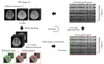 |
Jointly Denoise Diffusion-weighted Images Using a Weighted Nuclear Norm Minimization Approach
Yujiao Zhao1,2, Linfang Xiao1,2, Zhe Zhang3, Yilong Liu1,2, Hua Guo4, and Ed X. Wu1,2
1Laboratory of Biomedical Imaging and Signal Processing, The University of Hong Kong, Hong Kong, China, 2Department of Electrical and Electronic Engineering, The University of Hong Kong, Hong Kong, China, 3China National Clinical Research Center for Neurological Diseases, Beijing Tiantan Hospital, Capital Medical University, Beijing, China, 4Center for Biomedical Imaging Research, Department of Biomedical Engineering, School of Medicine, Tsinghua University, Beijing, China
Diffusion MRI intrinsically suffers from low signal-to-noise ratio (SNR), especially when spatial resolution or b-value is high. A typical diffusion MRI scanning session produces image sets with same geometries but different diffusion directions and b-values, thus these diffusion-weighted (DW) images often share strong structural similarities. In this study, we developed a joint denoising method for DW images based on low-rank matrix approximation. This denoising method exploits structural similarities of DW image set. Both simulation and in vivo brain experiments demonstrate significant noise reduction in all DW images, revealing more microstructural details in quantitative diffusion maps.
|
|||
1335.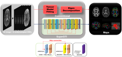 |
SuperDTI: Superfast Deep-learned Diffusion Tensor Imaging
Hongyu Li1, Zifei Liang2, Chaoyi Zhang1, Ruiying Liu1, Jing Li3, Weihong Zhang3, Dong Liang4, Bowen Shen5, Peizhou Huang6, Sunil Kumar Gaire1, Xiaoliang Zhang6, Yulin Ge2, Jiangyang Zhang2, and Leslie Ying1,6
1Electrical Engineering, University at Buffalo, State University of New York, Buffalo, NY, United States, 2Center for Biomedical Imaging, Radiology, New York University School of Medicine, New York, NY, United States, 3Radiology, Peking Union Medical College Hospital, Peking Union Medical College and Chinese Academy of Medical Sciences, Beijing, China, 4Paul C. Lauterbur Research Center for Biomedical Imaging, Medical AI research center, SIAT, CAS, Shenzhen, China, 5Computer Science, Virginia Tech, Blacksburg, VA, United States, 6Biomedical Engineering, University at Buffalo, State University of New York, Buffalo, NY, United States
The main factor that prevents diffusion tensor imaging (DTI) from being incorporated in clinical routines is its long acquisition time of a large number of diffusion-weighted images (DWIs) required for reliable tensor estimation. This abstract presents SuperDTI to learn the nonlinear relationship between DWIs (reduced in q-space and k-space) and the corresponding tensor-derived quantitative maps as well as fiber tractography. Experimental results show that the proposed method can generate fractional anisotropy and mean diffusivity maps, as well as fiber tractography, from as few as six undersampled raw DWIs with quality comparable to results from 90 DWIs using conventional tensor fitting.
|
|||
1336.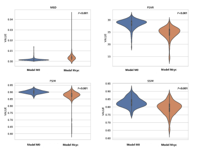 |
Deep learning for synthesizing high-b-value DWI of the prostate: A tentative study based on generative adversarial networks
lei hu1, jungong Zhao1,2, Caixia fu3, and Thomas Benkert4
1Department of Diagnostic and Interventional Radiology, Shanghai Jiao Tong University Affiliated Sixt, 上海, China, 2Department of Diagnostic and Interventional Radiology, Shanghai Jiao Tong University Affiliated Sixt, shanghai, China, 3MR Application Development, Siemens Shenzhen magnetic Resonance Ltd, shanghai, China, 4MR Application Predevelopment, Siemens Healthcare, Erlangen, Gernmany, Erlangen, Germany
A deep learning framework based on a generative adversarial network (GAN) to synthesize high-b-value DWI (syn-DWIb1500) with high quality using the acquired standard b-value DWI (a-DWIb800-1000) was developed. Reader ratings for image quality and PCa detection were performed on the a-DWI b1500, syn-DWIb1500, and optimized syn-DWIb1500 sets. Wilcoxon signed-rank tests and MRMC-ROC were used to compare the readers’ scores and diagnostic capabilities of each DWI set, respectively. Optimized syn-DWIb1500 resulted in significantly better image quality (all P≤0.001) and a higher mean AUC than a-DWIb1500 and cal-DWIb1500 (all P≤0.042).
|
|||
1337.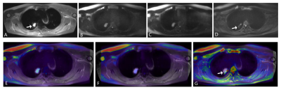 |
IVIM Imaging of Lung Cancer: A Comparison Between Gradient-and Spin-Echo, Turbo Spin-Echo and Echo-Planar Imaging Techniques
Tianyu Zhang1, Yishi Wang2, Chengxiu Yuan1, Xiaoyu Wang1, Jia Zhao1, and Huaqiang Sheng1
1The First Affiliated Hospital of Shandong First Medical University, Jinan, China, 2Philips Healthcare, Beijing, China
The aim of the study was to compare intravoxel incoherent motion diffusion-weighted imaging (IVIM-DWI) for evaluating lung cancer using single-shot gradient- and spin-echo (SS-GRASE), single-shot turbo spin-echo (SS-TSE) and single-shot echo-planar imaging (SS-EPI) on a 3.0T MRI scanner. The signal-to-noise ratio (SNR), contrast signal-to-noise ratio (CNR), image distortion of lesions, ADC and IVIM parameters from the three sequences were compared. We found that GRASE-IVIM had the characteristics of short scan time and small distortion, relatively low SNR but high CNR. Our study showed that GRASE-IVIM has great potential for lung cancer.
|
|||
1338.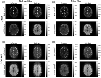 |
Flip-angle optimization for the diffusion-weighted SPLICE sequence for applications in brain imaging
Sofie Rahbek1, Tim Schakel2, Faisal Mahmood3,4, Kristoffer H. Madsen5,6, Marielle E.P. Philippens2, and Lars G. Hanson1,5
1Department of Health Technology, Technical University of Denmark, Kgs. Lyngby, Denmark, 2Department of Radiotherapy, University Medical Center Utrecht, Utrecht, Netherlands, 3Laboratory of Radiation Physics, Odense University Hospital, Odense, Denmark, 4Department of Clinical Research, University of Southern Denmark, Odense, Denmark, 5Danish Research Centre for Magnetic Resonance, Centre for Functional and Diagnostic Imaging and Research, Copenhagen University Hospital Hvidovre, Hvidovre, Denmark, 6Department of Applied Mathematics and Computer Science, Technical University of Denmark, Kgs. Lyngby, Denmark
The RARE-based diffusion-weighted MRI sequence, SPLICE, provides diffusion-weighted images free of geometric distortions. However, the image quality in terms of spatial resolution and SNR needs to be improved. We suggest a framework based on optimization of the flip-angles in the refocusing pulse train to improve the SNR for a chosen spatial point-spread-function. Simulations based on EPG calculations indicate superiority of the optimized flip-angle scheme and experimental recordings on a 1.5 T scanner demonstrate the improvements in a practical setting.
|
|||
1339.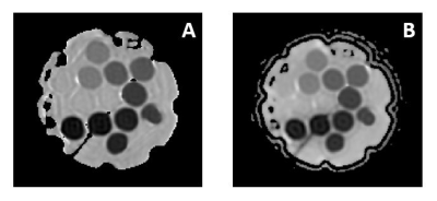 |
Validating the Accuracy of Multi-Spectral Metal Artifact Suppressed Diffusion-Weighted Imaging
John Neri1, Matthew F Koff1, Kevin M Koch2, and Ek Tan1
1Radiology and Imaging, Hospital for Special Surgery, New York, NY, United States, 2Medical College of Wisconsin, Wauwatosa, WI, United States
Diffusion-weighted MAVRIC is a novel pulse sequence that can obtain quantitative diffusion values in regions with high susceptibility. The goal of this work is to compare the quantitative accuracy of DWI-MAVRIC with sequences currently available on clinical MR scanners, using a diffusion phantom and images acquired at different orientation and spatial offsets in the scanner. Overall, good linearity with diffusivity was observed, with a large positive ADC bias at low diffusivity in both axial and coronal DWI-MAVRIC noted. Right and left spatial offsets increased ADC errors that can be mitigated by gradient nonlinearity correction.
|
|||
1340.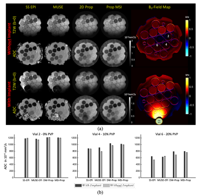 |
Quantitative Accuracy of Diffusion-Weighted Imaging Techniques as a Function of Susceptibility Artifact Resilience
Volkan Emre Arpinar1,2, Alexander D Cohen1, Sampada Bhave3, and Kevin M Koch1,2
1Radiology, Medical College of Wisconsin, Milwaukee, WI, United States, 2Center for Imaging Research, Medical College of Wisconsin, Milwaukee, WI, United States, 3Canon Medical Research USA, Cleveland, OH, United States
Diffusion weighted imaging(DWI) is a workhorse sequence for many clinical and research applications of MRI. Beyond the role in neuroimaging, DWI has also been deployed for use other anatomies outside the head. However, in some anatomies with air-tissue susceptibility boundaries or proximity to metallic implants, DWI assessment may be challenging due to substantial susceptibility artifacts. In this study, the diffusion quantification accuracy of several DWI sequences within increasing resilience to susceptibility artifacts were compared using an established DWI phantom. In addition, a controlled test of two sequences with substantial differences in susceptibility artifact reduction was performed on spinal cord DWI.
|
|||
1341.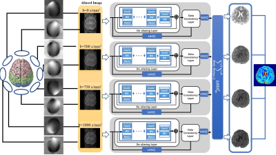 |
Simultaneous Reconstruction of High-resolution Multi b-value DWI with Single-shot Acquisition
Fanwen Wang1, Hui Zhang1, Fei Dai1, Weibo Chen2, Chengyan Wang3, and He Wang1,3
1Institute of Science and Technology for Brain-Inspired Intelligence, Fudan University, Shanghai, China, 2Philips Healthcare, Shanghai, China, 3Human Phenome Institute, Fudan University, Shanghai, China
Compared with the standard imaging method, i.e. single-shot echo planar imaging for diffusion MRI, Multi-Shot EPI (MS-EPI) is featured with high spatial resolution, though suffering from longer scanning time and severer phase variations. This study proposed a novel method to reconstruct four-shot high-resolution DWIs from one-shot data for multiple b-values simultaneously, enabling the physiological feature transformation through different b-values. Trained on healthy volunteers, we succeeded in recovering the aliasing images which violates the Nyquist-Shannon theorem from one-shot DWI both on healthy people and tumor patients, showing great clinical generalization.
|
|||
1342.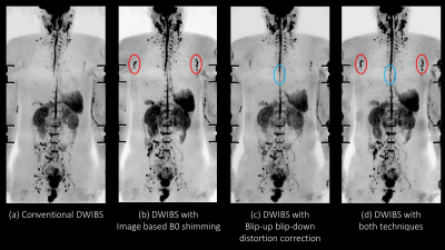 |
Robust method for Whole Body DWIBS applied both Image based B0 Shimming and Blip-up Blip-down Distortion Correction
Hiroshi Hamano1, Masami Yoneyama1, Yasutomo Katsumata2, Kazuhiro Katahira3, and Kenji Iinuma1
1Philips Japan, Tokyo, Japan, 2Philips Healthcare, Tokyo, Japan, 3Department of Radiology, Kumamoto Chuo Hospital, Kumamoto, Japan
DWIBS based on single shot echo-planar imaging (EPI) is a first-choice sequence in routine clinical examinations. However, it sometimes suffers from sever image distortion due to the presence of air within and/or at edge of the FOV, especially at the border of chest and abdomen. On the other hand, image based B0 shimming and blip-up blip-down distortion correction improved for image qualities of DWI. We demonstrated that whole body DWIBS applied both image based B0 shimming and blip-up blip-down distortion correction to provide higher robustness.
|
|||
1343. |
Is Perfect Filtering Enough Leading to Perfect Phase Correction?
Feihong Liu1,2, Junwei Yang2,3, Zhiming Cui2,4, Xiaowei He1,5, Jun Feng1,5, and Dinggang Shen2,6,7
1School of Information Science and Technology, Northwest University, Xi'an, China, 2School of Biomedical Engineering, ShanghaiTech University, Shanghai, China, 3Department of Computer Science and Technology, University of Cambridge, Cambridge, United Kingdom, 4Department of Computer Science, The University of Hong Kong, Hong Kong, China, 5State-Province Joint Engineering and Research Center of Advanced Networking and Intelligent Information Services, Xi'an, China, 6Shanghai United Imaging Intelligence Co., Ltd., Shanghai, China, 7Department of Artificial Intelligence, Korea University, Seoul, Korea, Republic of
We cautiously analyze phase correction procedures, unveiling why they falsely introduce dark holes to diffusion-weighted images, and calibrate them with the goal of unbiased white matter microstructure estimation. The calibrated procedures properly rotate image contents to be negative, hence, dark hole issue is effectively avoided.
|
|||
1344.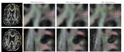 |
Optimal Diffusion Sampling Scheme for High Performance Gradients
Nastaren Abad1, Luca Marinelli1, Radhika Madhavan1, James Kevin DeMarco2, Robert Y Shih2,3, Vincent B Ho2,3, Gail Kohls2, and Tom K.F Foo1
1General Electric Global Research, Niskayuna, NY, United States, 2Walter Reed National Military Medical Center, Bethesda, MD, United States, 3Uniformed Services University of the Health Sciences, Bethesda, MD, United States
In order to establish a benchmark for future studies, we utilize a data driven approach towards optimizing diffusion sampling for an ultra-high-performance gradient MRI sub-system as a means to establish minimal discrepancy compared to a fully sampled, defined superset. This study focused on b-value contribution and the impact of decreased sampling on uncertainty of diffusion and kurtosis tensor estimates, and fiber orientation to resolve sub-voxel information.
|
|||
1345.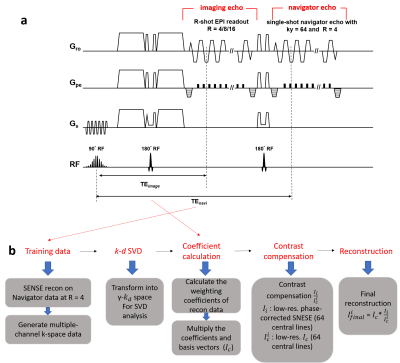 |
Highly-Accelerated Multi-shot Diffusion Imaging with High Angular Resolution Enabled by k-d SVD
Shihui Chen1 and Hing-Chiu Chang1
1Department of Diagnostic Radiology, The University of Hong Kong, Hong Kong, Hong Kong
High angular resolution diffusion imaging (HARDI) is a useful tool for neuroscience research, but the widespread clinical applications are limited by its long scan time and low spatial resolution. Multiple strategies have been proposed to achieve in-plane acceleration to improve the scan efficiency and geometric fidelity for HARDI. However, the feasible in-plane acceleration factor is still limited by the number of coils and the noise amplification associated with parallel imaging reconstruction. In this study, we proposed a reconstruction method based on SVD for HARDI to achieve superior reconstruction performance, even when acceleration factor is greater than number of coils.
|
|||
1346.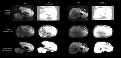 |
Improved multi-shot EPI ghost correction for high gradient strength diffusion MRI using Structured Low-Rank Modeling k-space reconstruction
Gabriel Ramos-Llordén1, Rodrigo A. Lobos2, Tae Hyung Kim1, Qiyuan Tian1, Slimane Tounetki1, Thomas Witzel3, Boris Keil4, Anatasia Yendiki1, Berkin Bilgic1,5, Justin P. Haldar2, and Susie Huang1,5
1Athinoula A. Martinos Center for Biomedical Imaging, Department of Radiology, Masachusetts General Hospital, Harvard Medical School, Charlestown, MA, United States, 2Ming Hsieh Department of Electrical and Computer Engineering, University of Southern California, Los Angeles, CA, United States, 3Q Bio Inc, San Carlos, CA, United States, 4Institute of Medical Physics and Radiation Protection, Mittelhessen University of Applied Sciences, Giessen, Germany, 5Harvard-MIT Division of Health Sciences and Technology, Massachusetts Institute of Technology, Cambridge, MA, United States
Multi-shot EPI diffusion MRI acquired using high diffusion-encoding gradient strengths suffers from severe ghosting artifacts, which can bias and confound the estimation of diffusion microstructural MRI measures at high b-values. In this work, we show that conventional EPI ghost correction techniques fall short in ghosting reduction when high diffusion-encoding gradient strengths ~250mT/m are used, and that advanced reconstruction algorithms based on structured low-rank matrix modeling can substantially reduce ghosting without introducing additional artifacts.
|
|||
1347.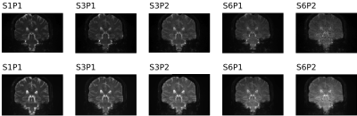 |
Quantitative Evaluation of Multiband Diffusion MRI Data
Arun Venkataraman1, Benjamin Risk2, Deqian Qiu3,4, Jianhui Zhong1,5, Feng (Vankee) Lin6,7, and Zhengwu Zhang8
1Physics and Astronomy, University of Rochester, Rochester, NY, United States, 2Biostatistics and Bioinformatics, Emory University, Atlanta, GA, United States, 3Radiology and Imaging Sciences, Emory University School of Medicine, Atlanta, GA, United States, 4Biomedical Engineering, Emory University, Atlanta, GA, United States, 5Imaging Sciences, University of Rochester Medical Center, Rochester, NY, United States, 6Brain and Cognitive Sciences, University of Rochester, Rochester, NY, United States, 7Neuroscience, University of Rochester Medical Center, Rochester, NY, United States, 8Biostatistics and Computational Biology, University of Rochester Medical Center, Rochester, NY, United States
We sought to understand the implications of slice and phase acceleration factors on diffusion MRI (dMRI) data quality. As part of an on-going study about the impact of acceleration on dMRI quality, reconstruction, and data analysis, we acquired data from young and old, healthy subjects as well as mild cognitive impairment (MCI) subjects. From our study results, we found that there appears to be a trade-off between SNR loss due to higher acceleration and SNR gain from reducing the impact/magnitude of motion. Future studies should examine how the costs and benefits of acceleration impact diffusion metrics, tractography, and reproducibility.
|
|||
1348.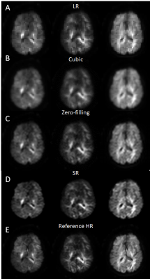 |
Deep Learning Based Super-resolution of Diffusion MRI Data
Zifei Liang1 and Jiangyang Zhang1
1Center for Biomedical Imaging, Dept. of Radiology, New York University School of Medicine, NEW YORK, NY, United States
Deep-learning/machine-learning based super-resolution techniques have shown promises in improving the resolution of MRI without additional acquisition. In this study, we examined the capability of deep-learning based super-resolution using a newly developed network at resolutions from 0.2 mm to 0.025 mm. We also investigated whether the networks were able to enhance data acquired with a different contrast. Our results demonstrated that the enhancement of deep learning based super-resolution, although better than cubic interpolation, remained limited. In order to achieve the best performance, the network needs to be trained using data acquired at the target resolution and share similar contrasts.
|
|||
1349.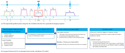 |
A General Framework for Automated and Accurate b-Matrix Calculation for dMRI Pulse Sequences
Lisha Yuan1, Qing Li2, Guojing Wei3, Hongjian He1, and Jianhui Zhong4
1Department of Biomedical Engineering, Center for Brain Imaging Science and Technology, Zhejiang University, Hangzhou, China, 2MR Collaborations, Siemens Healthcare Ltd., Shanghai, China, 3SHS DI MR R&D SZN LP, Siemens Shenzhen Magnetic Resonance Ltd., Shenzhen, China, 4Department of Imaging Sciences, University of Rochester, Rochester, NY, United States
Since both imaging and diffusion gradient pulses could lead to significant diffusion-related signal attenuation, one must account for the effect of all imaging and diffusion gradient pulses to achieve an accurate estimation of diffusion metrics and tensors. Based on a generalized definition of b matrix, a general framework for an automated and accurate b-matrix calculation was proposed in this study. Its correctness was verified in SE-diffusion sequence, and the importance of accurate inclusion of all gradient pulses was shown in both SE- and SPEN-diffusion sequence. The proposed method is suitable for accurate evaluation of diffusion effects in various sequences.
|
|||
1350.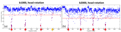 |
Automatic Phase Image Texture Analysis for Motion Detection in Diffusion MRI (APITA-MDD) with Adaptive Thresholding
Xiao Liang1, Pan Su2, Steve Roys1, Rao P Gullapalli1, Jerry L Prince3, and Jiachen Zhuo1
1Department of Diagnostic Radiology and Nuclear Medicine, University of Maryland School of Medicine, Baltimore, MD, United States, 2Siemens Medical Solutions USA Inc, Malvern, PA, United States, 3Department of Electrical and Computer Engineering, Johns Hopkins University, Baltimore, MD, United States
In this study, we propose a more robust PITA-MDD method with automatic brain ROI selection and adaptive thresholding for the motion threshold (termed APITA-MDD). Automatic brain ROI is initially identified by Otsu’s method and then improved by erosion and dilation. Haralick’s Homogeneity Index (HHI) of each slice is converted to a deviation score independent of image SNR and ROI size for motion detection. APITA-MDD was tested on brain dMRI data acquired with head motion and leg crossing motion and correctly detected motion slices missed by PITA-MDD due to insufficient coverage at edge slices and single thresholding.
|
|||
1351.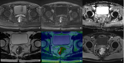 |
Diffusion-Weighted Imaging with Integrated Slice-Specific Dynamic-Shimming for Rectal Cancer Detection and Characterization
Jianxing Qiu1, Jing Liu1, Chenwen Liu2, Jinxia Zhu2, and Thomas Benkert3
1Peking University First Hospital, Beijing, China, 2MR Collaboration, Siemens Healthcare Ltd China, Beijing, China, 3MR Application Development, Siemens Healthcare GmbH, Erlangen, Germany
Diffusion-weighted imaging (DWI) with integrated slice-specific dynamic-shimming (iShim) could improve image quality compared with conventional 3D-Shimming single-shot echo-planar-imaging (SS-EPI) DWI in patients with rectal cancer. iShim DWI can help differentiate rectal cancer from normal walls and predict pathologic characterizations. iShim DWI could be an effective and promising approach and a good alternative to conventional 3D-Shimming SS-EPI DWI for patients with rectal cancer.
|
The International Society for Magnetic Resonance in Medicine is accredited by the Accreditation Council for Continuing Medical Education to provide continuing medical education for physicians.