Digital Posters
Diffusion Applications: Cancer
ISMRM & SMRT Annual Meeting • 15-20 May 2021

| Concurrent 4 | 13:00 - 14:00 |
3657.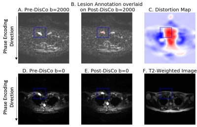 |
Correcting B0 inhomogeneity-induced distortions in whole-body diffusion MRI of bone metastases
Leonardino A. Digma1, Christine H. Feng1, Christopher C. Conlin2, Ana E. Rodriguez-Soto2, Kanha Batra3, Aaron Simon1, Roshan Karunamuni1, Joshua Kuperman2, Rebecca Rakow-Penner2, Michael E. Hahn2, Anders M. Dale2, and Tyler M. Seibert1,4
1Department of Radiation Medicine and Applied Sciences, UCSD School of Medicine, La Jolla, CA, United States, 2Department of Radiology, UCSD School of Medicine, La Jolla, CA, United States, 3Department of Electrical and Computer Engineering, UC San Diego, La Jolla, CA, United States, 4Department of Bioengineering, UC San Diego, La Jolla, CA, United States
Bone is among the most common sites of cancer metastases. Diffusion weighted imaging (DWI) can be used to detect these metastases. However, when acquired with echo-planar imaging, DWI suffers distortions due to static magnetic field inhomogeneities. In this study, we first used the reverse polarity gradient (RPG) technique to measure spatial distortions of bone metastases on DWI. Next, we demonstrated that RPG can be used to correct these distortions and produce diffusion images that more accurately reflect the underlying anatomy. Taken together, findings support the use of distortion correction techniques to improve localization of bone metastases on DWI.
|
|||
3658.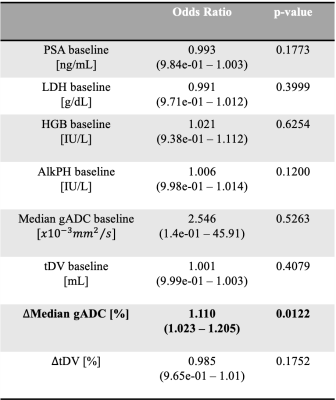 |
Developing a multi-parametric model of response based on biomarkers derived from Whole-Body Diffusion Weighted Imaging
Antonio Candito1, Matthew D Blackledge1, Fabio Zugni2, Richard Holbrey1, Sebastian Schäfer3, Matthew R Orton1, Ana Ribeiro4, Matthias Baumhauer3, Nina Tunariu1, and Dow-Mu Koh1
1The Institute of Cancer Research, London, United Kingdom, 2IEO, European Institute of Oncology IRCCS, Milan, Italy, 3Mint Medical, Heidelberg, Germany, 4The Royal Marsden NHS Foundation Trust, London, United Kingdom
We develop a model of treatment response in patients with advanced prostate cancer (APC), based on measurements of median Apparent Diffusion Coefficient (ADC) and tumour Diffusion Volume (tDV) derived using Whole-Body Diffusion Weighted Imaging (WBDWI) and blood biomarkers. The model showed that change in ADC is positively correlated with response in this patient population. Furthermore, our model predicted response with an accuracy of 87%, and sensitivity/specificity of 75/93% using a probability cut-off of 0.5.
|
|||
3659.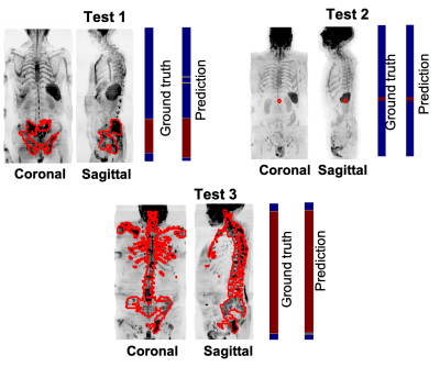 |
Developing a deep learning model to classify normal bone and metastatic bone disease on Whole-Body Diffusion Weighted Imaging
Antonio Candito1, Matthew D Blackledge1, Fabio Zugni2, Richard Holbrey1, Sebastian Schäfer3, Matthew R Orton1, Ana Ribeiro4, Matthias Baumhauer3, Nina Tunariu1, and Dow-Mu Koh1
1The Institute of Cancer Research, London, United Kingdom, 2IEO, European Institute of Oncology IRCCS, Milan, Italy, 3Mint Medical, Heidelberg, Germany, 4The Royal Marsden NHS Foundation Trust, London, United Kingdom
We employed a deep transfer-learning model to classify whether images from whole-body diffusion-weighted MRI (WBDWI) contain metastatic bone lesions. Our results demonstrate sensitivity/specificity of 0.87/0.89 on 8 test patients, who were not included in the model training. Such a model may accelerate radiological assessment of disease extent from WBDWI, which currently can be cumbersome to interpret due to the large quantity of data (approximately 200-250 images per patient).
|
|||
3660.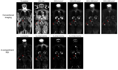 |
Improved assessment of prostate-cancer bone metastases through multicompartmental analysis of whole-body DWI data
Christopher C Conlin1, Christine H Feng2, Leonardino A Digma2, Ana E Rodriguez-Soto1, Joshua M Kuperman1, Dominic Holland3, Rebecca Rakow-Penner1, Tyler M Seibert1,2,4, Michael E Hahn1, and Anders M Dale1,3,5
1Department of Radiology, UC San Diego School of Medicine, La Jolla, CA, United States, 2Department of Radiation Medicine and Applied Sciences, UC San Diego School of Medicine, La Jolla, CA, United States, 3Department of Neurosciences, UC San Diego School of Medicine, La Jolla, CA, United States, 4Department of Bioengineering, UC San Diego Jacobs School of Engineering, La Jolla, CA, United States, 5Halıcıoğlu Data Science Institute, UC San Diego, La Jolla, CA, United States
Whole-body DWI is increasingly used to assess bone involvement in prostate cancer. Multicompartmental diffusion modeling can outperform conventional DWI techniques for evaluating tumors, but has yet to be applied to whole-body imaging. In this study, we determined an optimal multicompartmental model for describing whole-body diffusion and applied it to examine metastatic bone lesions in vivo. We found that a 4-compartment model best characterized whole-body diffusion. Compartmental signal-contributions revealed by this model show improved bone-lesion conspicuity and may help to assess microstructural changes that accompany prostate-cancer bone involvement.
|
|||
3661.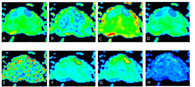 |
Assessment the Preponderant Diagnostic Performances of Oligometastatic Prostate Cancer Using Diffusion Kurtosis Imaging
Suhong Qin1, Ailian Liu1, SHUANG MENG1, Lihua Chen1, Qinhe Zhang1, Qingwei Song1, and Yunsong Liu1
1The First Affiliated Hospital of Dalian Medical University, Dalian, China
It remains a challenge to diagnose the oligometastatic prostate cancer (PCa) due to the ambiguous definition of oligometastatic PCa. Previous studies had shown that diffusion kurtosis imaging (DKI) is a non-gaussian diffusion weighted imaging (DWI) method that yields more accurate results on the microstructural complexity of prostate cancer tissue structure. This study indicated that performances of DKI cannot differentiate between oligometastatic and widely metastatic PCa, however it has the potential to assess tumor load and aggressiveness in metastatic PCa.
|
|||
3662.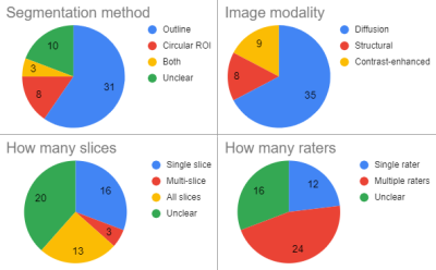 |
Methodological considerations on segmenting MRI data of rhabdomyosarcoma
Cyrano Chatziantoniou1,2, Reineke Schoot2, Roelof van Ewijk2, Simone ter Horst2, Rick van Rijn3, Hans Merks2, Alexander Leemans1, and Alberto de Luca1
1Image Science Institute, UMC Utrecht, Utrecht, Netherlands, 2Princess Máxima Center for Pediatric Oncology, Utrecht, Netherlands, 3Department of Radiology, Academic Medical Centre Amsterdam, Amsterdam, Netherlands Rhabdomyosarcoma is a rare form of cancer that is particularly prevalent in children. There is a pressing need for new imaging biomarkers for monitoring treatment response, such diffusion. No standards currently exist to annotate rhabdomyosarcoma on MRI and measure the diffusion. This work analyses segmentation strategies in recent literature on saromas and compares them on a toy example. It shows that there is a large inconsistency in the application of segmentation strategies and that the different methods for segmenting the tumor can yield high variation in measured diffusion. |
|||
3663.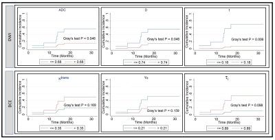 |
Non-gaussian IVIM DW-and fast exchange regime DCE- MRI for predicting of locoregional failure in nasopharyngeal carcinoma
Ramesh Paudyal1, Linda Chen2, Jung Hun Oh1, Kaveh Zakeri2, Vaios Hatzoglou3, Chiaojung Jillian Tsai2, Nancy Lee2, and Amita Shukla-Dave1,3
1Medical Physics, Memorial Sloan Kettering Cancer Center, New York, NY, United States, 2Radiation Oncology, Memorial Sloan Kettering Cancer Center, New York, NY, United States, 3Radiology, Memorial Sloan Kettering Cancer Center, New York, NY, United States
The study aims to assess quantitative imaging (QI) metrics from pre-treatment (TX) non-gaussian intravoxel incoherent DW- and fast exchange regime (FXR) DCE-MRI for predicting locoregional failure (LRF) in nasopharyngeal carcinoma (NPC) patients. Cumulative incidence analysis (CIA) was performed on the two subgroups dichotomized with Youden’s index. Competing-risks regression based on Fine and Gray’s (FG) proportional sub hazards model was used to estimate survival subdistribution hazard ratios (SHRs). The pre-TX ADC, D, f, and ti cutoff values from CIA analysis and K cutoff value from the competing risk regression analysis indicated these QI’s could predict the LRF in NPC patients.
|
|||
3664.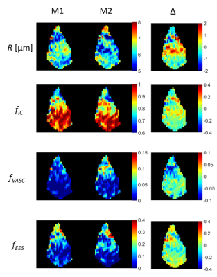 |
Repeatability of VERDICT diffusion MRI in a model of human neuroendocrine tumour
Lukas Lundholm1, Mikael Montelius1, Oscar Jalnefjord1, Eva Forssell-Aronsson1, and Maria Ljungberg1
1Department of Radiation Physics, Institute of Clinical Sciences, Gothenburg, Sweden
VERDICT dMRI allows for estimation of microstructural parameters in tumours which could facilitate planning and assessment of treatment. Due to the complexity of the model there is a risk for overfitting to data and there is hence a need to determine the reliability of the estimated parameters. A mouse model of human SI-NETs (n=5) was measured twice using the same dMRI protocol and the VERDICT model was fitted to data. Results showed an overall good repeatability of the tumour mean parameter values estimated by VERDICT. However, some local clusters of voxels showed larger differences between repeated scans.
|
|||
3665.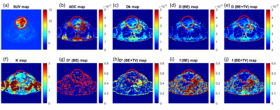 |
Qualitative and quantitative comparison between IVIM-DKI and PET/CT imaging in lymphoma
Archana Vadiraj Malagi1, Devasenathipathy Kandasamy2, Kedar Khare3, Deepam Pushpam4, Rakesh Kumar5, Sameer Bakhshi4, and Amit Mehndiratta1,6
1Centre for Biomedical Engineering, Indian Institute of Technology Delhi, New Delhi, India, 2Department of Radiodiagnosis, All India Institute of Medical Sciences Delhi, New Delhi, India, 3Department of Physics, Indian Institute of Technology Delhi, New Delhi, India, 4Department of Medical Oncology, Dr. B.R. Ambedkar Institute-Rotary Cancer Hospital (IRCH), All India Institute of Medical Sciences Delhi, New Delhi, India, 5Department of Nuclear Medicine, All India Institute of Medical Sciences Delhi, New Delhi, India, 6Department of Biomedical Engineering, All India Institute of Medical Sciences Delhi, New Delhi, India
PET/CT plays an important role in diagnosis and assessment of treatment response in lymphoma. The goal of this study was to evaluate the role of IVIM-DKI parameters in comparison to PET parameters in lymphoma. PET images were registered onto IVIM-DKI at b=0s/mm2 images for tumor ROI using 3D-multimodal affine registration. Qualitatively, IVIM parameters with state-of-the-art Total-Variation produced better quality parameter maps. Tumor appeared hyperintense in SUV and K maps and hypointense in diffusion and perfusion parameters. No correlation was observed between IVIM-DKI with PET parameters. IVIM with Total-Variation showed substantial reproducibility as compared to conventional IVIM, DKI and SUV parameters.
|
|||
3666. |
Preliminary Study on Monitoring Drug Resistance of Colon Cancer with Intravoxel Incoherent Motion MRI In Vivo
Qi Xie1, Wenjuan He1, Zhilin Tan1, Yajie Wang1, Jinbin Wu1, Xiyan Shao2, Yiming Yang3, Jing Zhang4, Kangwei Wang5, Guiqin Wang6, Qifeng Pan1, and Yunzhu Wu7
1Medical Imaging Department, Nansha Hospital, Guangzhou First People's Hospital, School of Medicine, South China University of Technology, Guangzhou, China, 2Ultrasound Imaging Department, Longgang District People’s Hospital, Shenzhen, China, 3Department of Radiology, The Second Affiliated Hospital of Guangzhou University of Chinese Medicine, Guangdong Provincial Hospital of Traditional Chinese Medicine, Guangzhou, China, 4Department of Pathology, Cancer Center, Sun Yat-sen University, Guangzhou, China, 5Department of Pathology, Nansha Hospital, Guangzhou First People's Hospital, School of Medicine, South China University of Technology, Guangzhou, China, 6Medical Record Department, Nansha Hospital, Guangzhou First People's Hospital, School of Medicine, South China University of Technology, Guangzhou, China, 7MR Scientific Marketing, SIEMENS Healthcare Ltd., Guangzhou, China
This study has demonstrated that ADC value of DWI with single-exponential model and diffusion coefficient D of IVIM are valuable for discriminating 5-FU-responsive and 5-FU-resistant colon cancer in vivo. IVIM is expected to be a simple and practical way to quantitatively monitor tumor resistance in vivo, and ADC &D value may be an imaging biomarker.
|
|||
3667.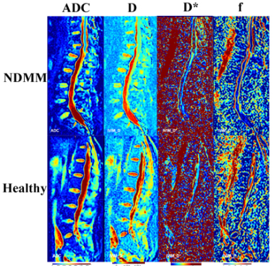 |
Evaluation of bone marrow infiltration in the newly diagnostic Multiple Myeloma with Intravoxel Incoherent Motion Diffusion-weighted MRI
Xiaojiao Pei1, Tao Jiang1, Zhenyu Pan1, Yufei Lian1, Yueluan jiang2, and qinglei Shi 3
1Radiology, Beijing Chaoyang Hospital Affiliated to Capital Medical University, Beijing, China, 2MR Scientific Marketing, Diagnosis Imaging, Siemens Healthineers China, Beijing, China, 3Scientific Marketing, Diagnosis Imaging, Siemens Healthineers China, Beijing, China
Intravoxel incoherent motion diffusion-weighted MR imaging (IVIM-DWI) can simultaneously acquire diffusion parameters and perfusion parameters without intravenous administration, compared with conventional diffusion imaging. This study investigated the application of IVIM in the newly diagnostic multiple myeloma (NDMM) patients.
|
|||
3668.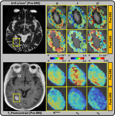 |
Diffusion and perfusion MRI predicts response preceding and shortly after stereotactic radiosurgery to brain metastases
Amaresha Shridhar Konar1, Akash Deelip Shah2, Ramesh Paudyal1, Jung Hun Oh1, Eve LoCastro1, David Aramburu Nuñez1, Nathaniel Swinburne2, Robert J. Young2, Andrei I. Holodny2, Kathryn Beal3, Vaios Hatzoglou2, and Amita Shukla-Dave1,2
1Medical Physics, Memorial Sloan Kettering Cancer Center, New York, NY, United States, 2Radiology, Memorial Sloan Kettering Cancer Center, New York, NY, United States, 3Radiation Oncology, Memorial Sloan Kettering Cancer Center, New York, NY, United States
In the clinical settings it is essential to accurately assess, whether or not a brain metastases has been successfully treated or whether it requires additional treatment, especially in high dose radiation therapy, such as stereotactic radiosurgery (SRS). The present prospective study aims to determine the ability of Diffusion Weighted (DW)- and Dynamic Contrast Enhanced (DCE)-MRI to predict the long-term response of brain metastases within 72 hours of SRS. The preliminary results are promising as it will inform the treating physicians at an early time point about which patients will benefit from SRS (or not).
|
|||
3669.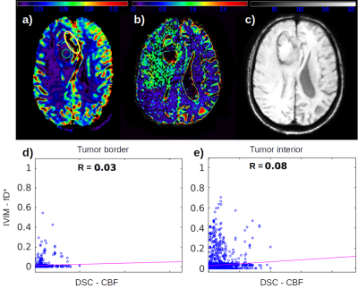 |
The utility of IVIM maps in the assessment of microvascular perfusion of brain glioma
Andre Monteiro Paschoal1,2, Raquel Andrade Moreno3,4, Antonio Carlos dos Santos5, and Renata Ferranti Leoni6
1LIM44, Instituto e Departamento de Radiologia, Faculdade de Medicina, Universidade de Sao Paulo, Sao Paulo, Brazil, 2InBrain Lab, University of Sao Paulo, Ribeirao Preto, Brazil, 3Instituto do Cancer do Estado de Sao Paulo, Sao Paulo, Brazil, 4Memorial Sloan-Kettering Cancer Center, New York, NY, United States, 5Departamento de Clinica Medica, Faculdade de Medicina de Ribeirao Preto, Universidade de Sao Paulo, Ribeirao Preto, Brazil, 6InBrain Lab - University of Sao Paulo, Ribeirao Preto, Brazil
The assessment perfusion in glioma is an important information in the tumor characterization. Traditional MRI methods employed to analyze glioma are based on the application of contrast agents to enhance the T1 signal and/or to measure perfusion within the tumor cells. The analysis of tumor microvasculature however is hampered by the size of contrast agent molecules. In that sense, SWI can be employed, although it does not provide quantitative information. IVIM maps showed to be an interesting alternative for such assessment, even showing potential of an early biomarker to assess early changes in perfusion, specially in low grade gliomas.
|
|||
 |
3670.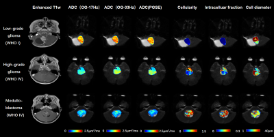 |
Diffusion-time-dependent diffusion MRI based microstructural mapping for grading and categorizing in pediatric brain tumor
ruicheng ba1, Hongxi Zhang2, Zhongwei Gu3, Yuhao Liao1, Xingwang Yong1, Zhiyong Zhao1, Yi Zhang1, and Dan Wu1
1Key Laboratory for Biomedical Engineering of Ministry of Education, Department of Biomedical Engineering, College of Biomedical Engineering & Instrument Science, Zhejiang University, Hang zhou, Zhejiang, China, 2Children's Hospital, Zhejiang University School of Medicne,Department of Radiology, Hang zhou, Zhejiang, China, 3Children's Hospital, Zhejiang University School of Medicne, Department of Pathology, Hangzhou, Zhejiang, China
Diffusion-time dependent diffusion MRI (dMRI) has been proposed to characterize tumor microstructure. This study aimed to evaluate diagnostic value of time-dependent dMRI based microstructural mapping to grade and categorize pediatric brain tumor at 3T. Oscillating and pulsed gradient dMRI was performed in 35 pediatric patients. Cell diameter (d), intracellular fraction (fin) and extracellular diffusivity (Dex) were fitted based on the IMPULSED model. Higher cellularity (fin/d) and fin and lower Dex were found in high-grade glioma than low-grade ones. The cellularity further increased in medulobastoma compared with glioma. Cellularity achieved the highest area-under-the-curve than the other dMRI metrics in grading glioma.
|
||
3671. |
Imaging attributes of H3K27M mutation in Diffuse Midline Gliomas on Multiparametric MRI
Richa Singh Chauhan1, Nihar Kathrani2, Jitender Saini1, Maya D Bhat1, Karthik Kulanthaivelu1, Vani Santosh3, Nishanth S4, and Subhas Konar4
1Neuroimaging and Interventional Radiology, NIMHANS, BENGALURU, India, 2Interventional Radiology, Paras Hospital, Gurgaon, India, 3Neuropathology, NIMHANS, BENGALURU, India, 4Neurosurgery, NIMHANS, BENGALURU, India
H3K27M mutant diffuse midline gliomas are a newly classified entity in the 2016 World health organization classification of CNS tumors. They are high grade tumors, with the mere presence of this mutation confers them a WHO grade IV designation, irrespective of their histologic morphology. Patients harboring these tumors have dismal prognosis and shorter overall survival. Furthermore, since these are deep-seated lesions involving the eloquent brain areas, biopsy can be challenging with substantial risk of morbidity. Our work proposes non-invasive multiparametric MRI-based imaging attributes to detect the H3K27M mutation pre-operatively. Results demonstrate a considerable accuracy on 123 patients.
|
|||
3672.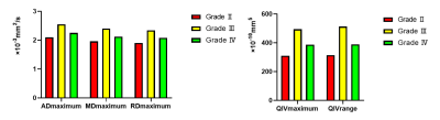 |
Grading of glioma with histogram analysis of multiparameter using advanced diffusion models
Gao Eryuan1, Gao Ankang1, Zhang Huiting2, Wang Shaoyu2, Yan Xu2, Bai Jie1, and Cheng Jingliang1
1Dept. of MRI, The First Affiliated Hospital of Zhengzhou University, Zhengzhou, China, Zhengzhou, China, 2MR Scientific Marketing, Siemens Healthcare, Shanghai, China, Shanghai, China
This study aimed to investigate the efficiency of four advanced diffusion models, including diffusion tensor imaging (DTI), diffusion kurtosis imaging (DKI), neurite orientation dispersion and density imaging (NODDI) and mean apparent propagator (MAP) in grading of glioma. Through histogram analysis of parameters, we found that axial diffusivity (AD)maximum, mean diffusivity (MD)maximum and radial diffusivity (RD)maximum from DTI, and Q-space inverse variance (QIV)maximum and QIVrange from MAP had significant differences and good diagnostic efficiency in all comparisons among different grading of glioma.
|
|||
3673.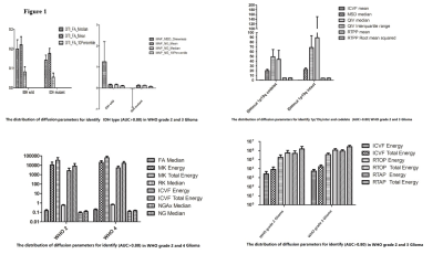 |
Histogram analysis in prediction of Isocitrate Dehydrogenase Genotype in Gliomas with MRI: The Gaussian versus non-Gaussian Diffusion Models
Gao Ankang1, Gao Eryuan1, Zhang Huiting2, Wang Shaoyu2, Yan Xu2, Bai Jie1, and Cheng Jingliang1
1Dept. of MRI, The First Affiliated Hospital of Zhengzhou University, Zhengzhou, China, 2MR Scientific Marketing, Siemens Healthcare, Shanghai, China, Shanghai, China
The Isocitrate dehydrogenase (IDH) genotyping and epigenetic 1p/19q codeletion as two key molecular markers are included in the glioma WHO 2016 classification. Gaussian or non-Gaussian diffusion models were recently proposed to provide additional microstructure information. In present work, we applied four diffusion models in glioma grading and genotyping, including DTI, DKI, MAP-MRI and NODDI models, which could be acquired within a single scan.
|
|||
3674.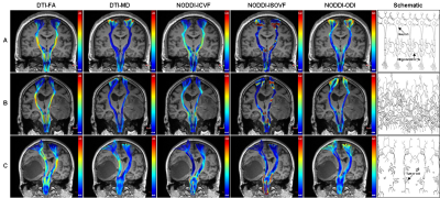 |
Neurite Orientation Dispersion and Density Imaging in Evaluation of Glioma-induced Corticospinal Tract Injury
Rifeng Jiang1, Kaiji Deng1, Yixin Guo2, and Zhongshuai Zhang3
1Fujian Medical University Union Hospital, Fuzhou, China, 2Fujian Medical University, Fuzhou, China, 3MR Scientific Marketing, Siemens Healthcare, Shanghai, China
This study found that NODDI seems to be a more potent approach in evaluating the early CST infiltration by HGG, and can evaluate the CST destruction with a similar performance to MD by providing additional information about neurite density for HGG-induced CST injury.
|
|||
3675.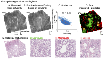 |
Beyond cellularity: Which microstructural features determine the mesoscopic mean diffusivity in meningiomas?
Jan Brabec1, Filip Szczepankiewicz2, Jaromír Šrámek3, Elisabet Englund4, Johan Bengzon5, Linda Knutsson1,6, Carl-Fredrik Westin7,8, Pia C Sundgren2,9, and Markus Nilsson2
1Medical Radiation Physics, Lund University, Lund, Sweden, 2Diagnostic Radiology, Lund University, Lund, Sweden, 3Institute of Histology and Embryology, First Faculty of Medicine, Charles University, Prague, Czech Republic, 4Division of Oncology and Pathology, Department of Clinical Sciences, Lund University, Lund, Sweden, 5Division of Neurosurgery, Department of Clinical Sciences, Lund University, Lund, Sweden, 6Russell H. Morgan Department of Radiology and Radiological Science, Johns Hopkins University School of Medicine, Baltimore, MD, United States, 7Harvard Medical School, Boston, MA, United States, 8Radiology, Brigham and Women’s Hospital, Boston, MA, United States, 9Lund University Bioimaging Center, Lund University, Lund, Sweden
Mean diffusivity (MD) in tumors is often too readily interpreted as cellularity. Here, we investigated which microstructural features explain the MD in meningioma tumors. We performed high-resolution MRI and histological imaging on excised mengioma tumor samples and coregistered MD to histology. We found that cellularity alone was a poor predictor of MD, and that the prediction improved markedly by considering also the microcyst density. A qualitative analysis also indicated that collagen fibers, stroma and psammoma bodies affect the MD.
|
|||
3676.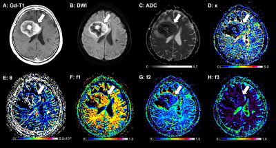 |
Diffusion MRI based on a gamma distribution model for the differentiation of primary central nervous system lymphomas and glioblastomas
Osamu Togao1, Akio Hiwatashi2, Toru Chikui3, Kazufumi Kikuchi2, Yukiko Kami3, Kenji Tokumori4, and Kousei Ishigami2
1Molecular Imaging & Diagnosis, Kyushu University, Fukuoka, Japan, 2Clinical Radiology, Kyushu University, Fukuoka, Japan, 3Oral and Maxillofacial Radiology, Kyushu University, Fukuoka, Japan, 4Clinical Radiology, Teikyo University, Omuta, Japan
The aim of the present study was to determine whether the gamma distribution (GD) model is useful in the differentiation of glioblastomas (GBs) and primary CNS lymphomas (PCNSLs). The GD model well described the histological features of PCNSLs and GBs, and its use enabled the significant differentiation of these tumors. The κ, f2, and f3 values were significantly smaller and the f1 values were significantly larger in the PCNSLs than in the GBs. The GD model-derived parameters correlated well with the IVIM-derived parameters. The GD model may therefore contribute to the characterization of brain tumor from the histological viewpoint.
|
The International Society for Magnetic Resonance in Medicine is accredited by the Accreditation Council for Continuing Medical Education to provide continuing medical education for physicians.