Digital Posters
Arterial Spin Labelling: Methods
ISMRM & SMRT Annual Meeting • 15-20 May 2021

Digital Posters
Arterial Spin Labelling: Methods
| Concurrent 4 | 13:00 - 14:00 |
2713.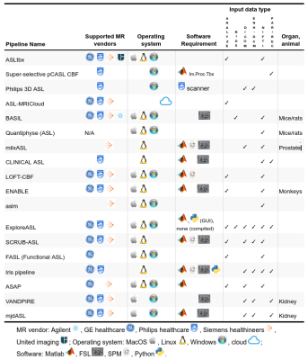 |
The Open Source Initiative for Perfusion Imaging (OSIPI): ASL Pipeline inventory
Sudipto Dolui1, Hongli Fan2, Paula L. Croal3,4, Charlotte Buchanan4, Lydiane Hirschler5, Udunna Anazodo6, David L. Thomas7, Henk J.M.M. Mutsaerts8, and Jan Petr9
1Department of Radiology, University of Pennsylvania, Philadelphia, PA, United States, 2The Johns Hopkins School of Medicine, Baltimore, BC, United States, 3Radiological Sciences, Division of Clinical Neuroscience, School of Medicine, University of Nottingham, Nottingham, United Kingdom, 4Sir Peter Mansfield Imaging Centre, School of Medicine, University of Nottingham, Nottingham, United Kingdom, 5Leiden University Medical Center, Leiden, Netherlands, 6Western University, London, ON, Canada, 7UCL Queen Square Institute of Neurology, University College London, London, United Kingdom, 8Department of Radiology and Nuclear Medicine, Amsterdam University Medical Center, location VU, Amsterdam, Netherlands, 9Helmholtz-Zentrum Dresden-Rossendorf, Dresden, Germany
As a part of the Open Source Initiative for Perfusion Imaging (OSIPI), we have created an inventory of software for automated processing of Arterial Spin Labeling (ASL) perfusion MRI data. We contacted the ASL community through different channels, inviting software developers to list their pipelines by completing a questionnaire covering different aspects and features of a desired pipeline. We received inputs from 18 developers and have summarized the main characteristics of their pipelines based on the information they provided. We expect that this inventory will facilitate ASL-related research, reduce duplicate development, and enable translation of ASL to clinical practice.
|
|||
2714.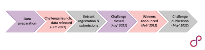 |
The Open Source Initiative for Perfusion Imaging (OSIPI) ASL MRI Challenge
Udunna Anazodo1,2, Joana Pinto3, Flora A. Kennedy McConnell4,5,6, Maria-Eleni Dounavi7, Cassandra Gould van Praag8, Henk Mutsaerts9, Aaron Oliver Taylor10, André Paschoal11, Jan Petr12, Diego Pineda-Ordóñez13, Joseph G. Woods14, Moss Y. Zhao15, and Paula L. Croal4,5
1Department of Medical Biophysics, University of Western Ontario, London, ON, Canada, 2Imaging Division, Lawson Health Research Institute, London, ON, Canada, 3Institute of Biomedical Engineering, Department of Engineering Science, University of Oxford, Oxford, United Kingdom, 4Sir Peter Mansfield Imaging Centre, University of Nottingham, Nottingham, United Kingdom, 5Radiological Sciences, Division of Clinical Neuroscience, School of Medicine, University of Nottingham, Nottingham, United Kingdom, 6Nottingham Biomedical Research Centre, Queens Medical Centre, Nottingham, United Kingdom, 7Department of Psychiatry, University of Cambridge, Cambridge, United Kingdom, 8Wellcome Centre for Integrative Neuroimaging, University of Oxford, Oxford, United Kingdom, 9Amsterdam University Medical Center, Amsterdam, Netherlands, 10Gold Standard Phantoms Limited, London, United Kingdom, 11Department of Radiology, LIM44 - HCFMUSP, Sao Paulo, Brazil, 12Helmholtz-Zentrum Dresden-Rossendorf, Dresden, Germany, 13Department of Radiology, Clinica Del Country, Bogotá, Colombia, 14Department of Radiology, University of California San Diego, San Diego, CA, United States, 15Department of Radiology, Stanford University, Stanford, CA, United States The OSIPI ASL MRI Challenge is a community-led initiative aiming to establish the range of approaches used for ASL image analysis and cerebral blood flow (CBF) quantification. Challenge data will consist of population-based and synthetic pseudo-continuous ASL images, with participants analysing the data and submitting resulting CBF maps and mean tissue CBF, along with documentation. Entries will be scored on accuracy, reproducibility and documentation quality. Through documenting the analysis choices made within the community, we will begin to better understand sources of variability, ultimately identifying an optimum pipeline, and moving towards the much-needed consensus of ASL image processing standards. |
|||
2715.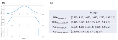 |
Optimization of post-labeling delays in multi-delay 3D pCASL by modeling arterial transit time distribution
Zhiyuan Zhang1,2, Timothy Macaulay3, and Lirong Yan1
1USC Stevens Neuroimaging and Informatics Institute, Keck School of Medicine, University of Southern California, Los Angeles, CA, United States, 2Department of Biomedical Engineering, University of Southern California, Los Angeles, CA, United States, 3Division of Biokinesiology and Physical Therapy, Ostrow School of Dentistry, University of Southern California, Los Angeles, CA, United States
The design of post-labeling delays (PLDs) directly affects the accuracy of CBF and ATT quantifications using multi-delay ASL. In this study, we optimized PLDs in 3D pCASL based upon different ATT distributions including normal distribution directly derived from in vivo ASL data and uniform distribution. Evenly spaced PLDs were also applied for comparison. Our results showed that optimal PLDs based on ATT normal distribution had the best performance in CBF and ATT quantifications with the smallest errors.
|
|||
2716.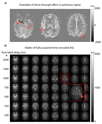 |
Investigation of angiographic shine-through in time-encoded pCASL
Lena Vaclavu1, Dilek Betül Arslan2, Lydiane Hirschler1, Carles Falcon3,4, Esin Ozturk-Isik2, Juan Domingo Gispert3,4, Paula Montesinos5, Kim van de Ven6, and Matthias JP van Osch1
1Department of Radiology, C.J. Gorter Center for High Field MRI, Leiden University Medical Center, Leiden, Netherlands, 2Biomedical Engineering Institute, Boğaziçi University, Istanbul, Turkey, 3BarcelonaBeta Brain Research Center (BBRC), Pasqual Maragall Foundation, Barcelona, Spain, 4CIBER-BBN, Madrid, Spain, 5Philips Healthcare Iberia, Madrid, Spain, 6Philips Healthcare, Best, Netherlands
Physiologic fluctuations can lead to the appearance of artefactual angiographic signal in the perfusion image of time-encoded pCASL previously dubbed “shine-through effect”. The aim of this study was to investigate the potential mechanisms inducing this artefact by characterizing the noise in time-encoded pCASL signal from individual Hadamard columns and rows as well as without labeling. We observed higher temporal standard deviation in the arteries compared to gray matter, and found that the shine-through effect was present even without labeling, suggesting that it is caused by the selective background suppression pulse applied close to acquisition time and the associated fresh inflow.
|
|||
2717.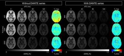 |
Verifying the effect of DANTE preparation pulse for separating spin-compartments in arterial spin labeling using T2-measurement
Shota Ishida1, Hirohiko Kimura2, Naoyuki Takei3, Yasuhiro Fujiwara4, Tsuyoshi Matsuda5, Yuki Matta1, Masayuki Kanamoto1, Nobuyuki Kosaka2, and Eiji Kidoya1
1Radiological center, University of Fukui Hospital, Eiheiji, Japan, 2Department of Radiology, Faculty of Medical Sciences, University of Fukui, Eiheiji, Japan, 3Global MR Applications and Workflow, GE Healthcare Japan, Hino, Japan, 4Department of Medical Image Sciences, Faculty of Life Sciences, Kumamoto University, Chuo-ku, Japan, 5Division of Ultra-high Field MRI, Institute for Biomedical Science, Iwate Medical University, Shiwa-gun, Japan
To verify the characteristics of DANTE for vascular suppression, T2 values of the ASL signal under the application of DANTE were determined. While T2 values without DANTE decreased as the PLD increased, T2 values with DANTE (T2_DANTE) did not change among the PLDs. Moreover, T2_DANTE were equivalent to those of the reference. The positive correlation between T2 values and the ATT without DANTE was not observed when DANTE was used. T2_DANTE were neither dependent on the ATT nor PLD. Therefore, DANTE separates the spin compartments in ASL by selective elimination of intra-vascular signals from total ASL signals.
|
|||
2718.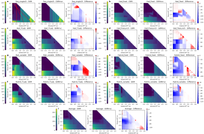 |
Systematic investigation of the sensitivity of optimised pCASL protocols to macrovascular contamination
Logan Xin Zhang1 and Michael A Chappell1,2,3
1Institute of Biomedical Engineering, University of Oxford, Oxford, United Kingdom, 2Radiological Sciences, Division of Clinical Neurosciences, School of Medicine, University of Nottingham, Nottingham, United Kingdom, 3Sir Peter Mansfield Imaging Centre, School of Medicine, University of Nottingham, Nottingham, United Kingdom
Optimised pCASL protocols may still be sensitive to macrovascular contamination (MVC), leading to high CBF estimation error. In this study, we conducted a systematic investigation of CBF sensitivity to MVC from a comprehensive set of optimal pCASL protocols. The general kinetic model was only robust for CBF estimation when tissue ATT was less than 1.5s. An extended kinetic model could reduce CBF sensitivity to up to 2.3s. The results of CBF sensitivity maps could be used for examining a protocol for possible MVC effects before scanning, or data interpretation afterwards.
|
|||
2719.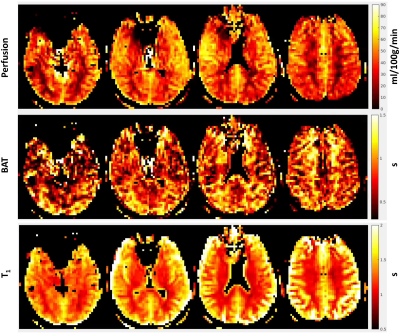 |
Perfusion Quantification using Velocity Selective Inversion pulses in a combined ASL-MRF Framework
Anish Lahiri1, Jeffrey Fessler1, and Luis Hernandez-Garcia2
1Electrical and Computer Engineering, University of Michigan, Ann Arbor, MI, United States, 2FMRI Laboratory and Biomedical Engineering, University of Michigan, Ann Arbor, MI, United States
This work proposes a quantitative perfusion imaging using Velocity Selective Inversion pulses combined with an MR Fingerprinting ASL framework that allows for the alleviation of several nuisance parameters in the model, and provides hemodynamic estimates over an extensive region of the brain in a single scan. Preliminary in-vivo experiments indicate that the obtained hemodynamic estimates in gray and white matter agree with values typically found in literature.
|
|||
 |
2720.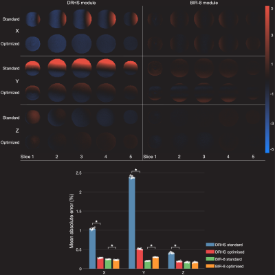 |
A general framework for eddy current minimization in Velocity Selective Arterial Spin Labeling
Joseph G. Woods1, Eric C. Wong1, David D. Shin2, and Divya Bolar1
1Department of Radiology, University of California San Diego, San Diego, CA, United States, 2GE Healthcare, Menlo Park, CA, United States
A novel framework that minimizes eddy current mismatch between tag and control acquisitions in velocity-selective (VS) arterial spin labeling is introduced. The framework is based on minimizing the 0th-order gradient moment in the presence of eddy currents and is applicable to any VS module design, without suffering a time penalty in module duration. Simulation and empirical data are presented and demonstrate reductions in eddy current artifacts in both phantom and in vivo data.
|
||
2721.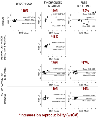 |
Reduction of motion effects in myocardial arterial spin labeling
Verónica Aramendía-Vidaurreta1,2, Pedro M. Gordaliza3,4, Marta Vidorreta5, Rebeca Echeverría-Chasco1,2, Gorka Bastarrika1,2, Arrate Muñoz-Barrutia3,4, and María A. Fernández-Seara1,2
1Radiology, Clínica Universidad de Navarra, Pamplona, Spain, 2IdiSNA, Instituto de Investigación Sanitaria de Navarra, Pamplona, Spain, 3Universidad Carlos III de Madrid, Madrid, Spain, 4Instituto de Investigación Sanitaria Gregorio Marañón, Madrid, Spain, 5Siemens Healthineers, Madrid, Spain
This work investigated the impact of three different breathing strategies, named breathhold (BH), synchronized-breathing (SB) and free-breathing (FB), together with motion detection and correction algorithms in myocardial arterial spin labeling (ASL) images. Results indicate the superiority of FB combined with pairwise registration, which showed higher accuracy (in synthetic images) and higher intrasession reproducibility together with lower variability across subjects (in in vivo images). BH and SB after motion detection provided similar results, but their practical application is more complicated as it demands the subject's collaboration to follow the respiratory pattern in SB or perform the apneas in BH.
|
|||
2722.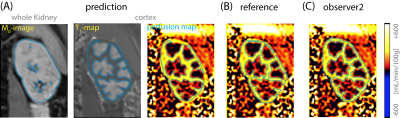 |
Clinically applicable automatic quantitative renal perfusion measurement using ASL-MRI and machine learning
Isabell Katrin Bones1, Clemens Bos1, Chrit Moonen1, Jeroen Hendrikse2, and Marijn van Stralen1
1Center for Image Sciences, University Medical Center Utrecht, Utrecht, Netherlands, 2Department of Radiology, University Medical Center Utrecht, Utrecht, Netherlands
ASL-MRI quantification involves kidney segmentation and cortex-medulla differentiation to obtain cortical renal blood flow, requiring time consuming manual interaction hampering clinical adoption. We applied machine learning to automat renal ASL-MRI quantification. A cascade of three U-nets was constructed to replace manual segmentation steps. Automatic segmentation yielded a dice score of 0.78, which was similar to the inter-observer variability of 0.77. Moreover, good agreement for cortical RBF was found between automatic and manual segmentations on group and individual level; 211±31 and 208±31mL/min/100g, respectively. Our proposed method automates quantification without compromising performance. This makes renal ASL-MRI more attractive for clinical application.
|
|||
2723.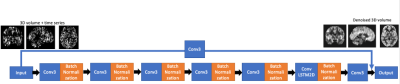 |
Arterial Spin Labeling Denoising with Convolutional Neural Network and Convolutional Long-Short-Term-Memory (ConvLSTM)
Qinyang Shou1, Chaitanya Gupte2, Danny JJ Wang1, and Hosung Kim2
1Laboratory of Functional MRI Technology (LOFT), Stevens Neuroimaging and Informatics Institute, University of Southern California, Los Angeles, CA, United States, 2Neuroimaging with Deep Learning Lab (NIDLL), Stevens Neuroimaging and Informatics Institute, University of Southern California, Los Angeles, CA, United States
The purpose of this study was to develop a deep learning algorithm for 3D spatial-temporal denoising of ASL perfusion image series. 162 datasets from the Pediatric Template of Brain Perfusion database were used for model training and testing. The results showed that the proposed method can achieve higher Peak Signal-to-Noise Ratio (PSNR) and higher Structural Similarity Index (SSIM) than averaging of the time series and using traditional Principal Component Analysis (PCA) denoising. This result was robust when reducing the input measurements to one quarter of the total measurements, which shows the potential to reduce the scan time for ASL imaging.
|
|||
2724.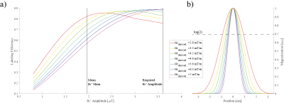 |
Gradient adjustments for improved pcASL exploiting a B1+ shimmed labeling train
Christian R. Meixner1, Sebastian Schmitter2,3, Jürgen Herrler4, Arnd Dörfler4, Michael Uder1, and Armin M. Nagel1,3
1Institute of Radiology, University Hospital Erlangen, Friedrich-Alexander-Universität Erlangen-Nürnberg, Erlangen, Germany, 2Physikalisch-Technische Bundesanstalt (PTB), Braunschweig und Berlin, Germany, 3Division of Medical Physics in Radiology, German Cancer Research Center (DKFZ), Heidelberg, Germany, 4Institute of Neuro-Radiology, Friedrich-Alexander Universität Erlangen-Nürnberg, Erlangen, Germany
Pseudo-continuous arterial spin labeling (pcASL) at 7T suffers from insufficient B1+-amplitudes and specific absorption rate (SAR) constraints. Even with B1+ phase-only or phase/amplitude shimming for the labeling the resulting inversion is suboptimal. In this work, we performed Bloch simulations to examine the impact of thicker pcASL labeling slices by adjusting the slice-selective gradient of a B1+-shimmed labeling train exploiting Variable-Rate Selective Excitation. The findings were evaluated experimentally in 5 healthy volunteers. Using lower slice-selective gradients and subsequently longer exposure of the spins to the pcASL labeling train, the perfusion images improved according to gray matter temporal signal-to-noise ratio by 35%.
|
|||
2725.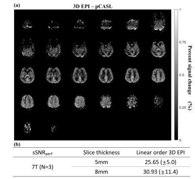 |
Whole-brain Perfusion Mapping at 7T by SAR-efficient Non-segmented 3D EPI-pCASL
Seon-Ha Hwang1, SoHyun Han2, Seong-Gi Kim2, Jaeseok Park3,4, and Sung-Hong Park1
1Department of Bio and Brain Engineering, Korea Advanced Institute of Science and Technology, Daejeon, Korea, Republic of, 2Center for Neuroscience Imaging Research, Institute of Basic Science, Suwon, Korea, Republic of, 3Department of Biomedical Engineering, Sungkyunkwan University, Suwon, Korea, Republic of, 4Department of Intelligent Precision Healthcare Convergence, Suwon, Korea, Republic of
Due to high specific absorption rate (SAR), it has not been easy to apply pseudo-continuous arterial spin labeling (pCASL) at 7T human MRI, especially with 3D readouts. In this study, non-segmented 3D-EPI-pCASL is proposed for whole-brain perfusion mapping. SAR was reduced by multiple low-flip-angle water-excitation rectangular RF pulses for 3D EPI. The proposed 3D-EPI-pCASL produced consistent perfusion maps at both 3T and 7T compared to 2D-EPI-pCASL which was available only at 3T because of SAR. The high temporal resolution of the proposed non-segmented 3D-EPI-pCASL enabled us to get a whole-brain 3D pCASL fMRI map at 7T for the first time.
|
|||
2726.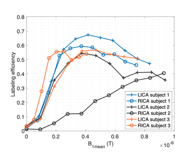 |
PCASL labeling efficiency measurement with B0 off-resonance compensation at 7T
Gael Saib1, Alan Koretsky1, and S Lalith Talagala2
1NINDS/LFMI, National Institutes of Health, Bethesda, MD, United States, 2NINDS/NMRF, National Institutes of Health, Bethesda, MD, United States
At 7T, it is known that B1+ and B0 inhomogeneities reduce the PCASL labeling efficiency. Further, the short T2 of blood also reduces the maximum blood labeling efficiency that can be achieved in practice. In this work, we employed a PCASL-prepared FLASH sequence to measure vessel-specific labeling efficiency of PCASL with B0 off-resonance correction. At 7T, experimental data indicates a maximal labeling efficiency of ~0.6. This study also shows that optimization of PCASL labeling efficiency is possible by tailoring the average gradient according to the blood velocity range and B1 at the feeding arteries.
|
|||
2727.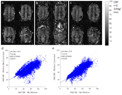 |
Retrospective Motion Correction of multi-shot 3D GRASE Arterial Spin Labelling using ESPIRiT reconstruction
Jack Highton1, Enrico De Vita2, and David Thomas3,4,5
1UCL Queen Square Institute of Neurology, University College London, London, United Kingdom, 2School of Biomedical Engineering and Imaging Sciences, Kings College London, London, United Kingdom, 3Dementia Research Centre, UCL Queen Square Institute of Neurology, University College London, London, United Kingdom, 4Neuroradiological Academic Unit, Department of Brain Repair and Rehabilitation, University College London, London, United Kingdom, 5Wellcome Centre for Human Neuroimaging, University College London, London, United Kingdom
A retrospective motion correction method is presented, for multi-shot 3D GRASE ASL. ESPIRiT is used to calculate coil sensitivity fields, using raw ASL k-space data. These are used to reconstruct complete images from the interleaved fractions of k-space sampled during each shot using SENSE. Therefore, typical image registration can be used to correct inter-shot motion. The method was tested using a simulation of 3D GRASE ASL and an equivalent experiment, where the subject was trained to nod or keep still. The motion correction reduced artefacts, and increased the correlation between cerebral blood flow measurements acquired with and without motion.
|
|||
2728.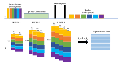 |
Optimization and Evaluation of Super-Resolution SMS ASL with Slice-Dithered Enhanced Resolution (SLIDER) technique
Qinyang Shou1, Xingfeng Shao1, and Danny JJ Wang1
1Laboratory of Functional MRI Technology (LOFT), Stevens Neuroimaging and Informatics Institute, University of Southern California, Los Angeles, CA, United States
The goal of this study is to optimize and evaluate super-resolution simultaneous multislice (SMS) Arterial Spin Labeling (ASL) using the Slice Dithered Enhanced Resolution (SLIDER). Different numbers (2/3/4) of shifted slices for SLIDER SMS ASL were evaluated for spatial and temporal SNR. The results showed that the SNR efficiency of SLIDER SMS ASL increased with a greater number of shifted slices which may be attributed to reduced g-factor for SMS imaging of thicker slices as well as the presence of physiological noise.
|
|||
2729.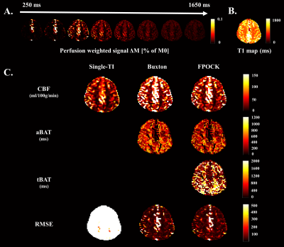 |
Saturated Look-Locker FAIR (SALL-FAIR) Sequence with FPOCK Model for Simultaneous Acquisition of CBF, aBAT, tBAT, and T1 Map
Zihan Ning1, Shuo Chen1, Zhensen Chen1, Huiyu Qiao1, Hualu Han1, Rui Shen1, Dandan Yang1, and Xihai Zhao1
1Center for Biomedical Imaging Research, Department of Biomedical Engineering, School of Medicine Tsinghua University, Beijing, China
A SAturated Look-Locker Flow-sensitive Alternating Inversion Recovery (SALL-FAIR) sequence combined with the Four-Phase One-Compartment Kety (FPOCK) kinetic model was proposed for simultaneous acquisition of CBF, aBAT, tBAT and T1 map with a single scan. The T1 maps of SALL-FAIR were verified by standard IR-SE on phantom, and higher accuracy of perfusion quantification from SALL-FAIR with FPOCK model was proved by comparing with the Buxton’s and the single-TI model in vivo.
|
|||
2730. |
BATS: the Boston ASL Template and Simulator – development and initial evaluation
Manuel Taso1, Fanny Munsch1, and David C Alsop1
1Division of MRI research, Department of Radiology, Beth Israel Deaconess Medical Center, Harvard Medical School, Boston, MA, United States
Neuroscientific and clinical research has seen tremendous advances over the past 25 years. While hardware, pulse-sequences and reconstruction developments have been an immense driver for this, image processing tools have also been crucial.While anatomical templates such as the MNI152 have become widely used and are a hallmark of neuroimaging analyses, functional contrast-specific templates for group studies are much scarcer but of great interest. We therefore report here the initial development of BATS – the Boston ASL Template and Simulator, highlighting its development and initial use.
|
|||
2731.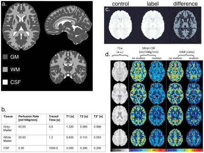 |
ASLDRO: Digital reference object software for Arterial Spin Labelling
Aaron Oliver-Taylor1, Thomas Hampshire1, Nadia A S Smith2, Michael Stritt3, Jan Petr4, Johannes Gregori3, Matthias Günther3,5, Henk J Mutsaerts6, and Xavier Golay1,7
1Gold Standard Phantoms Limited, London, United Kingdom, 2National Physical Laboratory, Teddington, United Kingdom, 3mediri GmbH, Heidelberg, Germany, 4Helmholtz-Zentrum Dresden-Rossendorf, Dresden, Germany, 5Fraunhofer MEVIS, Bremen, Germany, 6Amsterdam University Medical Center, Amsterdam, Netherlands, 7Institute of Neurology, University College London, London, United Kingdom
ASLDRO is digital reference object software for Arterial Spin Labelling. Here we present the development and demonstration of the DRO software, and its use in a sensitivity and uncertainty analysis of the single-subtraction equation for ASL perfusion quantification.The DRO software was written in python, and can generate synthetic ASL control, label and M0 data in ASL BIDS format. Pulsed and continuous labelling are supported, and patient motion and instrument noise are accurately simulated. It can be used both for testing and validation of image processing software, and for investigating ASL quantification models.
|
|||
2732.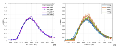 |
Reproducibility and repeatability of quantitative pCASL measurements in a 3D-printed perfusion phantom
Yiming Wang1, Limin Zhou1, Durga Udayakumar1,2, and Ananth J. Madhuranthakam1,2
1Radiology, UT Southwestern Medical Center, Dallas, TX, United States, 2Advanced Imaging Research Center, UT Southwestern Medical Center, Dallas, TX, United States
Arterial spin labelling (ASL) is a non-invasive and non-contrast perfusion imaging technique that can serve as an imaging biomarker to assess tissue blood flow characteristics. A perfusion phantom is valuable to evaluate newly developed ASL sequences, test consistency and to compare sequence reproducibility across various scanners. In this study, we performed a longitudinal assessment of the reproducibility and repeatability of perfusion measurement using 2D pCASL sequence with 20 PLDs over 5 weeks duration with a previously developed 3D printed perfusion phantom. Intra-class correlation coefficient (ICC) of measured perfusion and T1 are 0.96 and 0.94 respectively, indicating good reproducibility and repeatability.
|
The International Society for Magnetic Resonance in Medicine is accredited by the Accreditation Council for Continuing Medical Education to provide continuing medical education for physicians.