Digital Posters
Diffusion Tractography: Methods
ISMRM & SMRT Annual Meeting • 15-20 May 2021

Digital Posters
Diffusion Tractography: Methods
| Concurrent 4 | 19:00 - 20:00 |
4282.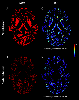 |
Where do streamlines come from? Seeding strategies impact on streamline distribution
Manon Edde1, Etienne St-Onge1, Antoine Théberge1,2, Guillaume Theaud1, Emmanuelle Renauld1, and Maxime Descoteaux1
1Sherbrooke Connectivity Imaging Lab (SCIL), Université de Sherbrooke, Sherbrooke, QC, Canada, 2Videos & Images Theory and Analytics Laboratory (VITAL), Université de Sherbrooke, Sherbrooke, QC, Canada
Tractography algorithms require a seeding map in which each point initiates one streamline. We can determine how many streamlines end in each voxel from one of their ends, but their initial point is unknown. We propose a method to compute the initial streamline points (IPS) in each voxel and evaluate their distribution depending on two strategies of seeding. With the surface-based interface, IPSs maps are more uniform along the cortex independently of the cortical volume, allowing a greater cortical coverage and specificity at the level of the cortical regions both at the whole-brain and bundle level.
|
|||
4283.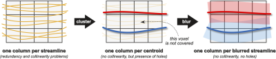 |
Blurred streamlines: a new concept to improve tractography accuracy by spatially blurring signal contributions
Alessandro Daducci1, Francesco Gobbi1, Nicola Febbrari1, Matteo Battocchio1, and Simona Schiavi1
1University of Verona, Verona, Italy
Tractography is a powerful tool to study brain connectivity but it suffers from an intrinsic trade-off between sensitivity and specificity. The former can be increased by constructing more streamlines, while filtering techniques can improve the latter. However, creating many streamlines may introduce redundancy in the tractograms and negatively affect the performances of filtering methods, especially those based on linear optimization. Here, we present the “blurred streamlines”, a novel concept based on a combination of streamline clustering and spatial blurring of their signal contributions. Preliminary results show the potential of this formulation and open new perspectives for improving tractography accuracy.
|
|||
4284.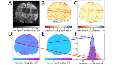 |
Robust residual bootstrapping algorithm for accurate SH representation of DW MRI signal that contains outliers
Viljami Sairanen1,2 and Chantal M. W. Tax3,4
1Department of Computer Science, University of Verona, Verona, Italy, 2Translational Imaging in Neurology, Department of Medicine and Biomedical Engineering, University of Basel, Basel, Switzerland, 3CUBRIC, Cardiff University, Cardiff, United Kingdom, 4Image Sciences Institute, University Medical Center Utrecht, Utrecht, Netherlands
Diffusion MRI measurements are affected by noise which propagates into the model estimation. Residual bootstrapping has been proposed to assess the uncertainty of parameter estimates, but do not consider the confounding effect of measurement outliers (e.g. due to subject motion), limiting their usage on clinical data. We present a robust bootstrapping algorithm and demonstrate its performance on the estimation and uncertainty quantification of rotationally invariant spherical harmonic (RISH) features with simulations and clinical data. Our algorithm can improve the reliability of cross-scanner harmonization relying on RISH and probabilistic tractography.
|
|||
4285.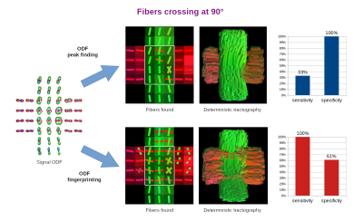 |
ODF-Fingerprinting Improves Reconstruction of Fibers Crossing at Shallow Angles: A Study on Diffusion Phantom
Patryk Filipiak1, Lee Basler2, Anthony Zuccolotto2, Ying-Chia Lin1, Dimitris G. Placantonakis3, Timothy Shepherd1, Walter Schneider4, Fernando E. Boada1, and Steven H. Baete1
1Center for Advanced Imaging Innovation and Research (CAI2R), NYU School of Medicine, New York, NY, United States, 2Psychology Software Tools, Inc., Pittsburgh, PA, United States, 3Department of Neurosurgery, Perlmutter Cancer Center, Neuroscience Institute, Kimmel Center for Stem Cell Biology, NYU School of Medicine, New York, NY, United States, 4University of Pittsburgh, Pittsburgh, PA, United States
ODF peak finding typically fails to reconstruct fibers crossing at angles below 45 degrees. We aim to break this barrier with ODF-Fingerprinting. Our approach replaces the peak finding mechanism with pattern matching, allowing to use all the information stored in ODFs. In this work, we study the ability of ODF-Fingerprinting to reconstruct fibers crossing at 90, 45, and 30 degrees in a diffusion phantom composed of textile tubes with 0.8µm diameter, approaching the anatomical scale of axons. Our approach reaches much higher sensitivity (84-100%) than the ODF peak finding (0-33%), especially at the shallow angles.
|
|||
4286.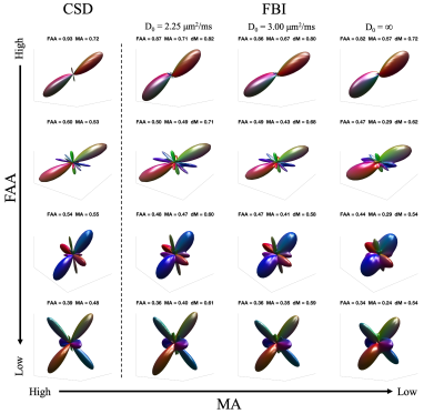 |
Quantitative comparison of fiber orientation distribution functions obtained with constrained spherical deconvolution and fiber ball imaging
Hunter Moss1 and Jens Jensen1
1Neurosciences, Medical University of South Carolina, Charleston, SC, United States
The fiber orientation distribution function (fODF) can be estimated with diffusion MRI and gives the angular probability density of axon orientations in white matter. Here fODFs, as estimated with constrained spherical deconvolution (CSD) and fiber ball imaging (FBI), are compared quantitatively in order to assess their consistency. An important distinction between these two approaches is that CSD relies on a globally defined, empirical response function while FBI does not. For two measures of anisotropy, similar results are found for the CSD and FBI fODFs. However, a distance metric reveals significant differences in fODF peak directions and fine structure.
|
|||
4287.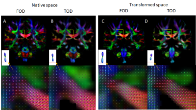 |
Group Average Tractography of the Human Brain using Direct Streamline Mapping
Zifei Liang1 and Jiangyang Zhang1
1Center for Biomedical Imaging, Dept. of Radiology, New York University School of Medicine, NEW YORK, NY, United States
Diffusion MRI based tractography is widely used to examine structural connectivity in the brain. Due to noises and motions, tractography in individual subject may contain erroneous results, which may be removed by averaging over a group. Although several group average tractography (GAT) approaches are available, their accuracy has not been thoroughly examined. In this study, we compared GAT based on spatial normalization of fiber orientation distribution maps and direct streamline mapping. Our results suggest that direct streamline mapping better preserve small and secondary axonal projections and is better-suited for studying group average tractography of the brain.
|
|||
4288.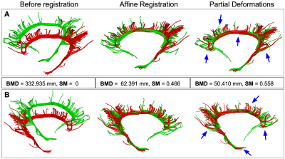 |
StND: Non-Rigid Partial Deformation Tractography Registration
Bramsh Qamar Chandio1 and Eleftherios Garyfallidis1
1Intelligent Systems Engineering, Indiana University Bloomington, Bloomington, IN, United States
StND is a method for non-rigid registration of white matter tracts in streamline-space. StND performs two-step registration to align the moving bundle with a static (reference) bundle. In the first step, it affinely registers the two given tracts, and in the second step, it partially deforms the moving bundle to better align it with the static bundle. This partial deformation allows us to improve registration by deforming only areas where there is higher correspondence between streamlines of the tracts. This also helps in preserving the original anatomical structure of the moving bundle.
|
|||
4289.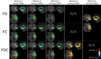 |
On false positive control in Fixel-Based Analysis
Robert Smith1,2, Daan Christiaens3,4, Ben Jeurissen5, Maximillian Pietsch3, David Vaughan1,2,6, Graeme Jackson1,2,6, and J-Donald Tournier3
1Florey Institute of Neuroscience & Mental Health, Melbourne, Australia, 2Florey Department of Neuroscience and Mental Health, The University of Melbourne, Melbourne, Australia, 3School of Biomedical Engineering & Imaging Sciences, King's College London, London, United Kingdom, 4Department of Electrical Engineering, KU Leuven, Leuven, Belgium, 5Department of Physics, University of Antwerp, Antwerp, Belgium, 6Department of Neurology, Austin Health, Melbourne, Australia
While the Fixel-Based Analysis (FBA) framework provides familywise error control across a whole-brain template accounting for the presence of crossing fibres in the white matter, its typical usage fails to correct for multiple hypothesis tests due to the utilisation of multiple quantitative metrics. We demonstrate different methods that can be employed to provide more comprehensive false positive control in this context.
|
|||
4290.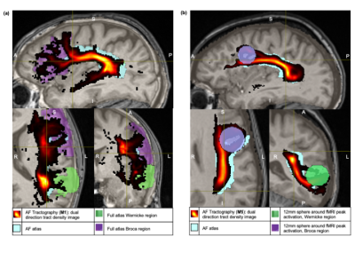 |
Probabilistic Tractography of the Arcuate Fasciculus: Sensitivity and Specificity of Standardised fMRI and Atlas-based Approaches
Jane Ansell1,2, Irène Brumer3, Jonathan Ashmore4, Enrico de Vita5, Josef Jarosz2, and Marco Borri2
1King's College London, London, United Kingdom, 2Department of Neuroradiology, King's College Hospital, London, United Kingdom, 3Computer Assisted Clinical Medicine, Medical Faculty Mannheim, Heidelberg University, Mannheim, Germany, 4Department of Medical Physics and Bioengineering, NHS Highland, Inverness, United Kingdom, 5Department of Biomedical Engineering, School of Biomedical Engineering and Imagine Sciences, King's College London, London, United Kingdom
Atlas and fMRI-based approaches to standardise seed and end regions for probabilistic tractography of the arcuate fasciculus are investigated. fMRI-based approaches use spheres centered on language task peak activation. Within this pilot cohort, an atlas-based approach demonstrates the greatest sensitivity. fMRI-based approaches are more specific, but sensitivity can be increased by enlarging sphere size. Within each approach, a trade-off between sensitivity and specificity is seen as seed/end region size increases. For patients with abnormalities or lesions, where atlas approaches might be compromised, fMRI methods may be preferred. Further work to optimise fMRI-based approach is warranted, alongside application to patient data.
|
|||
4291.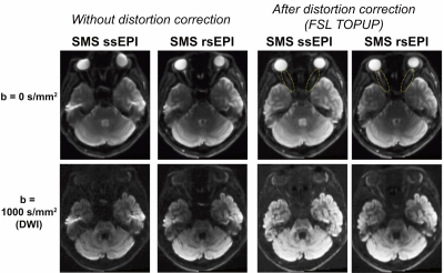 |
Advantage of simultaneous multi-slice readout-segmented echo-planar imaging on diffusion MRI measurements of the human optic nerve
Hiromasa Takemura1,2, Wei Liu3, Hideto Kuribayashi4, and Ikuhiro Kida1,2
1Center for Information and Neural Networks (CiNet), National Institute of Information and Communications Technology, Suita-shi, Japan, 2Graduate School of Frontier Biosciences, Osaka University, Suita-shi, Japan, 3Siemens Shenzhen Magnetic Resonance Ltd, Shenzhen, China, 4Siemens Healthcare K.K., Tokyo, Japan
Diffusion MRI measurements of the human optic nerve remain challenging due to susceptibility-induced artifacts, despite their importance in neuro-ophthalmology research. Here, we demonstrate that simultaneous multi-slice (SMS) readout-segmented echo-planar imaging (rsEPI) acquisition has advantages in measuring the human optic nerve over conventional SMS single-shot EPI (ssEPI) with respect to image quality. This result was confirmed with statistical comparisons demonstrating higher fractional anisotropy along the optic nerve in SMS rsEPI data compared with that in SMS ssEPI data. Taken together, this study demonstrates the utility of SMS rsEPI for diffusion MRI measurements of tissue properties of the optic nerve.
|
|||
4292.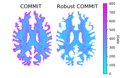 |
Enhancing tractography filtering by accounting for outliers and partial voluming in diffusion weighted measurements
Viljami Sairanen1,2, Mario Ocampo-Pineda1, Cristina Granziera2, Simona Schiavi1, and Alessandro Daducci1
1Department of Computer Science, University of Verona, Verona, Italy, 2Translational Imaging in Neurology, Department of Medicine and Biomedical Engineering, University Hospital Basel and University of Basel, Basel, Switzerland
The white matter structures of the human brain can be represented via diffusion tractography. Unfortunately, tractography is prone to find false-positive streamlines causing a severe decline in its specificity and limiting its clinical feasibility. Filtering algorithms have been proposed to reduce these invalid streamlines. We augmented the COMMIT filtering algorithm to adjust for two typical artifacts present in diffusion-weighted images: partial voluming and signal drop-outs due to subject motion. We demonstrate that our robust algorithm is capable to properly filter tractography reconstructions despite these artifacts and could be useful especially for clinical studies with uncooperative patient groups such as neonates.
|
|||
4293.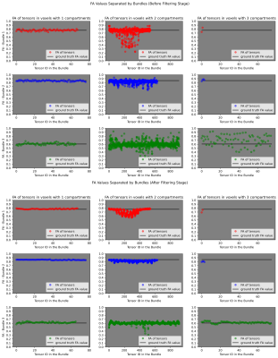 |
Robust Estimation of Fascicle-based Fractional Anisotropy on Fiber Crossings
Erick Hernandez-Gutierrez1, Ricardo Coronado-Leija2, Gabrielle Grenier3, Maxime Descoteaux3, and Alonso Ramirez-Manzanares1
1Computer Science, CIMAT A.C., Guanajuato, Mexico, 2New York University School of Medicine, New York, NY, United States, 3Université de Sherbrooke, Sherbrooke, QC, Canada
In this work, a novel multi-shell modelling method is proposed to compute fascicle-based fractional anisotropy that is shown accurate and robust in the centrum semiovale of human brain clinical multi-shell datasets. It does not require tractography and it is informed by the local modelling estimations provided by the method called MRDS. The effectiveness of our methodology is also shown on synthetic experiments.
|
|||
4294.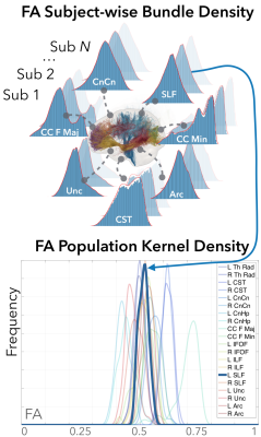 |
White Matter Inter-tract Connectivity Using Cosine Similarity between Population Kernel Densities of Diffusion Measures
David Lee1, Ashish Sahib1, Antoni Kubicki1, Katherine Narr1, and Shantanu Joshi1
1UCLA, Los Angeles, CA, United States
Pairwise inter-tract correlations of the mean diffusion measures have been used to infer structural connectivity among major white matter (WM) fiber tract pathways. Instead of using the mean, we propose a novel approach of constructing kernel density distributions to represent population variability of fractional anisotropy. We also propose the cosine similarity metric between the full shapes of the kernel densities to investigate inter-tract connectivity among the fiber tracts. Our representation and the shape similarity measure is a better predictor of the short- and long-range connectivity disruptions in WM tracts in major depressive disorder patients compared to the conventional mean-based approach.
|
|||
4295.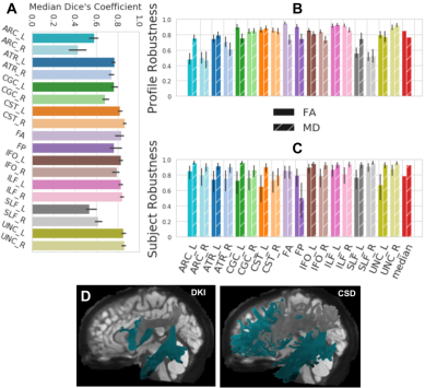 |
The test-retest reliability and robustness of diffusion-MRI based tractometry
John Kruper1,2, Jason D Yeatman3,4, Adam Richie-Halford2, David Bloom1,2, Mareike Grotheer5,6, Sendy Caffarra3,4,7, Greg Kiar8, Iliana I. Karipidis9, Ethan Roy3, and Ariel Rokem1,2
1Department of Psychology, University of Washington, Seattle, WA, United States, 2eScience Institute, University of Washington, Seattle, WA, United States, 3Graduate School of Education, Stanford University, Stanford, CA, United States, 4Division of Developmental-Behavioral Pediatrics, Stanford University School of Medicine, Stanford, CA, United States, 5Center for Mind, Brain and Behavior - CMBB, Hans-Meerwein-Straße 6, Marburg, Germany, 6Department of Psychiatry, University of Marburg, Marburg, Germany, 7Basque Center on Cognition (BCBL), Brain and Language, Donostia‐San Sebastián, Spain, 8Department of Biomedical Engineering, McGill University, Montreal, QC, Canada, 9Center for Interdisciplinary Brain Sciences Research, Department of Psychiatry and Behavioral Sciences, Stanford University School of Medicine, Stanford, CA, United States
Tractometry from diffusion MRI estimates the tissue properties along the length of major white matter tracts, using: computational tractography; tract segmentation using atlases or other classification methods; and microstructural modeling in voxels along the length of the estimated tracts. Given previous concerns about the sensitivity of dMRI-based analysis to variations in methodology, we tested: the reliability of tractometry results within individuals across measurements; and within measurement, across variations in the analysis methods. We found that although there are variations that arise from differences in tractography methods, bundle segmentation methods, microstructural modeling, and different software implementations, tractometry is overall quite robust.
|
|||
4296. |
Improving tractography Reconstruction using Local-phase Features
Alireza Shirazinodeh1,2 and Hamidreza Saligheh Rad1,2
1Department of Medical Physics and Biomedical Engineering, Tehran university of Medical Science, Tehran, Iran (Islamic Republic of), 2Quantitative MR Imaging and Spectroscopy Group, Research Center for Molecular and Cellular Imaging, Tehran, Iran (Islamic Republic of)
Tractography of the brain fibers from diffusion signals in magnetic resonance imaging needs to be more sensitive to structures of diffusion images to reduce the number of ODF used to predict tracts and avoid determining false-positive cases in pixels whose values may have changed due to noise or other unwanted factors. The local-based methods can be improved tractography results. In this abstract, we designed a mask based on the local structures of the diffusion phantom images in 64 directions. Using this mask we reduced the number of gradients and false-positive predicted results from the outputs of the tractography algorithm.
|
|||
4297.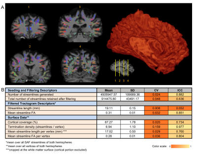 |
Surface-based Short Association Fibre Tractography
Dmitri Shastin1,2,3, Sila Genc1, Greg Parker1, Kristin Koller1, Chantal M.W. Tax1,4, John Evans1, Khalid Hamandi1,3,5, Derek Jones1,3, William Gray1,2,3, and Maxime Chamberland1
1Cardiff University Brain Research Imaging Centre (CUBRIC), Cardiff University, Cardiff, United Kingdom, 2Department of Neurosurgery, University Hospital of Wales, Cardiff, United Kingdom, 3BRAIN Biomedical Research Unit, Health & Care Research Wales, Cardiff, United Kingdom, 4Image Sciences Institute, University Medical Center Utrecht, Utrecht, Netherlands, 5Department of Neurology, University Hospital of Wales, Cardiff, United Kingdom
We propose a method of utilising mesh representations of cortical surfaces for generating tractograms of short association fibres facilitating a focused investigation into superficial white matter in health and disease. The method additionally allows to relate streamline metrics to surface topology in native space before registration for comparisons within and between subjects is performed. Our approach is applied to state-of-the-art test-retest data and shows good-to-excellent repeatability.
|
|||
4298.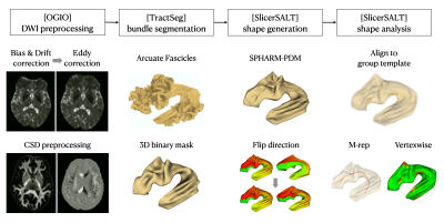 |
Surface-based Shape Morphometry Analysis of Human White Matter Tracts
Yi-Chia Kung1, Chu-Chung Huang2, Chih-Chin Heather Hsu1,3, Shin Tai Chong1, Ching-Po Lin1, and Chun-Hung Yeh4,5
1Institute of Neuroscience, National Yang-Ming University, Taipei, Taiwan, 2Institute of Cognitive Neuroscience, School of Psychology and Cognitive Science, East China Normal University, Shanghai, China, 3Center for Geriatrics and Gerontology, Taipei Veterans General Hospita, Taipei, Taiwan, 4Institute for Radiological Research, Chang Gung University and Chang Gung Memorial Hospital, Taoyuan, Taiwan, 5Department of Child and Adolescent Psychiatry, Chang Gung Memorial Hospital, Linkou Medical Center, Taoyuan, Taiwan
This study proposes a novel framework for quantifying the alteration in shape morphometry of white matter (WM) fiber tracts. The surface of each WM bundle is segmented from diffusion MRI data using TractSeg, followed by surface reconstruction, parameterization, and modelling. With the M-rep parameterization and vertex-wise model distance, the proposed method was able to differentiate the level of morphological lateralization between bilateral arcuate fasciculi and cortical spinal tracts.
|
|||
4299.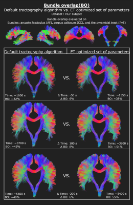 |
Improving white matter bundle recovery: A fast & practical ensemble tractography pipeline
Alexandre Joanisse1, Guillaume Theaud1, Jon Haitz Legarreta1, François Rheault1,2, and Maxime Descoteaux1
1Computer Sciences, Université de Sherbrooke, Sherbrooke, QC, Canada, 2Electrical Engineering, Vanderbilt University, Nashville, TN, United States
In most tractography settings, users choose a single tracking algorithm and a single set of tracking parameters, which may limit the quality and impact of the bundle reconstruction results. The present ensemble tractography processing pipeline aims at producing diverse whole brain tractograms faster by tuning and automatically concatenating multiple tractograms from different combinations of parameters and algorithms. Compared to classical tractography, our optimized ensemble tractography obtains improved or similar white matter bundle recovery but in 62% of the processing time.
|
|||
4300. |
GPU-accelerated diffusion MRI tractography in DIPY
Ariel Rokem1, Mauro Bisson2, Josh Romero2, Thorsten Kurth2, Massimiliano Fatica2, Pablo Damasceno3, Xihe Xie3, Adam Richie-Halford4, Serge Koudoro5, and Eleftherios Garyfallidis5
1Psychology, University of Washington, Seattle, WA, United States, 2NVIDIA, Menlo Park, CA, United States, 3University of California, San Francisco, San Francisco, CA, United States, 4University of Washington, Seattle, WA, United States, 5Indiana University, Bloomington, IN, United States
Tractography based on diffusion-weighted MRI provides non-invasive in vivo estimates of trajectories of long-range brain connections. These estimates are important in research that measures individual differences in brain connections and in clinical use-cases. But the computational demands of tractography present a barrier to progress. Here, we present a GPU-based tractography implementation that accelerates tractography algorithms implemented as part of the Diffusion Imaging in Python (DIPY) project. This implementation speeds up tractography by at least a factor of ~200X, providing tractographies that closely match CPU-based solutions. These speedups enable applications of tractography in clinical data, and in very large datasets.
|
|||
4301.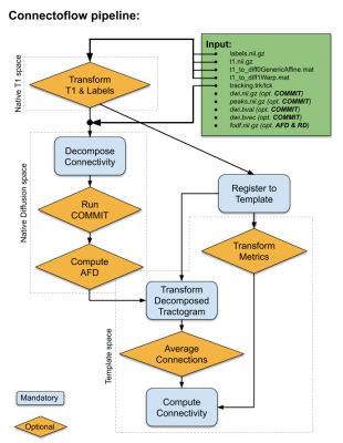 |
Connectoflow: A cutting-edge Nextflow pipeline for structural connectomics
Francois Rheault1,2, Jean-Christophe Houde2, Jasmeen Sidhu3, Sami Obaid2,4, Guido Guberman5, Alessandro Daducci6, and Maxime Descoteaux2
1Electrical Engineering, Vanderbilt University, Nashville, TN, United States, 2Computer Sciences, Université de Sherbrooke, Sherbrooke, QC, Canada, 3Faculté de médecine et des sciences de la santé, Université de Sherbrooke, Sherbrooke, QC, Canada, 4Centre de Recherche du Centre Hospitalier, Université de Montréal, Montréal, QC, Canada, 5Department of Neurology and Neurosurgery, McGill University, Montréal, QC, Canada, 6Computer Science, University of Verona, Verona, Italy
Tractography involves complicated processing and connectomics include even more complexity. To facilitate structural connectome reconstruction we present: Connectoflow. Connectoflow requires simple inputs, has simple options and provides simple outputs, all with cutting-edge processing. By leveraging the simplicity of Nextflow and Docker/Singularity, Connectoflow is robust and efficient. By combining Tractoflow with Connectoflow, one can go from raw DW-images to structural connectomes in a few simplified steps. The proposed pipeline innovates by including connection-wise cleaning/filtering, provides connection weights that go beyond streamline count (COMMIT) as well as advanced connection-wise metrics (similarity and AFD).
|
The International Society for Magnetic Resonance in Medicine is accredited by the Accreditation Council for Continuing Medical Education to provide continuing medical education for physicians.