Digital Posters
Diffusion Tractography: Applications
ISMRM & SMRT Annual Meeting • 15-20 May 2021

Digital Posters
Diffusion Tractography: Applications
| Concurrent 4 | 19:00 - 20:00 |
4302.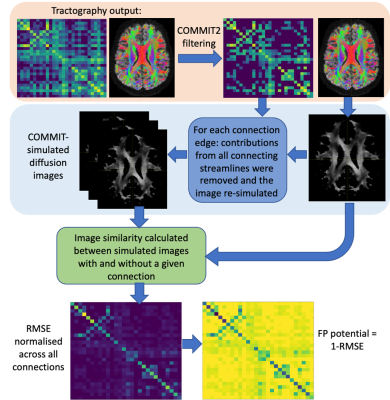 |
False-positive potential of tractography-derived connections improves network reconstruction for disease spreading models
Anna Schroder1, Neil P. Oxtoby1, Marco Palombo1, Simona Schiavi2, Alessandro Daducci2, and Daniel C. Alexander1
1Centre for Medical Image Computing, Department of Computer Science, University College London, London, United Kingdom, 2Department of Computer Science, University of Verona, Verona, Italy
Network spreading models of disease propagation utilise functional or anatomical connectivity to predict regional pathology in-vivo. Models based on anatomical connectivity are likely to be disrupted by the inevitable presence of errors from tractography. To mitigate this problem, we propose a method to evaluate the potential of each tractography-derived connection to be false-positive. By incorporating this false-positive potential into a network spreading model, we are able to predict a pattern of tau accumulation more closely aligned with the pathology observed in a cohort of Alzheimer’s Disease patients.
|
|||
4303.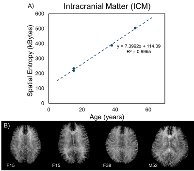 |
Spatial Entropy Mapping of the Connectome using White Matter Fibrography: Quantifying the Effects of Age.
Ryan McNaughton1, Hernan Jara1,2, Ning Hua2,3, Andy Ellison2,3, Osamu Sakai2, Lee Goldstein1,2,3, and Stephan Anderson2,3
1Boston University, Boston, MA, United States, 2Boston University Medical Center, Boston, MA, United States, 3Center for Translational Neuroimaging, Boston, MA, United States
Purpose: To develop algorithms for mapping the spatial entropy (SE); extending white matter fibrography (WMF) for quantifying whole-brain information content. Methods: SE algorithms were applied to WM texture images of four individuals of increasing age, optimized for maximum information content, and implemented to calculate the whole-brain WM complexity. Results: SE was positively correlated with subject age. Conclusion: SE is a promising quantitative metric for objectively distinguishing the state of WM architecture from fibrograms, particularly as it relates to age effects.
|
|||
4304.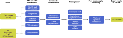 |
Automatic tractography for brain tumor surgery: towards clinical application
Stephan Meesters1,2, Maud Landers2, Geert-Jan Rutten2, and Luc Florack1
1TU/e Eindhoven University of Technology, Eindhoven, Netherlands, 2ETZ Elisabeth-TweeSteden hospital, Tilburg, Netherlands
We have introduced an automatic pipeline based on diffusion-weighted tractography for the reconstruction of four white-matter structures of interest. These anatomical reconstructions are visualized during brain tumour surgery to aid in the prevention of sensorimotor, visual and language deficits. The automatic tractography pipeline produces robust results, owing in part to the deep-learning based segmentation of brain regions which is robust to deformations of the brain due to brain shift, and the post-tractography filtering method which can remove spurious fibers.
|
|||
4305.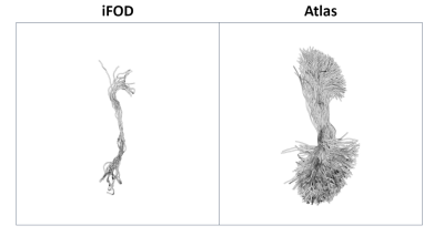 |
Quantitative evaluation of white matter tracts reconstruction with an anatomically curated atlas in patients with brain tumours
Umberto Villani1,2, Erica Silvestri1,2,3, Colpo Maria2, D'Avella Domenico3, Della Puppa Alessandro4, Maurizio Corbetta1,3, Cecchin Diego5, and Alessandra Bertoldo1,2
1Padova Neuroscience Center, University of Padova, Padova, Italy, 2Department of Information Engineering, University of Padova, Padova, Italy, 3Department of Neuroscience, University of Padova, Padova, Italy, 4Department of Neurosurgery, University of Firenze, Firenze, Italy, 5Department of Medicine, Unit of Nuclear Medicine, University of Padova, Padova, Italy
We propose an application of the open-source whitematteranalysis software which moves a step towards the goal of reproducible and quantitative evaluation of anatomical tracts reconstruction by dMRI tractography. We quantify the tractogram in 11 patients suffering from brain tumours with four different algorithms, perform the automated spectral clustering procedure implemented in the software and evaluate simple metrics to compare the tractograms to an available anatomically curated atlas. Independently from the tumour position, the four investigated algorithms failed to properly reconstruct certain anatomical tracts. Evaluating the overall streamline representation of all tracts, the iFOD2 algorithm was found to perform best.
|
|||
4306. |
Assessment of structural connectivity matrices sensitivity to tractography termination criteria in glioma patients
Maria Colpo1, Erica Silvestri1,2,3, Umberto Villani1,2, Domenico D'Avella3, Alessandro Della Puppa4, Diego Cecchin2,5, Maurizio Corbetta2,3, and Alessandra Bertoldo1,2
1Department of Information Engineering, University of Padova, Padova, Italy, 2Padova Neuroscience Center, University of Padova, Padova, Italy, 3Department of Neuroscience, University of Padova, Padova, Italy, 4Department of Neurosurgery, University of Firenze, Firenze, Italy, 5Department of Medicine, Unit of Nuclear Medicine, University of Padova, Padova, Italy
Varying termination criteria values of tractography algorithms produce tangible effects on the reconstructed tractograms. While these effects are extensively documented in the state-of-the-art literature for healthy subjects, little is known about streamline reconstruction differences in presence of brain tumours. In this work we apply a statistical framework to assess the impact of the cut-off value on the resulting structural connectivity matrices in 11 patients suffering from gliomas. Results show how varying the cut-off value in the studied range does not produce significant changes in the matrix structure, thus validating literature recommendations for healthy subjects in the case of glioma patients.
|
|||
4307. |
DTI fiber tracking reveals positive effects of motor training on stroke recovery
Bastian Maus1,2, Lydia Wachsmuth1,2, Maike Hoppen3, Jens Minnerup3, Antje Schmidt-Pogoda3, and Cornelius Faber1,2
1Radiology, University Hospital Münster, Münster, Germany, 2Medical Faculty, Experimental Magnetic Resonance, Westfälische-Wilhelms-Universität Münster, Münster, Germany, 3Department of Neurology with Institute for Translational Neurology, University Hospital Münster, Münster, Germany
Ex vivo diffusion tensor imaging (DTI) was used to study the effects of motor training on interhemispheric connectivity after ischemic stroke in mice. Training increased the number of fibers of the corpus callosum by one third. No overall effect of lesion size on DTI parameters and general interhemispheric connectivity was observed. Trained animals with large lesions, however, had higher fiber counts and axial diffusivity compared to non-trained animals with similarly large lesions. A larger benefit of motor training on animals with more severe stroke is implied.
|
|||
4308.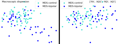 |
Classification of bipolar patients using a macroscopic dispersion measure of the white matter tractogram
Ali Demir1,2, Mehmed Özkan1, and Aziz Müfit Uluğ1,3
1Institute of Biomedical Engineering, Boğaziçi University, Istanbul, Turkey, 2Istanbul Medipol University, Istanbul, Turkey, 3Coretech Labs Inc., San Diego, CA, United States
Imaging based bio-markers are important in classification of psychiatric disorders for accurate diagnosis. We evaluated the performance of using a macroscopic dispersion measure of the white matter tractogram in the random forest classifier for identifying bipolar subjects. The macroscopic dispersion of the white matter tractogram enables increased performance of the bipolar/normal classification. Multidimensional scaling plots support that the macroscopic changes along the brain white matter improves the discrimination of bipolar and normal groups.
|
|||
4309.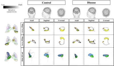 |
Comparison of differences in brain structure area and differences through tractography analysis: patients with subclinical depression
Sang Jin Im1, Jeong hwan Lee2, and Sie kyeong Kim3
1Gacheon University, Incheon, Korea, Republic of, 22Department of Psychiatry, Chungbuk National University College of Medicine, Cheongju, Korea, Republic of, 3Department of Psychiatry, Chungbuk National University College of Medicine, Cheongju, Korea, Republic of
Structural and functional changes in the subcortical area affect cognitive function and can cause increased vulnerability to mood symptoms such as anxiety and depression. However, studies on changes in subcortical structures show inconsistencies. This research shows the structural differences in segmented subcortical regions between control and depression groups and visualizes the pathway between structures according to the connection strength through tractography analysis to confirm functional differences.
|
|||
4310. |
Diffusion MRI and fibertracking of brachial plexus to diagnose injury
Nyoman Dana Kurniawan1, Jin Jin2, Clare Berry3, Wei Liu4, Aiman Al-Najjar1, Katie L McMahon5, Tom Lloyd6, Silvia Manzanero7, James Powell8, Michael Schuetz7, Trevor Gervais9, and Paul McEniery9
1Centre for Advanced Imaging, The University of Queensland, St Lucia, Australia, 2Siemens Healthcare, Brisbane, Australia, Brisbane, Australia, 3Metro North Hospital and Health Service, Brisbane, Australia, 4Application Department, Siemens Shenzhen Magnetic Resonance Ltd, Shenzhen, China, 5School of Clinical Sciences, Queensland University of Technology, Brisbane, Australia, 6Department of Radiology, Princess Alexandra Hospital, Brisbane, Australia, 7Jamieson Trauma Institute, Metro North Hospital and Health Service, Brisbane, Australia, 8Orthopaedics Department, Royal Brisbane and Women’s Hospital, Brisbane, Australia, 9Orthopedics Department, Royal Brisbane and Women’s Hospital, Brisbane, Australia
We describe the utility, optimization of a diffusion weighted imaging protocol, and the post-processing for fibertracking reconstruction of the brachial plexus nerve at 3T using a prototype Simultaneous Multi-Slice RESOLVE Diffusion-Weighted Imaging (SMS RESOLVE DWI) sequence. Best tractography profile was obtained for the acquisition acquired at 2.7 mm isotropic 3D resolution, using 7 segmentations, and 30 diffusion encoding directions at b-value of 800 s/mm2, combined with tractography reconstruction using a probabilistic tensor algorithm.
|
|||
4311.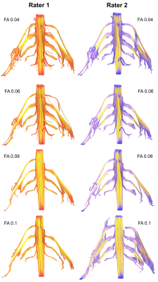 |
Fractional Anisotropy Thresholding for Deterministic Tractography of the Roots of the Brachial Plexus
Ryckie George Wade1, Irvin Teh1, Gustav Andersson2, Fang-Cheng Yeh3, Mikael Wiberg2, and Grainne Bourke1
1University of Leeds, Leeds, United Kingdom, 2Umeå University, Umeå, Sweden, 3University of Pittsburgh, Pittsburgh, PA, United States
We acquired high-angular resolution DTI from 10 healthy adults. We investigated how altering the fractional anisotropy threshold changed tractograms and tract-related metrics. At thresholds above 0.06, the propagation of valid tracts reduced significantly. The fractional anisotropy thresholds required to generate valid tractograms of the roots of the brachial plexus are lower than thresholds conventionally used in the brain. We provide estimates of the probability of generating valid tracts for each spinal nerve root of the brachial plexus, at different fractional anisotropy thresholds.
|
|||
 |
4312. |
High angular resolution diffusion MRI tractography-based thalamic parcellation of postmortem fetal brain in the second trimester
Sheng-Min Huang1, Kuan-Hung Cho1, Koping Chang2, Pei-Hsin Huang2,3, and Li-Wei Kuo1,4
1Institute of Biomedical Engineering and Nanomedicine, National Health Research Institutes, Miaoli, Taiwan, 2Department of Pathology, National Taiwan University Hospital, Taipei, Taiwan, 3Graduate Institute of Pathology, National Taiwan University College of Medicine, Taipei, Taiwan, 4Institute of Medical Device and Imaging, National Taiwan University College of Medicine, Taipei, Taiwan
Proper fiber connections between cerebral areas are particularly essential to build brain functions. As a central relay, the thalamus plays an important role in regulating diverse brain functions. Here, we aimed to investigate the thalamo-cortical connections and thalamic parcellation of ex vivo fetal brains in the second trimester using high-resolution postmortem diffusion MRI tractography on 3T. Diffusion images with 333 µm isotropic voxel size were acquired and tractography-based thalamic parcellation was performed. The segregated thalamic patterns were found as similar as adult’s ones reported previously. Our results showed the thalamic development and demonstrated the capability of postmortem diffusion MRI tractography.
|
||
4313. |
Exploring the thalamus of the human brain using tractography analysis in 3Tesla MRI
Sang Jin Im1 and Hyeon-Man Baek2
1Gacheon University, Incheon, Korea, Republic of, 2Lee Gil Ya Cancer and Diabetes Institute, Incheon, Korea, Republic of
Although there are discrepancies between studies, it can be deduced that the thalamus region has a clear effect on neurological disorders due to a strong relationship between the thalamus and neurological functions such as emotional control and processing. In this study, MPRAGE and DTI data were acquired using 3Tesla MRI, and thalamus regions were segmented by subdivisions based on the THOMAS atlas 4. In addition, tractography analysis was performed to investigate the connectivity between the thalamus subregions.
|
|||
4314.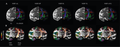 |
Mapping white matter bundle tracts and cortical myelin from multi-contrast imaging in the awake macaque monkey
Rakshit Dadarwal1,2, Fabien Balezeau3, Marcus Haag4, Michael C. Schmid3,4, Susann Boretius1,2, and Michael Oritz-Rios1,3
1Functional Imaging Laboratory, German Primate Center, Göttingen, Germany, 2Georg August Universität Göttingen, Göttingen, Germany, 3Biosciences Institute, Newcastle University, Newcastle upon Tyne, United Kingdom, 4Faculty of Science and Medicine, University of Fribourg, Fribourg, Switzerland
In-vivo whole-brain multi-contrast awake imaging enables the detection of structural and functional features in brain networks without anesthesia. In this work, we demonstrated the feasibility and robustness of multi-contrast MRI data acquisitions across sessions and subjects in awake macaque monkeys.
|
|||
4315.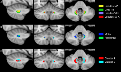 |
Cognitive and motor topography of human dentate nuclei identified with tractography and clustering methods
Fulvia Palesi1,2, Matteo Ferrante3, Marta Gaviraghi4, Anastasia Misiti4, Giovanni Savini5, Alessandro Lascialfari3, Egidio D'Angelo1,2, and Claudia A. M. Gandini Wheeler-Kingshott1,2,6
1Department of Brain and Behavioral Sciences, University of Pavia, Pavia, Italy, 2Brain Connectivity Center Research Department, IRCCS Mondino Foundation, Pavia, Italy, 3Department of Physics, University of Pavia, Pavia, Italy, 4Department of Electrical, Computer and Biomedical Engineering, University of Pavia, Pavia, Italy, 5Advanced Imaging and Radiomics Center, Neuroradiology Department, IRCCS Mondino Foundation, Pavia, Italy, 6NMR Research Unit, Queen Square MS Centre, Department of Neuroinflammation, UCL Queen Square Institute of Neurology, Faculty of Brain Sciences, University College London (UCL), London, United Kingdom
The dentate nuclei (DNs) represent the main output relay of the cerebellum and yet are often unexplored especially in human studies. For the first time, we used both tractography and an unsupervised fuzzy c-means classification algorithm to identify DNs topography on the basis of their connectivity to cerebellar and cerebral areas as well as their microstructural features. Our findings indicate that DNs can be parcellated in two main areas: one predominant non-motor representation and one motor representation. Furthermore, connectivity-based and microstructure-based atlases provide complementary information. These results represent a step-forward that could help interpretating pathological conditions involving cerebro-cerebellar circuits.
|
|||
4316.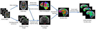 |
Patient specific tracts-based analysis of diffusion compartment models: application to multiple sclerosis patients with acute optic neuritis
Olivier Commowick1, Renaud Hédouin1, Charlotte Laurent2, and Jean-Christophe Ferré1,3
1Univ Rennes, Inria, CNRS, Inserm, IRISA UMR 6074, Empenn - ERL U1228, Rennes, France, 2Ophthalmology department, CHU Rennes, Rennes, France, 3Radiology department, CHU Rennes, Rennes, France
Multiple sclerosis is a complex disease where voxel-based, group-based statistics of the brain microstructure have shown their limits in explaining patient evolution. This is first due to too simple diffusion models, mixing information. Voxel-based studies also lack knowledge on brain structural connectivity. Finally, group-based analysis does not describe well the specific patient status (a crucial point for clinicians). We propose an atlas-based framework, combined with advanced diffusion compartment models, for patient specific analysis of microstructural disease burden on major fiber bundles. We apply our framework to the analysis of optic radiations of MS patients with acute optic neuritis.
|
|||
4317.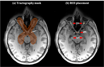 |
Anatomical assessment of retinogeniculate visual pathway tractography: a comparison of multiple tractography methods
Jianzhong He1,2, Fan Zhang2, Guoqiang Xie2,3, Shun Yao2,4, Yuanjing Feng1, Dhiego C.A. Bastos2, Yogesh Rathi2, Nikos Makris2, Ron Kikinis2, Alexandra J. Golby2, and Lauren J. O'Donnell2
1Zhejiang University of Technology, HangZhou, China, 2Harvard Medical School, Boston, MA, United States, 3Nuclear Industry 215 Hospital of Shaanxi Province, XianYang, China, 4The First Affiliated Hospital, GuangZhou, China
In this work, we investigate the performance of four widely used tractography methods (SD-Stream, iFOD1, UKF-1T, and UKF-2T) for the complete retinogeniculate visual pathway (RGVP) reconstruction. Anatomical measurement and expert judgement results indicate that UKF-2T and iFOD1 produce the best performance. The percentage of decussating fibers in the iFOD1 method was more similar to the known percentage from anatomical studies, while the UKF-2T method produced reconstructed RGVPs that were judged to better correspond to the anatomical course of the RGVP.
|
|||
4318.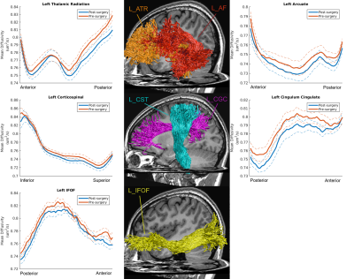 |
Microstructural Properties of Major White Matter Tracts in Constant Exotropia before and after Strabismus Surgery
Yanming Wang1, Xiaoxiao Wang1, Hongmei Shi2,3, Lin Xia2, Jiong Dong2, Benedictor Alexander Nguchu1, Jean De Dieu Uwisengeyimana1, Yanpeng Liu1, Du Zhang1, Lixia Feng2, and Bensheng Qiu1
1Hefei National Lab for Physical Sciences at the Microscale and the Centers for Biomedical Engineering, University of Science and Technology of China, Hefei, China, 2Department of Ophthalmology, The First Affiliated Hospital of Anhui Medical University, Hefei, China, 3Department of Ophthalmology, The People's Hospital of Bozhou, Bozhou, China
This study apply AFQ, in conjunction with MRtrix3 and ConTrack, to identify 24 major white matter fiber bundles in HCs, patients with XT before and after strabismus surgery. Meanwhile, we evaluate ocular dominance (OD) and stereo acuity of post-operative patients. We found that microstructures (MD) of vision-related fibers changed after surgery. Moreover, these changes were associated with indicators of OD. We infer that microstructural changes of the visual spatially related fiber bundles might contribute to the restoration of stereopsis, and the balanced binocular input may be more conducive to the improvements and restoration of binocular visual function.
|
|||
4319.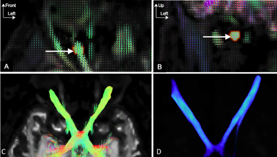 |
TractLearn: comparison with the General Linear Model for optic pathways exploration
Clement Jean1, Arnaud Attyé1,2, Alexandre Krainik1, Sylvie Grand1, Christophe Chiquet3, Olivier Casez4, Laurent Lamalle5, Felix Renard6, and Fernando Calamante2,7
1Neuroradiology, Grenoble University Hospital, Grenoble, France, 2School of Biomedical Engineering, The University of Sydney, Sydney, Australia, 3Ophthalmology, Grenoble University Hospital, Grenoble, France, 4Neurology, Grenoble University Hospital, Grenoble, France, 5IRMaGe, Inserm US 17, CNRS UMS 3552, Grenoble, France, 6Pixyl Medical, Grenoble, France, 7Sydney Imaging, Sydney, Australia
TractLearn was recently proposed for tract-based MRI quantitative analyses, based on Riemannian distances between anatomical structures. It allows to detect joint quantitative variations in a group of voxels, and in theory to decrease the number of false negatives compared with the General Linear Model (GLM). TractLearn also takes advantage of a manifold approach to capture controls variability as standard reference. Here we aim at comparing the performance of TractLearn with the GLM in detecting optic nerve voxel alteration, using the side of visual impairment as clinical reference.
|
|||
4320.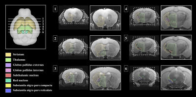 |
Tractography analysis of dopamine transporter genetic mouse models in Parkinson's disease
Sang-Jin Im1 and Hyeon-Man Baek1
1Lee Gil Ya Cancer and Diabetes Institute, Incheon, Korea, Republic of
Parkinson's disease is characterized by degeneration of dopaminergic nigrostriatal neurons with dysfunctional cortico–striatal–thalamic loops mainly in the basal ganglia. However, Parkinson’s disease studies on the neural connections between brain structural regions have not reached a clear consensus on how Parkinson’s disease effects the mouse brain. In this study, probabilistic tractography analysis was performed on important mouse brain structures related to Parkinson's disease mechanism, and pathways between each domain were visualized.
|
|||
| 4321. | The frontal aslant tract and its role in executive functions: a lesion-symptom study and awake electrical mapping study
Maud Landers1, Stephan Meesters2, Wouter de Baene3, and Geert-Jan Rutten1
1Department of Neurosurgery, Elisabeth TweeSteden Hospital, Tilburg, Netherlands, 2Department of mathematics and Computer Science, University of Technology, Eindhoven, Netherlands, 3Department of Cognitive Neuropsychology, University of Tilburg, Tilburg, Netherlands
Currently there is insufficient knowledge about the right frontal aslant tract’s exact cognitive importance. The aim of this study was to investigate the role of the right frontal aslant tract in executive functions via a lesion-symptom approach. The results suggest that the right frontal aslant tract is involved in shifting attention and phonemic fluency. As a follow-up the main findings were tested and validated in an awake mapping study where stimulation of the right frontal aslant tract lead to deficits in executive functioning.
|
The International Society for Magnetic Resonance in Medicine is accredited by the Accreditation Council for Continuing Medical Education to provide continuing medical education for physicians.