Digital Posters
Arterial Spin Labelling: Applications
ISMRM & SMRT Annual Meeting • 15-20 May 2021

Digital Posters
Arterial Spin Labelling: Applications
| Concurrent 4 | 13:00 - 14:00 |
2733. |
A quantitative multiparametric 18F-FPIA PET/MRI study for the characterization of primary brain gliomas
Marianna Inglese1, Shah Islam1, Matthew Grech-Sollars1,2, Giulio Anichini3, James Davies4, Azeem Saleem4,5, Matthew Williams6,7, Kevin S O'Neill3, Adam D Waldman8, and Eric O Aboagye1
1Surgery and Cancer, Imperial College London, London, United Kingdom, 2Imaging, Imperial College London Healthcare NHS Trust, London, United Kingdom, 3Imperial College London Healthcare NHS Trust, London, United Kingdom, 4Invicro Imperial College London, London, United Kingdom, 5Hull York Medical School, Faculty of Health Sciences, University of Hull, Hull, United Kingdom, 6Computational Oncology Group, Department of Surgery and Cancer, Imperial College London, London, United Kingdom, 7Institute for Global Health Innovation, Imperial College London, London, United Kingdom, 8Centre for Clinical Brain Sciences, University of Edinburgh, Edinburgh, United Kingdom 18F-FPIA PET/MRI integrates two imaging modalities that can provide valuable insight into the characterization and classification of brain tumours. 10 patients with primary brain gliomas were recruited to this study. Static and dynamic 18F-FPIA PET, together with perfusion/diffusion MRI data were post-processed for the extraction of 3D parametric maps for each subject. Correlations among parameters were evaluated with Spearman test. Tumour grade prediction was assessed with a machine learning model. A strong correlation was found between uptake and influx rate constant of FPIA and MRI perfusion parameters. The PET/MRI methodology provided 100% accuracy in differentiating low from high grade tumours. |
|||
2734. |
Identification of IDH1 mutation status in glioblastoma using multi-delay 3D arterial spin labeling perfusion MRI: a pilot study
Huilou Liang1,2, Lianwang Li3, Yuchao Liang3, Siqi Cai4, Jing An5, Yan Zhuo1,2,6, Lijuan Zhang4, Danny JJ Wang7, and Rong Xue1,2,8
1State Key Laboratory of Brain and Cognitive Science, Beijing MRI Center for Brain Research, Institute of Biophysics, Chinese Academy of Sciences, Beijing, China, 2University of Chinese Academy of Sciences, Beijing, China, 3Department of Neurosurgery, Beijing Tiantan Hospital of Capital Medical University, Beijing, China, 4Shenzhen Institutes of Advanced Technology, Chinese Academy of Sciences, Shenzhen, China, 5Siemens Shenzhen Magnetic Resonance Ltd, Shenzhen, China, 6CAS Center for Excellence in Brain Science and Intelligence Technology, Chinese Academy of Sciences, Beijing, China, 7Mark & Mary Stevens Neuroimaging and Informatics Institute, Keck School of Medicine, University of Southern California, Los Angeles, CA, United States, 8Beijing Institute for Brain Disorders, Beijing, China
Arterial spin labeling (ASL) perfusion MRI with single post-labeling delay (PLD) has been used to noninvasively predict the IDH1 mutation status in glioblastoma patients. However, single-delay ASL can make inaccurate estimations of cerebral blood flows (CBF) due to the variability of arterial transit times (ATT) among individuals. In this study, we applied multi-delay 3D ASL technique with multiple hemodynamic parameters including quantitative ATT, ATT-corrected CBF and arterial cerebral blood volume (aCBV) in glioblastoma. Our results show that aCBV-based relative perfusion parameters may provide a better identification of IDH1 mutation status and is worthy of further verification in future studies.
|
|||
2735. |
Intrasession reliability of arterial spin labeled MRI measured perfusion in GBM at 3T
Limin Zhou1, Yiming Wang1, Marco Da Cunha Pinho1,2, Edward Pan3,4,5, Yin Xi1,6, Joseph A Maldjian1,2, and Ananth J Madhuranthakam1,2
1Department of Radiology, University of Texas Southwestern Medical Center, Dallas, TX, United States, 2Advanced Imaging Research Center, University of Texas Southwestern Medical Center, Dallas, TX, United States, 3Department of Neurology and Neurotherapeutics, University of Texas Southwestern Medical Center, DALLAS, TX, United States, 4Department of Neurological Surgery, University of Texas Southwestern Medical Center, DALLAS, TX, United States, 5Harold C. Simmons Cancer Center, University of Texas Southwestern Medical Center, DALLAS, TX, United States, 6Department of Population and Data Sciences, University of Texas Southwestern Medical Center, Dallas, TX, United States
A 3D TSE using Cartesian acquisition with spiral profile reordering (CASPR) in combination with pseudo-continuous arterial spin labeling (pCASL) was developed to improve the robustness of ASL measured perfusion to increased B0 inhomogeneities in GBM patients. Previous results showed high intrasession reliability of this method in GBM patients. In this study, we compared the 3D TSE-CASPR measured perfusion with clinically available 3D GraSE at 3T. With 24 GBM imaging sessions, the results showed that 3D pCASL with TSE-CASPR is more robust to B0 inhomogeneities and has higher intrasession reliability than the clinical sequence, 3D pCASL with GraSE at 3T.
|
|||
2736. |
The feasibility of 3D pcASL MRI with Multiple post-labeling delay times in evaluating subtypes of parotid gland tumors
Lu Chen1, Guo-Yi Su1, Weiqiang Dou2, Yong Shen3, Fei-Yun Wu1, and Xiao-Quan Xu1
1Radiology, The First Affiliated Hospital of Nanjing Medical University, Nanjing, China, 2GE Healthcare, MR Research China, Beijing, P.R. China, Beijing, China, 3GE Healthcare, MR Enhanced Application China, Beijing, P.R. China, Beijing, China
In this study, we aimed to investigate 3D pcASL with multiple PLDs in evaluating subtypes of parotid gland tumors. By quantitatively measuring tumor blood flow (TBF) and comparing the values within and between subgroups, we found 3D pcASL with multiple PLDs can differentiate parotid gland tumors and reflect changes of TBF. With these findings, 3D pcASL MRI, especially with short PLD was suggested to evaluate patients with parotid gland tumors in routine clinical practice.
|
|||
2737.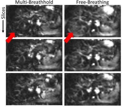 |
Robust Implementation of a 3D Pulsed ASL Sequence for Assessment of Liver Perfusion
Jörn Huber1, Daniel Hoinkiss1, and Matthias Günther1,2
1Fraunhofer MEVIS, Bremen, Germany, 2University of Bremen, Bremen, Germany
Liver perfusion can give valuable functional information about diseases like carcinoma and cirrhosis. Arterial Spin Labeling allows non-invasive assessment of liver perfusion without exogenous contrast agents being especially helpful in patients with renal failure. ASL in abdominal organs like liver faces increased challenges due to low perfusion rates, breathing motion and strong off-resonance as well as B1-inhomogeneity. In this work, a robust implementation of a pulsed ASL (PASL) sequence with FAIR labeling and 3D GRASE readout is provided, addressing these difficulties.
|
|||
2738.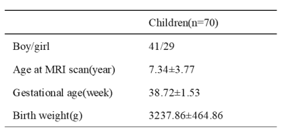 |
Depicting the developmental trajectories of brain cerebral blood flow using 3D ASL in children aged 28 days to 15 years.
Peiyao Chen1, Chao Jin1, Xianjun Li1, Miaomiao Wang1, Congcong Liu1, Xiaoyu Wang1, Fan Wu1, Yuli Zhang1, Cong Tian1, Mengxuan Li1, Xiaocheng Wei2, and Jian Yang1
1First Affiliated Hospital of Xi 'an Jiaotong University, Xi'an, Shaanxi, China, 2MR Research China, GE Healthcare, Beijing, China
Adequate cerebral blood flow (CBF) is essential for development of brain structure and function. However, little is known about the developmental trajectory of CBF across the broad age range in childhood. This study aims to investigate the spatiotemporal evolution of CBF in cerebral cortex and basal ganglia in healthy term children aged from 28 days to 15 years. Our results indicated that the estimated age with highest CBF was earliest in the occipital lobe, followed by temporal and parietal lobe, at last in the frontal lobe. The perfusion of basal ganglia showed a U-shaped curve, which slowly increased with age.
|
|||
2739.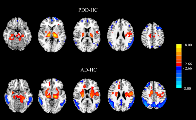 |
Brain perfusion in dementia with Parkinson's disease and Alzheimer’s disease: an arterial spin labeling MRI study
Hongri Chen1, Weiqiang Dou2, and Wei yin Liu2
1Dalian Medical University, Northern Jiangsu People’s Hospital, Yangzhou, China, Yangzhou, China, 2GE Healthcare, MR Research China, Beijing, P.R. China, Beijing, China
The present study was to explore regional perfusion changes in patients with Parkinson's disease dementia (PDD) and Alzheimer’s disease (AD) using 3D arterial spin labeling MRI and further assess the corresponding difference between PDD and AD patients. The results revealed that the perfusion pattern in PDD group was distinct from that in AD group despite of overlapped perfusion regions. The resultant normalized cerebral blood flow (CBF) can distinguish PDD from AD. Therefore, normalized CBF provided sensitive imaging-based markers that contribute to the diagnosis and differential diagnosis of the two dementia.
|
|||
 |
2740.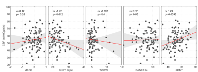 |
Relationship between global grey matter perfusion, damage and disability in multiple sclerosis
Daniele Mascali1, Antonio Maria Chiarelli1, Ilona Lipp2,3, Anna Digiovanni4, Valentina Tomassini1,3,4, and Richard Geoffrey Wise1,3
1Institute for Advanced Biomedical Technologies,Department of Neuroscience, Imaging and Clinical Sciences, "G. D'Annunzio University" of Chieti-Pescara, Chieti, Italy, 2Department of Neurophysics, Max Planck Institute for Human Cognitive and Brain Sciences, Leipzig, Germany, 3Cardiff University Brain Research Imaging Centre (CUBRIC) School of Psychology, Cardiff University, Cardiff, United Kingdom, 4MS Centre, Neurology Unit, SS. Annunziata University Hospital, Chieti, Italy
We investigated the relationship between grey matter perfusion and clinical and conventional MRI measures in patients with relapsing multiple sclerosis (MS) to test the hypothesis that an impaired energy supply is associated with the development of brain damage and clinical disability. Using multi-inversion time pulsed ASL we demonstrated that MS patients have significantly lower cerebral grey matter perfusion when compared to healthy controls. In the patients, lower perfusion correlates with greater tendency to develop irreversible tissue damage and with worse clinical scores, suggesting that altered energy supply may directly contribute to damage and disability in MS.
|
||
2741.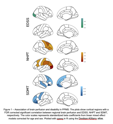 |
ASL perfusion and disability in primary progressive MS: an observational cohort study
Clara Delacour1, Ahmed-Ali El Ahmadi1, Gilles Brun1, Nadine Girard1,2, Christoph Heesen3,4, Arzu Ceylan Has3,4, and Jan-Patrick Stellmann2,3,4,5
1Neuroradiology, APHM La Timone, Marseille, France, 2Aix-Marseille Univ, CNRS, CRMBM, UMR 7339, Marseille, France, 3Institute of Neuroimmunology and MS (INIMS), University Medical Centre Hamburg-Eppendorf, Hamburg-Eppendorf, Germany, 4Neurology, University medical centre Hamburg-Eppendorf, Hamburg-Eppendorf, Germany, 5APHM La Timone, CEMEREM, Marseille, France
Disability progression in Multiple Sclerosis (MS) is driven by inflammation and neurodegeneration. Arterial spin labelling (ASL) is a non-invasive MRI method for the assessment of brain perfusion without the need for gadolinium. Here, we explored ASL perfusion as a biomarker for diseases progression and disability in a cohort of 77 patients with primary progressive MS (PPMS). While brain perfusion seemed rather stable during the follow-up of up to 5 years, we observed an association between higher regional perfusion rates and cognitive performance and hand functioning. Altered perfusion in PPMS seems thus not closely related to the major pathomechanism neurodegeneration.
|
|||
2742.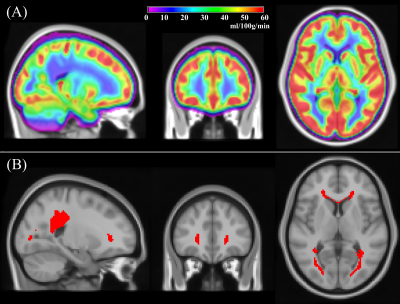 |
Reliability of Arterial Spin Labeling derived Cerebral Blood Flow measurements in Periventricular White Matter
Sudipto Dolui1, Audrey P. Fan2,3, Moss Y. Zhao3, Greg Zaharchuk3, and John A. Detre1,4
1Department of Radiology, University of Pennsylvania, Philadelphia, PA, United States, 2Departments of Biomedical Engineering and Neurology, University of California, Davis, CA, United States, 3Department of Radiology, Stanford University, Palo Alto, CA, United States, 4Department of Neurology, University of Pennsylvania, Philadelphia, PA, United States
Periventricular white matter (PVWM) is exclusively perfused by small vessels, and hence PVWM Cerebral Blood Flow (CBF) can provide a biomarker of cerebral small vessel functional integrity. Although PVWM is weakly perfused, we demonstrate that PVWM-CBF derived from state-of-the-art Arterial Spin Labeling (ASL) acquired twice ~15minutes apart in a cohort of 16 subjects showed high reproducibility that was comparable to other larger regions of interest. Further there was high correlation between PVWM-CBF derived from ASL and concurrently acquired [15O]-water Positron Emission Tomography data. These findings suggest that PVWM CBF can be reliably measured with advanced ASL methods.
|
|||
2743.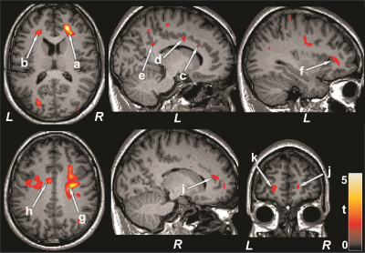 |
Reduced Cerebral Blood Flow in Patients with Pulmonary Arterial Hypertension
Bhaswati Roy1, Susana Vacas1, Kathy McCloy2, Rajan Saggar2, and Rajesh Kumar1,3,4,5
1Anesthesiology, University of California Los Angeles, Los Angeles, CA, United States, 2Medicine, University of California Los Angeles, Los Angeles, CA, United States, 3Bioengineering, University of California Los Angeles, Los Angeles, CA, United States, 4Radiological Sciences, University of California Los Angeles, Los Angeles, CA, United States, 5Brain Research Institute, University of California Los Angeles, Los Angeles, CA, United States
Pulmonary arterial hypertension (PAH) patients show cognitive and mood impairments, and brain tissue injury in those areas. However, the underlying cause of tissue damage in PAH patients remain unclear. Altered cerebral blood flow (CBF) may contribute to develop brain tissue injury and cognitive and mood deficits. We evaluated CBF in PAH patients over controls, and found changes in the prefrontal cortices, insula, cingulate, frontal cortex, corona radiate, temporal, occipital, and parietal gyrus. Significant correlations also emerged between CBF and functional deficits in PAH, including mood and cognition symptoms, implying altered hemodynamic contributing to brain changes and functional deficits.
|
|||
2744.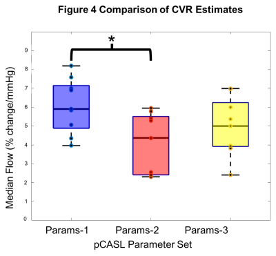 |
Moving Towards Robust Quantification of Cerebrovascular Reactivity (CVR) using Pseudocontinuous Arterial Spin Labelling (pCASL)
Colette C. Milbourn1, Thomas W. Okell2, and Nicholas P. Blockley1
1The School of Life Sciences, University of Nottingham, Nottingham, United Kingdom, 2Wellcome Centre for Integrative Neuroimaging, FMRIB, Nuffield Department of Clinical Neurosciences, University of Oxford, Oxford, United Kingdom
New clinical tools are needed for the diagnosis and prognosis of cerebrovascular diseases. Pseudocontinuous Arterial Spin Labelling (pCASL) is recommended for quantifying tissue cerebral blood flow due to its superior signal-to-noise. Stressors to the brain like high carbon dioxide (hypercapnia), used to map Cerebrovascular Reactivity (CVR), are known to reduce the labelling efficiency of pCASL and therefore underestimate CVR. In this study a systematic difference in CVR was observed across measurements made with three different sets of pCASL parameters. Ultimately this work will help to determine pCASL parameters that minimise systematic error and produce robust quantitative estimates of CVR.
|
|||
2745.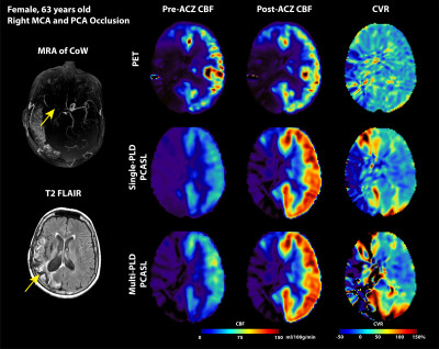 |
Arterial Spin Labeling Can Identify Cerebrovascular Reactivity Deficit in Patients with Vasculopathy: A Pilot Study Using Simultaneous PET/MRI
Moss Y Zhao1, Audrey P Fan2, David Chen3,4, Jia Guo5, Yosuke Ishii6, David Shin7, Mohammad Mehdi Khalighi1, Dawn Holley1, Kim Halbert1, Andrea Otte1, Brittney Williams1, Jun-Hyung Park1, Bin Shen1, Gary Steinberg8,
and Greg Zaharchuk1
1Radiology, Stanford University, Stanford, CA, United States, 2Biomedical Engineering and Neurology, University of California Davis, Davis, CA, United States, 3Medical Imaging, Taipei Medical University – Shuan-Ho Hospital, New Taipei City, Taiwan, 4Department of Radiology, School of Medicine, College of Medicine, Taipei Medical University, Taipei, Taiwan, 5Bioengineering, University of California Riverside, Riverside, CA, United States, 6Neurosurgery, Tokyo Medical and Dental University, Tokyo, Japan, 7GE Healthcare, Melo Park, CA, United States, 8Neurosurgery, Stanford University, Stanford, CA, United States
Cerebrovascular reactivity (CVR) reflects the capacity of the brain to respond to external stress. Impaired CVR leads to a higher risk of stroke. The standard CVR measuring tool relied on PET imaging but is inaccessible to most patients. Here we investigate the CVR of Moyamoya patients using arterial spin labeling (ASL), a non-invasive and quantitative MRI technique. Results showed that significant lower CVR was found in flow territories with severe stenosis or occlusion, making ASL a potential diagnostic tool to predict the risk of stroke for patients with vasculopathy.
|
|||
2746.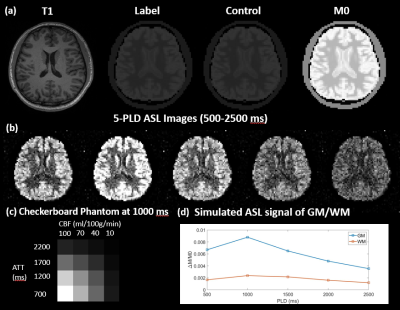 |
A digital brain perfusion phantom for validation of ASL data post-processing software
Chenyang Zhao1, Ze Wang2, and Danny Wang1
1Laboratory of Functional MRI Technology (LOFT), Mark & Mary Stevens Neuroimaging and Informatics Institute, University of Southern California, Los Angeles, CA, United States, 2Department of Diagnostic Radiology and Nuclear Medicine, School of Medicine, University of Maryland, Baltimore, MD, United States
A digital brain perfusion phantom was designed using a representative 5-delay 3D pCASL protocol and applied to validate the quantification accuracy of 4 ASL post-processing software packages, including ASLtbx, ASL_MRICloud, BASIL, and Cereflow. The 4 tested software can produce generally correct quantification results under normal SNR conditions, however their accuracy may be affected by the limitation occurred in low perfusion regions and low SNR condition. The digital phantom provides a reliable reference with high flexibility to validate ASL post-processing software.
|
|||
2747.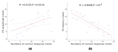 |
Association of Arterial Spin Labeling Global Metrics and Attentional Processes
Shichun Chen1, Yakun Zhang1, Zongpai Zhang1, Wenna Duan1, George Weinschenk1, Brandon E. Gibb2, Wenming Luh3, and Weiying Dai1
1Department of Computer Science, State University of New York at Binghamton, Binghamton, NY, United States, 2Department of Psychology, State University of New York at Binghamton, Binghamton, NY, United States, 3National Institute on Aging, National Institutes of Health, Baltimore, MD, United States
We investigated the relationship between brain global activity or global topology of brain networks and attentional processes. The former measures were evaluated by dynamic arterial spin labeling (ASL) perfusion fMRI. The latter measures were assessed by P3 amplitude and latency in event-related potentials (ERPs). The P3 properties were significantly correlated with numbers of correct response. We found positive associations between global perfusion and P3 amplitude and between characteristic path length and P3 latency, and negative associations between global perfusion, global efficiency, mean functional connectivity and P3 latency, indicating close correlation of brain global behavior and attention.
|
|||
2748.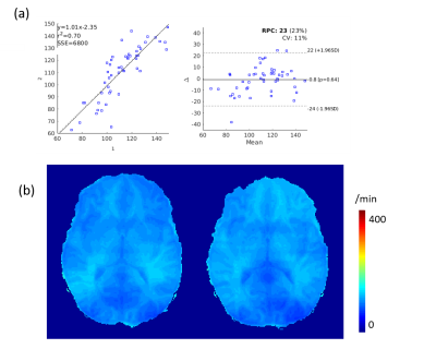 |
Mapping water exchange rate change after caffeine uptake using 3D diffusion prepared arterial spin labeled perfusion MRI
Qihao Zhang1, Jana Ivanidze2, Thanh Nguyen2, Pascal Spincemaille2, and Yi Wang1
1Cornell University, New York, NY, United States, 2Weill Cornell Medical College, New York, NY, United States
We propose to exam the water exchange rate (kw) change after caffeine uptake based on an optimized diffusion prepared arterial spin labeling (ASL) sequence and reconstruction method in a previous work1. 5 subjects went through two scans to test the reproducibility of the reconstruction pipeline. Another 5 subjects are scanned before and half an hour after caffeine uptake. Cerebral blood flow (CBF) and are reconstructed from the ASL images. A 26% decrease in CBF and 13% decrease of kw is observed.
|
|||
2749.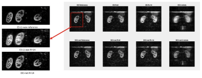 |
High spatio-temporal resolution 3D ASL renal perfusion with variable-density FSE and deep-learning reconstruction
Manuel Taso1, Uri Wollner2, Arnaud Guidon3, Rafi Barda2, Christopher J Hardy4, Sangtae Ahn4, and David C Alsop1
1Division of MRI research, Department of Radiology, Beth Israel Deaconess Medical Center, Harvard Medical School, Boston, MA, United States, 2GE Research, Herzliya, Israel, 3Global MR Applications and Workflow, GE Healthcare, Boston, MA, United States, 4GE Research, Niskayuna, NY, United States
Arterial spin labeling (ASL) has proven to be a powerful research and clinical technique for functional imaging of tissues. The combination of undersampled acquisitions and compressed sensing reconstruction shows promise for increased speed, resolution, and robustness but conventional CS reconstructions are slow and may not be satisfactory, especially for low SNR data. This work explores the feasibility and performance of Deep-Learning based reconstruction of similarly sampled data to realize the full potential of these ASL acquisitions using DCI-net, an unenrolled iterative CS network. We show DCI-net performance at high acceleration rates and potential for fast volumetric ASL perfusion imaging.
|
|||
2750.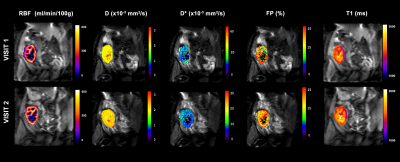 |
Reproducibility of multiparametric MRI in transplanted kidneys
Rebeca Echeverria-Chasco1,2, Marta Vidorreta3, Veronica Aramendia-Vidaurreta2,4, David Cano 1, Gorka Bastarrika2,4, Nuria Garcia-Fernandez2,5, Paloma L. Martin Moreno2,5, and Maria A. Fernandez-Seara2,4
1Radiology, Clínica Universidad de Navarra, Pamplona, NE, Spain, 2IdiSNA, Instituto de Investigación Sanitaria de Navarra, Pamplona, Spain, 3Siemens Healthineers, Madrid, Spain, 4Radiology, Clínica Universidad de Navarra, Pamplona, Spain, 5Nephrology, Clínica Universidad de Navarra, Pamplona, Spain The goals of this work were to test a multiparametric MRI protocol to measure perfusion, diffusion and T1 and to assess inter-session reproducibility in a group of transplanted patients with stable renal function. 18 patients were imaged in 2 exams employing pseudo-continuous arterial spin labeling technique, intravoxel incoherent motion technique and T1 mapping sequence. Reproducibility was assessed using Bland-Altman analysis, within-subject coefficient of variation (CVws) and intra-class correlation coefficient (ICC). Results of the 3 techniques were promising showing a CVws <20% in most of the cases (especially for T1 and D (<6%)) and high ICC coefficients for PCASL and T1. |
|||
2751.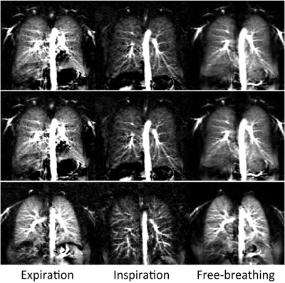 |
Measurement of Pulmonary Perfusion under Expiratory and Inspiratory Breathing Conditions using PCASL-bSSFP Imaging at 1.5 Tesla
Petros Martirosian1, Rolf Pohmann2, Martin Schwartz1,3, Thomas Kuestner4, Manuel Kolb4, Ahmed Othman4, Cecilia Zhang4, Klaus Scheffler2,5, Konstantin Nikolaou4, Fritz Schick1, and Ferdinand Seith4
1Section on Experimental Radiology, University of Tübingen, Tübingen, Germany, 2Max Planck Institute for Biological Cybernetics, Tübingen, Germany, 3Institute of Signal Processing and System Theory, University of Stuttgart, Stuttgart, Germany, 4Department of Diagnostic and Interventional Radiology, University of Tübingen, Tübingen, Germany, 5Department of Biomedical Magnetic Resonance, University of Tübingen, Tübingen, Germany
Pseudo-continuous-arterial-spin-labeling (PCASL) has been successfully applied in the lung providing high quality perfusion images. The pulmonary blood flow and the respiratory system interact closely: the intrathoracic pressure has impact on the venous return. Therefore, in this work, we evaluate the effects of intrathoracic pressure on lung perfusion by using PCASL imaging in end-expiratory and end-inspiratory breath-hold. PCASL imaging is able to detect changes of parenchymal lung perfusion caused by alterations of the intrathoracic pressure. Perfusion signal measured under end-inspiratory condition were noticeably reduced as compared to end-expiratory breath-hold. This correlated significantly with measured blood flow volume through the pulmonary trunk.
|
|||
2752.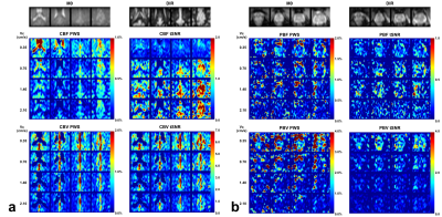 |
Prostate Perfusion Mapping using Fourier-Transform based Velocity-Selective Pulse Trains: Choice of Cutoff Velocity and Comparison with Brain
Dapeng Liu1,2, Dan Zhu3, Wenbo Li1,2, and Qin Qin1,2
1Department of Radiology, Johns Hopkins University School of Medicine, Baltimore, MD, United States, 2F.M. Kirby Research Center for Functional Brain Imaging, Kennedy Krieger Institute, Baltimore, MD, United States, 3Department of Biomedical Engineering, Johns Hopkins University School of Medicine, Baltimore, MD, United States
Prostate perfusion mapping using velocity-selective arterial spin labeling (VSASL) is desired for higher SNR and insensitivity to arterial transit delay. The choice of cutoff velocity (Vc) determines its sensitivity to perfusion-weighted signal (PWS). The utility of Fourier-transform based velocity-selective (FT-VS) pulse trains have been demonstrated in cerebral blood flow and cerebral blood volume mapping. In this study, FT-VS prepared blood flow and blood volume mapping sequences were performed in parallel in both brain and prostate to investigate their Vc dependence to PWS. The results suggest that lower Vc is demanded for prostate VSASL.
|
The International Society for Magnetic Resonance in Medicine is accredited by the Accreditation Council for Continuing Medical Education to provide continuing medical education for physicians.