Digital Posters
Flow, Volume & Permeability: DSC-MRI & Non-Contrast
ISMRM & SMRT Annual Meeting • 15-20 May 2021

Digital Posters
Flow, Volume & Permeability: DSC-MRI & Non-Contrast
| Concurrent 6 | 13:00 - 14:00 |
1073.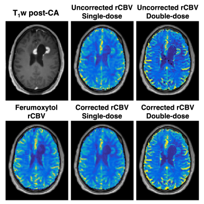 |
Comparison of Gadolinium- and Iron-Oxide-based Perfusion Imaging Metrics in Glioblastoma
Ashley M. Stokes1, Sudarshan Ragunathan1, Laura Bell1, Ekokobe Fonkem2, John Karis3, and C. Chad Quarles1
1Neuroimaging Research, Barrow Neurological Institute, Phoenix, AZ, United States, 2Neuro-Oncology, Barrow Neurological Institute, Phoenix, AZ, United States, 3Neuroradiology, Barrow Neurological Institute, Phoenix, AZ, United States
Relative cerebral blood volume (rCBV) maps are adversely impacted by contrast agent leakage effects when acquired using gadolinium-based contrast agents, and these effects can be corrected using standard leakage correction methods. In a cohort of high-grade glioma patients, we compared leakage-corrected rCBV maps acquired using gadolinium-based contrast agents to rCBV acquired using an intravascular iron-oxide-based contrast agent. The rCBV maps from both contrast agents were similar, with high voxel-wise agreement, suggesting that leakage effects are appropriately removed from rCBV maps using standard leakage correction methods.
|
|||
1074.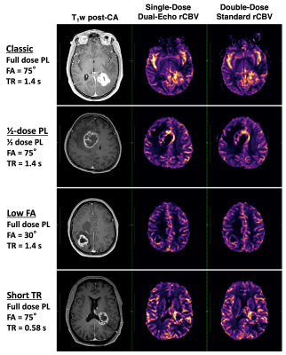 |
Do You Double-Dose? Clinical Validation of Single-Dose, Dual-Echo Perfusion Protocols in Glioma Patients
Ashley M. Stokes1, Taylor Olvey1, Leland S. Hu2, Leslie C. Baxter2, Laura C. Bell1, C. Chad Quarles1, and Lea Alhilali3
1Neuroimaging Research, Barrow Neurological Institute, Phoenix, AZ, United States, 2Mayo Clinic Arizona, Phoenix, AZ, United States, 3Neuroradiology, Barrow Neurological Institute, Phoenix, AZ, United States
Relative cerebral blood volume (rCBV) obtained from dynamic susceptibility contrast (DSC) MRI is adversely impacted by contrast agent leakage effects in enhancing brain tumors, often necessitating multiple contrast doses. The purpose of this study is to compare dual-echo rCBV maps (single-dose) with standard rCBV maps (double-dose) in patients with high-grade gliomas using commercially available software. High agreement was observed between rCBV maps with single- and double-doses, with substantial flexibility across pulse sequence parameters. This protocol could be used to lower costs and contrast dose in clinical settings, as well as eliminate variability in perfusion methods related to contrast timing schemes.
|
|||
1075.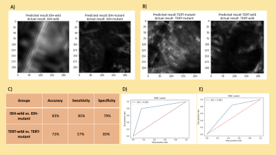 |
Identification of IDH and TERT Mutation Status in Glioma Patients using Dynamic Susceptibility Contrast MRI
Buse Buz Yalug1, Ayca Ersen Danyeli2,3, Cengiz Yakicier3,4, M. Necmettin Pamir3,5, Koray Ozduman3,5, Alp Dincer3,6, and Esin Ozturk-Isik1,3
1Institute of Biomedical Engineering, Bogazici University, Istanbul, Turkey, 2Department of Medical Pathology, Acibadem Mehmet Ali Aydinlar University, Istanbul, Turkey, 3Center for Neuroradiological Applications and Reseach, Acibadem Mehmet Ali Aydinlar University, Istanbul, Turkey, 4Department of Molecular Biology and Genetics, Acibadem Mehmet Ali Aydinlar University, Istanbul, Turkey, 5Department of Neurosurgery, Acibadem Mehmet Ali Aydinlar University, Istanbul, Turkey, 6Department of Radiology, Acibadem Mehmet Ali Aydinlar University, Istanbul, Turkey
The main purpose of this study was to identify isocitrate dehydrogenase (IDH) and telomerase reverse transcriptase (TERT) promoter mutations in glioma patients using classical machine learning and convolutional neural networks (CNN) on dynamic susceptibility contrast MRI (DSC-MRI). Relative cerebral blood volume (rCBV) maps of glioma patients with different genotypes including IDH-mutant, IDH-wildtype, TERT-mutant, and TERT-wildtype were compared in tumor areas. Classical machine learning classification results were over 85% for both IDH and TERT mutations. On the other hand, CNNs were able to classify IDH mutation status with 83% and TERT mutation status with 72% accuracies.
|
|||
1076.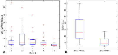 |
Application of Multiband SENSE SAGE EPI towards DSC MRI of Brain Tumors
Sudarshan Ragunathan1, Laura C Bell1, Ashley M Stokes1, Ekokobe Fonkem1, John P Karis1, and C Chad Quarles1
1Barrow Neurological Institute, Phoenix, AZ, United States
Multi-echo acquisition strategies have the advantage of simultaneous quantification of T1/T2* values in DCR MRI, and when combined with multiband strategies, could provide comparable spatiotemporal coverage as a single shot EPI dynamic scan. While there has been some work on SMS – SAGE acquisitions, this is potentially the first work that utilizes a SENSE reconstruction framework. We demonstrated that the CNR of MBSAGE with SENSE is similar to SAGE acquisitions, and that the dynamic CNR with respect to NAWM is on the order of or slightly higher than reported by some of the SMS-SAGE implementations in literature.
|
|||
 |
1077.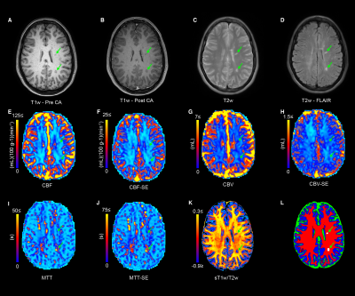 |
Investigating Relationships Between Multi-Scale Perfusion and Myelin Integrity in Patients with Relapsing-Remitting MS
Nicholas J Sisco1, Aimee Borazanci1, Richard Dortch1, and Ashley M Stokes1
1Neuroimaging Research, Barrow Neuroimaging Innovation Center, Phoenix, AZ, United States
The objective of this study is to develop magnetic resonance imaging (MRI) biomarkers that can probe complementary vascular function and myelin content. The development of biomarker assays to quantitatively probe both perfusion and myelin content is critical to assessing acute inflammatory activity and regenerative potential. We anticipate that this biomarker will give insight into the underlying pathophysiology of reversible and irreversible myelin damage.
|
||
1078. |
The relative cerebral blood volume in normal-appearing white and grey matter remains almost constant following radio(chemo)therapy
Katharina Witzmann1,2, Felix Raschke1,2, Tim Wesemann3, Mechthild Krause1,2,4,5,6, Jennifer Linn3, and Esther G.C. Troost1,2,4,5,6
1Helmholtz-Zentrum Dresden-Rossendorf, Institute of Radiooncology – OncoRay, Dresden, Germany, 2OncoRay - National Center for Radiation Research in Oncology, Faculty of Medicine and University Hospital Carl Gustav Carus, Technische Universität Dresden, Helmholtz-Zentrum Dresden-Rossendorf, Dresden, Germany, 3Institute of Neuroradiology, University Hospital Carl Gustav Carus and Medical Faculty of Technische Universität Dresden, Dresden, Germany, 4Department of Radiotherapy and Radiation Oncology, Faculty of Medicine and University Hospital Carl Gustav Carus, Technische Universität Dresden, Dresden, Germany, 5National Center for Tumor Diseases (NCT), Partner Site Dresden, Germany: German Cancer Research Center (DKFZ), Heidelberg, Germany; Faculty of Medicine and University Hospital Carl Gustav Carus, Technische Universität Dresden, Dresden, Germany, and; Helmholtz Association / Helmholtz-Zentrum Dresden - Rossendorf (HZDR), Dresden, Germany, 6German Cancer Consortium (DKTK), Partner Site Dresden, and German Cancer Research Center (DKFZ), Heidelberg, Germany
In adjuvant radio(chemo)therapy of glioma patients, irradiation of tumor-surrounding normal tissue is unavoidable, potentially leading to long-term side-effects. We analyzed radiation-induced perfusion changes measured as relative cerebral blood volume (rCBV) in supraventricular grey and white matter regions of 17 glioma patients before and 3, 6 and 9 months after radiotherapy. After treatment, perfusion remained constant in the entire regions and after dose-separation, except for a statistically significant rCBV decrease in low-dose white matter volumes 6 months after the end of radiotherapy (p=0.008) and a trend towards increasing white and grey matter perfusion in high-dose volumes at 9 months (p<0.1).
|
|||
1079. |
Aberrant cerebral perfusion pattern in amnestic mild cognitive impairment and Parkinson’s disease with mild cognitive impairment
Song'an Shang1, Weiqiang Dou2, and Jingtao Wu3
1Nanjing First Hospital, Nanjing Medical University, Nanjing, China, 2GE Healthcare, MR Research China, Beijing, China, 3Northern Jiangsu People’s Hospital, Yangzhou, China
We investigated the aberrations in regional perfusion properties among amnestic mild cognitive impairment (aMCI) patients, patients with Parkinson’s disease with MCI (PD-MCI), and healthy control (HC) by using 3D arterial spin labeling (ASL) imaging. Our results showed that normalized cerebral blood flow (CBF) as measured by 3D ASL revealed different patterns of perfusion between aMCI and PD-MCI, probably linked to distinct neural mechanisms. Therefore, this preliminary study demonstrates that normalized CBF might provide specific perfusion information for further pathological and neuropsychological studies.
|
|||
1080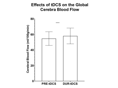 |
Simultaneous Effects of tDCS on Cerebral Blood Flow and Metabolic Response in Healthy Controls Video Permission Withheld
Marco Muccio, MSc1, Lillian Walton Masters1, Giuseppina Pilloni, PhD1, Lauren Krupp, MD1, Abhishek Datta, PhD2, Marom Bikson, PhD3, Leigh Charvet, PhD1, and Yulin Ge, MD1
1Department of Radiology, NYU Grossman School of Medicine, New York City, NY, United States, 2Soterix Medical Inc., New York City, NY, United States, 3Department of Biomedical Engineering, City College of New York, New York City, NY, United States
The underlying biological mechanisms of transcranial direct current stimulation (tDCS) remain relatively uncharacterized. In this report, MRI measures for cerebral blood flow (CBF) and cerebral metabolic rate of oxygen (CMRO2) of healthy controls was acquired before and during tDCS to better understand real-time neuronal response. We observed a significant increase in global CBF suggesting an increase of neuronal activity during compared to before tDCS. CMRO2 showed an increase of ~7% during tDCS compared to before tDCS. We suggest that the reported cortical excitability increase may also serve as a potential biomarker for neuronal reactivity or plasticity in many neurodegenerative diseases.
|
|||
|
1081.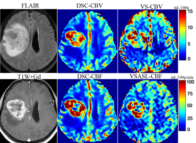 |
Evaluation of Brain Glioma Using Non-Contrast Cerebral Blood Volume Mapping: Correlations between Vascular proliferation and Perfusion MR
Yaoming Qu1, Qin Qin1,2, Haitao Wen1, Xiaochan Ou1, Yingjie Mei3, Weibo Chen4, and Zhibo Wen1
1Radiology, Zhujiang hospital of southern medical university, Guangzhou, China, 2F.M. Kirby Research Center for Functional Brain Imaging, Kennedy Krieger Institute, Baltimore, MD, United States, 3Philips Healthcare, Guangzhou, China, 4Philips Healthcare, Shanghai, China
Cerebral blood volume (CBV) mapping employing velocity-selective saturation pulse trains is an emerging velocity-selective arterial spin labeling (VSASL) based method for quantifying perfusion with higher SNR. Its utility was assessed for glioma patients at 3T and corelation between histopathologic vascular proliferation and perfusion MR Imaging. VSASL based CBV mapping, in good agreement with DSC-PWI and VSASL based cerebral blood flow (CBF) mapping, showed great promise for accurate quantitative assessment and preoperative grading of brain gliomas, and further potential in the IDH genotype and vascular proliferation status predicting.
|
||
1082.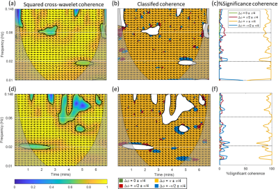 |
Characterization of Microcirculatory Regulation in Skeletal Muscle using Wavelet Coherence Analysis of Resting-State BOLD MRI
Jingting Yao1, Benjamin Risk1, Marijn Brummer1, Adam Daniel Singer1, Jeanie Park1, and David Reiter1
1Emory University, Atlanta, GA, United States
Microcirculatory regulation in the musculoskeletal system ensures tissue oxygenation and nutrient supply that are essential for maintaining normal muscular functions. BOLD MRI-derived mapping indices M0 and T2* have previously been established as markers of blood volume and blood oxygenation in healthy subjects. This study assesses dynamic resting-state microcirculation in the calf of healthy subjects using the wavelet coherence analysis, a time-frequency approach. Quantitative wavelet-based metrics characterizing the dynamic relationship between M0 and T2* may serve as markers of blood perfusion control and useful for characterizing degraded peripheral microvascular control in diseased population.
|
|||
1083.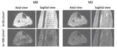 |
Evaluating the Effects of Motion Compensation to IVIM Fitting in In-Vivo DW-MRI of the Muscle
Jaume Coll-Font1,2,3, Or Perlman2,3, Shi Chen1, Robert A Eder1, Christian T. Farrar2,3, and Christopher T. Nguyen1,2,3
1Cardiovascular Research Center, Cardiology Division, Massachusetts General Hospital, Charlestown, MA, United States, 2Athinoula A. Martinos Center for Biomedical Imaging, Charlestown, MA, United States, 3Harvard Medical School, Boston, MA, United States
In order to acquire diffusion-weighted images (DWI) of the heart, motion compensated (M2) techniques are needed. However, these reduce the sensitivity to perfusion (D*) and hence hamper the accurate estimation of IVIM parameters. Here, we evaluate the effects of M2 to the IVIM estimates in the lower leg muscle and assess feasibility of an approach to combine DWI acquired with and without M2 to fit IVIM models. Our results show that M2 underestimates D* in the IVIM model. On the other hand, the combined approach resulted in IVIM parameter estimates similar to those obtained with standard DW MR.
|
|||
1084.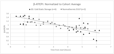 |
Comparing Preservation of Ex-Vivo Lungs for Transplantation, Using 31P MRS: Extended Ex-Vivo Lung Perfusion and Cold Static Storage
Jonathan Snow1, Mehrdad Pourfathi1, Ian Duncan1, Harrilla Profka1, Stephen Kadlecek1, Yi Xin1, Mostafa Ismail1, Sarmad Siddiqui1, Luis Loza1, Tahmina Achekzai1, Xiaoling Jin1, Hooman Hamedani1, Faraz Amzajerdian1, Federico
Sertic1, Kai Ruppert1, Ryan Baron1, Gabriel Unger1, Maurizio Cereda1, Shampa Chatterjee1, and Rahim Rizi1
1University of Pennsylvania, Philadelphia, PA, United States
Due to a shortage of transplantable lungs, careful preservation of viable donor lungs is of paramount importance. Ex-Vivo Lung Perfusion (EVLP) and cold static storage are two clinical techniques used to preserve donor lungs prior to transplantation. In this study, magnetic resonance spectroscopy was used to compare the ability of un-ventilated normothermic EVLP vs. cold static storage to preserve rat lungs’ ATP energy status over a 5-hour ischemic period. The EVLP model was slightly better than cold static storage at sustaining the lungs’ ATP.
|
|||
1085.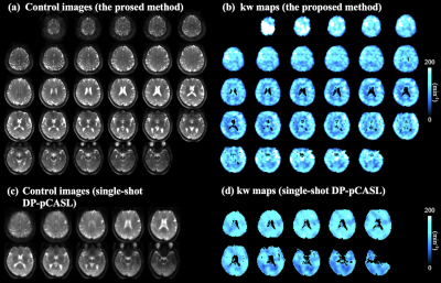 |
High resolution BBB water exchange rate mapping with multi-delay diffusion prepared pseudo-continuous arterial spin labeling
Xingfeng Shao1 and Danny JJ Wang1,2
1Laboratory of FMRI Technology (LOFT), Mark & Mary Stevens Neuroimaging and Informatics Institute, Keck School of Medicine, University of Southern California, Los Angeles, CA, United States, 2Department of Neurology, University of Southern California, Los Angeles, CA, United States
A segmented 3D diffusion prepared pseudo-continuous arterial spin labeling (DP-pCASL) sequence is proposed to acquire high resolution whole brain map of water exchange rate (kw) across the blood-brain barrier. And a new acquisition protocol is proposed which acquires DP-pCASL signals at multiple long post-labeling delays (PLDs) and allows kw to be quantified with a robust regression step and without the need for signal division or additional physiological parameter as inputs.
|
|||
1086.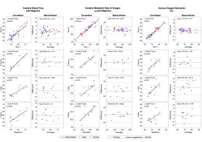 |
Short-term reproducibility of cerebral metabolism using magnetic resonance imaging
Ulrich Lindberg1, Signe Sloth Madsen2, Karsten Skovgaard Olsen2, Kirsten Møller2,3, Mohammad Sohail Asghar2, Henrik Bo Wiberg Larsson1,3, and Mark Bitsch Vestergaard1
1Department of Clinical Physiology, Nuclear Medicine and PET, Rigshospitalet, Copenhagen, Denmark, 2Department of Neuroanaesthesiology, Rigshospitalet, Copenhagen, Denmark, 3Department of Clinical Medicine, University of Copenhagen, Copenhagen, Denmark
The aim of this study was to investigate the short-term physiological variation in global cerebral perfusion, oxygen consumption and metabolite concentration, measured by MRI. Global cerebral oxygen consumption as well as N-acetyl-aspartate concentration remain stable over a one-week period. Global cerebral perfusion and lactate concentration was correlated, though showed decreasing within-subject correlation with increasing delay between scan session during the one-week follow-up period. These results should be carefully considered when performing longitudinal studies. In conclusion MRI does provide robust reliable measurements of global cerebral metabolism.
|
|||
1087.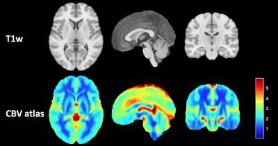 |
The use of a DSC-MRI perfusion atlas for cerebral blood volume normalization and its impact in improving prognostic estimation.
Elies Fuster-Garcia1, Javier Juan Albarracín2, Ivar T Hovden3, Maria del Mar Ávarez-Torres2, Alex Rovira4, Laura Oleaga5, Antonio J. Revert6, Silvano Filice7, Juan Miguel García-Gómez2, and Kyrre Eeg Emblem8
1Diagnostic Phyisics, Oslo University Hospital, Oslo, Norway, 2Universitat Politècnica de València, València, Spain, 3Oslo University Hospital, Olso, Norway, 4Hospital Universitari Vall d'Hebron, Barcelona, Spain, 5Hospital Clínic, Barcelona, Spain, 6Hospital de Manises, Manises, Spain, 7Azienda Ospedaliero-Universitaria di Parma, Parma, Italy, 8Diagnostic Physics, Oslo University Hospital, Oslo, Norway
In this work we present a method to improve the normalization of CBV maps by using a CBV atlas of the brain. This CBV atlas was generated using 134 non-linearly registered CBV maps from NCT03439332 clinical study. The proposed normalization method generates CBV values that better correlate with the patients’ OS, reaching smaller more significant p-values (p=0.034) based on Cox regression analysis, that better differentiate between high and low survivors (AUC=0.60, p=0.007) based on Kaplan Meier analysis.
|
|||
1088.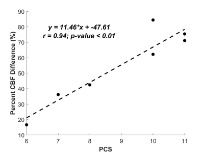 |
Prediction of Pial Collaterals using Delay and Dispersion Corrected MR Perfusion in Ischemic Stroke
Yong Ik Jeong1, Gregory Christoforidis1, Niloufar Saadat1, Steven Roth2, Marek Niekrasz1, and Timothy Carroll1
1University of Chicago, Chicago, IL, United States, 2University of Illinois College of Medicine, Chicago, IL, United States
After the onset of an ischemic stroke, blood flow may be restored to the affected territory via pial collateral blood vessels. In this study we investigate whether delay and dispersion corrected MR DSC perfusion can accurately measure the additional blood flow to estimate pial collaterals. The percent difference between non-corrected and corrected CBF is compared against measured pial collateral scores. The percent difference in CBF is found to be predictive of pial collateral scores. With more research, corrected MR DSC CBF may be used in the clinical setting for stroke patient management.
|
|||
1089.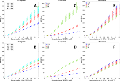 |
Dependency of R2(*) Relaxation on Gd-DTPA Concentration in Arterial Blood: Influence of Hematocrit and Magnetic Field Strength
Daniëlle van Dorth1, Krishnapriya Venugopal2, Dirk H. J. Poot2, Marion Smits2, Jeroen H. J. M. de Bresser3, Juan A. Hernandez-Tamames2, and Matthias J. P. van Osch1
1C. J. Gorter Center for High-Field MRI, Radiology, Leiden University Medical Center, Leiden, Netherlands, 2Radiology and Nuclear Medicine, Erasmus University Medical Center, Rotterdam, Netherlands, 3Radiology, Leiden University Medical Center, Leiden, Netherlands
In quantitative dynamic susceptibility contrast (DSC) MRI measurement of the arterial input function (AIF) is required. Generally, a linear relation between the R2(*) relaxation and the arterial contrast agent concentration is assumed (or the AIF is measured outside of an artery), although this is invalid for gradient-echo. In this study an open-source simulation tool is adapted to determine how R2(*) depends on contrast concentration for different physiological and MR parameters and this tool is validated by previously acquired in vitro data. The results show that sequence type (gradient-echo versus spin-echo), hematocrit and field strength all affect the relation.
|
|||
1090. |
Comparison of the arterial-voxel discriminatory power of DSC-MRI signal features and creation of a framework for determining optimal thresholds
Rashed Sobhan1, Donnie Cameron1,2, and Glyn Johnson1
1Norwich Medical School, University of East Anglia, Norwich, United Kingdom, 2C.J. Gorter Centre for High Field MRI, Department of Radiology, Leiden University Medical Centre, Leiden, Netherlands
To estimate brain perfusion from DSC-MRI, tissues’ arterial inputs are detected considering different features of the concentration time curve (CTC) collectively. No study has explored the individual AV-discriminatory power and optimal tissue-voxel-elimination thresholds for these features. Here, the area-under-the-receiver-operating-characteristic(ROC)-curves evaluated the former, while ROC cut-offs gave the latter. Three features were more effective than others: their optimal thresholds discarded tissue-voxels with high specificity and sensitivity. The knowledge of individual AV-discriminatory powers will allow Radiologists to make more informed choices while assessing the arterial candidacy of a CTC; other sites can use this threshold detection technique as a general framework.
|
|||
1091.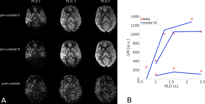 |
Blood-brain barrier water exchange estimation using optimised contrast-enhanced ASL
Elizabeth Powell1, Ben Dickie2, Yolanda Ohene2, Geoff JM Parker1,3,4, and Laura M Parkes2
1Centre for Medical Image Computing, Department of Computer Science, University College London, London, United Kingdom, 2Division of Neuroscience and Experimental Psychology, Faculty of Biology, Medicine and Health, The University of Manchester, Manchester, United Kingdom, 3Department of Neuroinflammation, Queen Square Institute of Neurology, University College London, London, United Kingdom, 4Bioxydyn Limited, Manchester, United Kingdom
Contrast-enhanced ASL (CE-ASL) has been proposed as a method for measuring blood-brain barrier (BBB) water exchange. Altering the T1 of blood using a gadolinium-based contrast agent should allow the label location to be identified as a function of post-labelling delay time; however, the shorter T1 of blood water leads to a trade-off between contrast dose and SNR. We show using simulations that there is an optimal T1 of blood post-contrast that balances this trade-off. Using this information we estimate the expected accuracy and precision of BBB water exchange measurements using CE-ASL. Finally, we demonstrate the method in vivo.
|
|||
1092.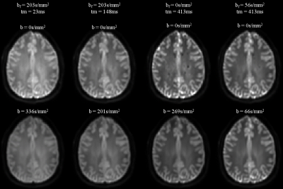 |
Diffusion-filtered exchange measurements of blood-brain barrier permeability to water
Elizabeth Powell1, Marco Battiston2, and Geoff JM Parker1,3,4
1Centre for Medical Image Computing, Department of Computer Science, University College London, London, United Kingdom, 2Queen Square MS Centre, Department of Neuroinflammation, UCL Queen Square Institute of Neurology, Faculty of Brain Sciences, University College London, London, United Kingdom, 3Department of Neuroinflammation, Queen Square Institute of Neurology, University College London, London, United Kingdom, 4Bioxydyn Limited, London, United Kingdom
We propose a method for quantifying water exchange across the blood-brain barrier (BBB) using diffusion-filtered exchange imaging. Careful design of the diffusion filter mitigates confounding effects from in-flowing blood water spins, and use of a two-compartment model aims to avoid known biases in the apparent exchange rate approximation. Using this approach, we optimise an acquisition protocol to provide maximum sensitivity to BBB water exchange and estimate the expected accuracy and precision of the method using simulations. The technique is then validated in a human volunteer.
|
The International Society for Magnetic Resonance in Medicine is accredited by the Accreditation Council for Continuing Medical Education to provide continuing medical education for physicians.