Digital Posters
Flow, Volume & Permeability: DCE-MRI
ISMRM & SMRT Annual Meeting • 15-20 May 2021

Digital Posters
Flow, Volume & Permeability: DCE-MRI
| Concurrent 6 | 13:00 - 14:00 |
1093.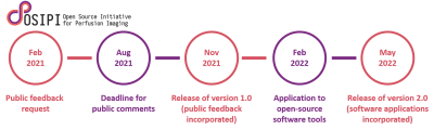 |
The Open Source Initiative for Perfusion Imaging (OSIPI): Contrast-based perfusion lexicon and reporting recommendations
Ina Nora Kompan1, Ben Dickie2, Steven Sourbron3, Petra van Houdt4, Laura Bell5, Rianne van der Heijden6, Andrey Fedorov7, Jonathan Arvidsson8, Charlotte Debus9, David Clunie10, Ingomar Gutmann11, Chad Quarles5, Zaki Ahmed12,
Ralf Floca1, and David Buckley13
1Medical Image Computing, German Cancer Research Center, Heidelberg, Germany, 2Division of Neuroscience and Experimental Psychology, University of Manchester, Manchester, United Kingdom, 3Department of Infection, Immunity and Cardiovascular Disease, University of Sheffield, Sheffield, United Kingdom, 4Department of Radiation Oncology, Netherlands Cancer Institute, Amsterdam, Netherlands, 5Barrow Neurological Institute, Phoenix, AZ, United States, 6Department of Radiology, Erasmus MC, Rotterdam, Netherlands, 7Brigham and Women’s Hospital, Boston, MA, United States, 8Department of Radiation Physics, University of Gothenburg, Gothenburg, Sweden, 9Steinbuch Centre for Computing, Karlsruhe Institute for Technology, Karlsruhe, Germany, 10PixelMed Publishing, LLC, Bangor, PA, United States, 11University of Vienna, Vienna, Austria, 12Mayo Clinic, Rochester, MN, United States, 13University of Leeds, Leeds, United Kingdom
As part of the Open Source Initiative for Perfusion Imaging (OSIPI), the aim of this work is to develop guidelines for reporting of contrast-based perfusion analysis in order to improve reproducibility, reusability and interoperability of perfusion analysis. For this, a perfusion analysis lexicon is developed, which provides standardized nomenclature for perfusion parameters and analysis processes, as well as a framework for reporting of analysis pipelines. The lexicon is intended to be a dynamically growing inventory updated by the perfusion community. For that, a variety of public feedback and review cycles are planned during the development process.
|
|||
 |
1094.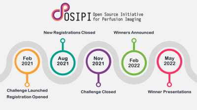 |
The Open Source Initiative for Perfusion Imaging (OSIPI): DCE-MRI Challenge
Anahita Fathi Kazerooni1, Laura C. Bell2, Floris Van den Abeele3, Ruben Verhack3, Xinze Zhou4, Salman Rezaei5, Zaki Ahmed6, Rianne Van Der Heijden7, Seyed Ali Nabavizadeh4, Leland S. Hu8, Hamidreza Saligheh Rad5, and Steven Sourbron9
1Department of Radiology, UPenn, Philadelphia, PA, United States, 2Division of Neuroimaging Research, Barrow Neurological Institute, Phoenix, PA, United States, 3Hyperfusion.ai, Gent, Belgium, 4University of Pennsylvania, Philadelphia, PA, United States, 5Quantitative MR Imaging and Spectroscopy Group, Research Center for Molecular and Cellular Imaging, Tehran University of Medical Sciences, Tehran, Iran (Islamic Republic of), 6Mayo Clinic, Rochester, MN, United States, 7Department of Radiology & Nuclear Medicine, Erasmus MC University Medical Center, Rotterdam, Netherlands, 8Neuroradiology Division, Department of Radiology, Mayo Clinic, Phoenix, AZ, United States, 9University of Sheffield, Sheffield, United Kingdom
Dynamic contrast enhanced (DCE-) MRI is widely acquired as a part of neuroimaging protocol for evaluation of glioblastoma tumors before the start of therapy or monitoring and assessment of treatment response in longitudinal scans. Nonetheless, lack of a standardized quantification method has limited its application in clinical settings, multi-institutional studies and clinical trials. These challenges have motivated efforts to validate DCE-MRI using benchmark biomedical image analysis methods. The Open-Source Initiative for Perfusion Imaging (OSIPI) has designed the OSIPI-DCE challenge to evaluate and compare DCE tools in terms of accuracy, repeatability, and reproducibility of Ktrans estimation in the brain.
|
||
 |
1095.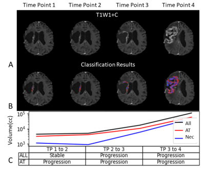 |
Differentiation Between Active Tumor and Radiation Necrosis in Patients with Glioblastoma Based on Model Free DCE Analysis
Idan Bressler1,2, Dafna Ben Bashat1,3,4, Orna Aizenstein3,5, Dror Limon3,6, Felix Bokestein3,7, Deborah T. Blumenthal3,7, Uri Nevo2,4, and Moran Artzi1,3,4
1Sagol Brain Institute, Tel Aviv Sourasky Medical Center, Tel Aviv, Israel, 2The Iby and Aladar Fleischman Faculty of Engineering Tel Aviv University, Tel Aviv, Israel, 3Sackler Faculty of Medicine, Tel Aviv University, Tel Aviv, Israel, 4Sagol School of Neuroscience, Tel Aviv University, Tel Aviv, Israel, 5Division of Radiology, Tel Aviv Sourasky Medical Center, Tel Aviv, Israel, 6Division of Oncology, Tel Aviv Sourasky Medical Center, Tel Aviv, Israel, 7Neuro-Oncology Service, Tel Aviv Sourasky Medical Center, Tel Aviv, Israel
A DCE based method for differentiation between active tumor tissue and radiation necrosis in patients with Glioblastoma is proposed. The study included 31 MRI scans of patients with Glioblastoma (6 patients with 4 longitudinal scans and 7 scans with biopsy results). The system is comprised of automatic, feature based discrimination of DCE dynamic data based on population analysis. Differentiation results show correlation to theoretical DCE models, and agreement with biopsy results in in the majority of cases. RANO based assessment of the differentiated results demonstrate potential for early detection of tumor progression.
|
||
1096. |
Effect of Normalization of DCE-MRI derived Tracer Kinetic Parameters on Glioma Grading
Dinil Sasi S1, Rakesh K Gupta2, Rana Patir2, Suneeta Ahlawat2, and Anup Singh1,3
1Indian Institute of Technology Delhi, New Delhi, India, 2Fortis Memorial Research Institute, Gurugram, India, 3All India Institute of Medical Science, New Delhi, India
Arterial-Input-Function(AIF) is a pre-requisite in fitting generalized-tracer-kinetic-model(GTKM) to dynamic-contrast-enhanced(DCE) MRI data for computing tracer-kinetic-parameters(TKP). TKP are highly sensitive to peak and shape of AIF, and this results in variation in computed parameter values across studies. These variations can reduce accuracy of TKP in glioma grading. We hypothesize that propagation of these AIF related errors to TKP can be mitigated using normalization w.r.t. corresponding average TKP values of healthy tissue and normalized TKP might improve glioma grading. The proposed normalization w.r.t. healthy gray-matter tissue has significantly reduced variations of TKP and improved accuracy of glioma grading particularly using Ktrans.
|
|||
1097.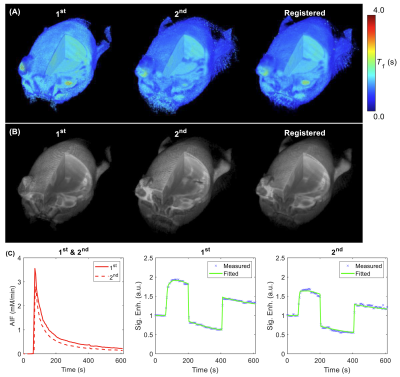 |
Repeatability of contrast kinetic parameters of whole tumor with isotropic resolution measured by 3D-UTE-GRASP method
Jin Zhang1, Ayesha Bharadwaj Das1, James Tranos2, Karl Kiser1, Youssef Zaim Wadghiri2, and Gene Kim1,2
1Weill Cornell Medicine, New York, NY, United States, 2NYU Langone Health, New York, NY, United States
The repeatability of dynamic contrast enhanced (DCE)-MRI has not been fully studied, particularly with contrast kinetic parameters including intracellular water lifetime (τi). The purposes of this study were: (1) to investigate the repeatability of DCE-MRI with the newly proposed technique, 3D-UTE-GRASP (Golden angle Radial Sparse Parallel), which can provide isotropic high-resolution images for quantitative pharmacokinetic model analysis; and (2) to investigate the repeatability of intracellular water lifetime estimation using the two-flip angle DCE-MRI approach.
|
|||
1098.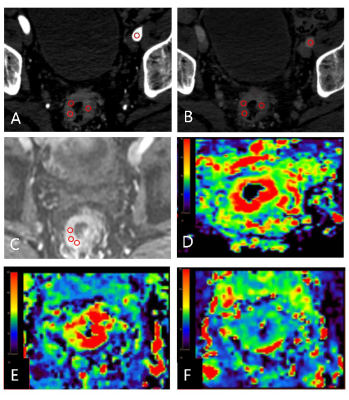 |
Combination study of spectral CT and DCE-MRI quantitatively predicting vascular invasion of rectal cancer preoperatively
Wan Dong1, Ailian Liu1, Anliang Chen1, Yuhui Liu1, and Lizhi Xie2
1Radiology, the First Affiliated Hospital of Dalian Medical University, Dalian, China, 2GE Healthcare, MR Research, Beijing, China
Vascular tumor thrombus has been proved to be closely related to the poor prognosis of rectal cance, of which the current gold standard for the diagnosis of is postoperative pathology. In this work, we explored the feasibility of spectral CT and DCE-MRI in quantitatively predicting vascular invasion of rectal cancer. Results showed that the arterial NIC combined with Ktrans can differentiate the vascular invasion from normal status more accurately. Therefore, spectral CT combined with DCE-MRI may serve as a feasible and non-invasive way in predicting vascular invasion of rectal cancer preoperatively, that is of great significance for clinical diagnosis.
|
|||
|
1099.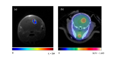 |
Tumor intracellular water residence time measured by DCE-MRI negatively correlates with the standard update value of 18F-FDG-PET
Karl Kiser1, Jin Zhang1, and S. Gene Kim1,2
1Radiology, Weil Cornell Medical College, New York, NY, United States, 2Center for Biomedical Imaging (CBI), Center for Advanced Imaging Innovation and Research (CAI2R), New York University School of Medicine, New York, NY, United States
Intracellular water residence time (τi) is an important property of solid tumors, with implications in cellular energy turnover. Measurement of τi using the active contrast encoding MRI method offers insight into tumor microenvironment heterogeneity and potentially metabolic activity. Our study compares τi, measured using ACE-MRI, in mouse gliomas with the standardized uptake value (SUV) from 18F-FDG PET in order to investigate the feasibility of using τi as an imaging marker for cellular metabolic activity.
|
||
1100.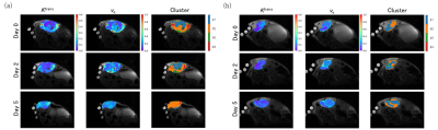 |
A new image clustering method for assessing tumor heterogeneity induced by a drug treatment in DCE-MRI
Kazuyuki Makihara1, Kazuya Sakaguchi1, Masayuki Yamaguchi2, Ken Ito3, Yusaku Hori3, Taro Semba3, Yasuhiro Funabashi3, Hirofumi Fujii2, and Yasuhiko Terada1
1Faculty of Pure and Applied Sciences, University of Tsukuba, Tsukuba, Ibaraki, Japan, 2National Cancer Center, EPOC, Division of Functional Imaging, Kashiwa, Chiba, Japan, 3Eisai Co.,Ltd. Oncology Business Group, Bunkyo City, Tokyo, Japan
Dynamic contrast-enhanced (DCE) -MRI has been widely used to assess tumor responses to anticancer drugs. Ktrans, one of the pharmacokinetic parameters of DCE-MRI, reflects blood perfusion and permeability in tumor tissue and is a good indicator of perfusion assessment. However, conventional analyses of DCE-MRI do not reflect drug response well, since tumor is extremely heterogeneous. In this study, we have developed a new method for quantitatively assessing the heterogeneous response of tumors during anticancer treatment, and applied it to examine the drug response of E7130, a novel anti-cancer agent with spatiotemporal variation in human breast cancer xenograft model.
|
|||
|
1101.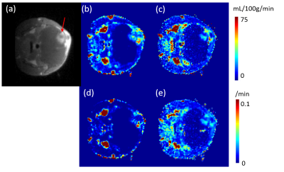 |
Two compartment quantitative transport mapping: evaluating tracer exchange rate between vascular and extravascular space
Qihao Zhang1, Jin Zhang2, Gene Kim2, John Morgan2, Thanh Nguyen2, Pascal Spincemaille2, and Yi Wang1
1Cornell University, New York, NY, United States, 2Weill Cornell Medical College, New York, NY, United States
In this study, we introduce compartment modeling into quantitative transport mapping (QTM), with the goal of mapping both the blood flow and the tracer exchange between vascular space and extravascular space. The nonlinear optimization problem is solved using the Levenberg–Marquardt method. We validate two compartment QTM (2c-QTM) in a porous media simulation and apply this method to dynamic PET data.
|
||
1102. |
tMB Whole Heart Perfusion Imaging with Iterative Reconstruction at 3.0T
Lixian Zou1,2, Changjun Tie1, Jian Xu3, Hairong Zheng1, Xin Liu1, and Yuan Zheng3
1Paul C. Lauterbur Research Centre for Biomedical Imaging, Shenzhen Institutes of Advanced Technology, Chinese Academy of Science, Shenzhen, China, 2Shenzhen College of Advanced Technology, University of Chinese Academy of Sciences, Shenzhen, China, 3UIH America Inc., Houston, TX, United States
Auto-calibrated multiband CAIPIRINHA with through-time encoding (tMB) has been proposed to acquire multiple slices simultaneously without extra reference scans. Reference images are estimated from the consecutive cardiac phases at a lower temporal resolution for subsequent slice separation. We implemented the tMB method in a myocardial perfusion sequence and demonstrated the feasibility of whole heart perfusion imaging with tMB and iterative reconstruction using in a clinical setting.
|
|||
1103.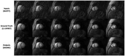 |
High-resolution Spiral First-pass Myocardial Perfusion Imaging using DEep learning-based rapid Spiral Image REconstruction (DESIRE)
Junyu Wang1, Daniel Weller2, Patricia Rodriguez Lozano3, and Michael Salerno1,3,4
1Biomedical Engineering, University of Virginia, Charlottesville, VA, United States, 2Electrical and Computer Engineering, University of Virginia, Charlottesville, VA, United States, 3Medicine, University of Virginia, Charlottesville, VA, United States, 4Radiology, University of Virginia, Charlottesville, VA, United States
First-pass contrast-enhanced myocardial perfusion imaging is valuable for evaluating coronary artery disease (CAD). Spiral perfusion imaging techniques, using a motion-compensated L1-SPIRiT based reconstruction, are capable of whole-heart high-resolution perfusion imaging. However, this reconstruction is performed off-line and takes ~1 hour per slice. To address this limitation, we developed a DEep learning-based Spiral Image REconstruction technique (DESIRE) for spiral first-pass myocardial perfusion imaging, for both single-slice (SS) and simultaneous multi-slice (SMS) MB=2 acquisitions, to provide fast and high-quality image reconstruction and make rapid online reconstruction feasible. High image quality was demonstrated using the proposed technique for healthy volunteers and patients.
|
|||
1104.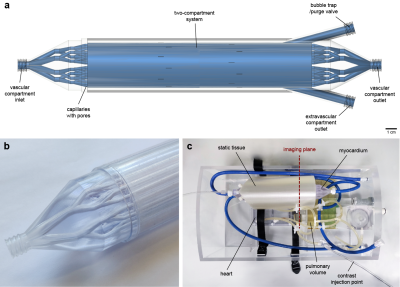 |
Calibration of myocardial perfusion quantification using a dedicated two-compartment cardiac phantom
Xenios Milidonis1, Richard Crawley1, and Amedeo Chiribiri1
1School of Biomedical Engineering & Imaging Sciences, King's College London, London, United Kingdom
Myocardial perfusion quantification by cardiovascular MR imaging has shown great promise in the detection of coronary artery disease. However, the lack of true standardization of methods across centers hinders the effective comparison and pooling of measurements. We sought to develop a novel cardiac phantom mimicking dynamic contrast exchange between two myocardial compartments and use it for the calibration of perfusion measured using three common quantification methods. Without calibration, perfusion measurements differed significantly between methods. Calibration led to accurate and non-significantly different measurements, suggesting that it could be an effective and reliable approach for the universal standardization of quantification methods.
|
|||
1105.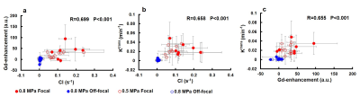 |
Associate Focused Ultrasound-induced MR Signal Changes With Gd-Enhancement and Ktrans
Tzu-Ming Hung1, Yu-Ting Jiang1, Cheng-Tao Ho1, Po-Hung Hsu2, Hao-Li Liu3, Chih-Kuang Yeh1, and Hsu-Hsia Peng1
1Department of Biomedical Engineering and Environmental Sciences, National Tsing Hua University, Hsinchu, Taiwan, 2Center for Advanced Molecular Imaging and Translation, Chang Gung Memorial Hospital, Taoyuan, Taiwan, 3Department of Electrical Engineering, Chang-gung University, Taoyuan, Taiwan
Blood brain barrier (BBB) can be opened transiently by the cavitation effect with the assist of focused ultrasound (FUS) and microbubble (MB). A previous study proved that FUS-induced MR signal changes can monitor cavitation effect in in vitro phantom experiments. However, whether MR signal changes can reflect BBB opening levels in vivo remains uncertain. In this study, a cavitation index (CI) map was employed to evaluate MR signal changes during FUS sonication in in vivo experiments. We aim to examine the relationship between MR signal change and BBB opening levels by associating CI map with Gd-enhancement and Ktrans maps.
|
|||
1106. |
Assessment of Fatty liver disease in rats using quantitative transport mapping (QTM) method against pathology validation
Qihao Zhang1, Xianfu Luo2, Thanh Nguyen2, Pascal Spincemaille2, and Yi Wang1
1Cornell University, New York, NY, United States, 2Weill Cornell Medical College, New York, NY, United States
We propose to assess the severity of Nonalcoholic fatty liver disease (NAFLD) using quantitative transport mapping (QTM), a recently introduced flow quantification method. A numerical simulation was performed to compare QTM with traditional kinetic modeling. QTM successfully reconstructed blood flow with high accuracy (relative root mean square error = 0.27). Using DCE MRI in 5 adult rats with methionine choline-deficient diet-induced NAFLD (grade F3) and in 13 untreated control rats, only the QTM derived velocity |u| showed a significant difference between NAFLD and healthy controls.
|
|||
1107.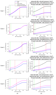 |
Use of Reference Region Model to Improve Arterial Input Function Selection for Estimating Kidney Function with DCE-MRI
Cemre Ariyurek1, Onur Afacan1, Jeanne Chow1, Simon K Warfield1, and Sila Kurugol1
1Radiology, Boston Children's Hospital and Harvard Medical School, Boston, MA, United States
Estimating glomerular filtration rate (GFR) is crucial for diagnostic purposes. DCE-MRI is capable of measuring tracer kinetic parameters of kidney function. Tracer kinetic models require arterial input function to find tracer kinetic parameters such as filtration rate. However, determining the arterial input function concentration is challenging and important for estimating GFR accurately. Here, we propose to use reference region model to improve arterial input function selection to estimate GFR more accurately. Comparing the GFR values estimated using the improved arterial input function with the ground truth GFR, we have observed a significant decrease in the mean absolute error in GFR.
|
|||
1108. |
Personalized DCE-MRI Parametric Mapping using GRASP and Iterative Joint Estimation of Arterial Input Function and Pharmacokinetic Parameters
Yousef Mazaheri1, Nathanael Kim1, Yuliya Lakhman2, Ramin Jafari1, Hebert Vargas Alvarez2, and Ricardo Otazo1
1Medical Physics, Memorial Sloan Kettering Cancer Center, New York, NY, United States, 2Radiology, Memorial Sloan Kettering Cancer Center, New York, NY, United States
To develop a personalized DCE-MRI parametric mapping technique using high spatial and temporal resolution golden-angle radial sparse parallel MRI (GRASP) and patient specific iterative estimation of the arterial input function (AIF) and pharmacokinetic parameters.
|
|||
1109.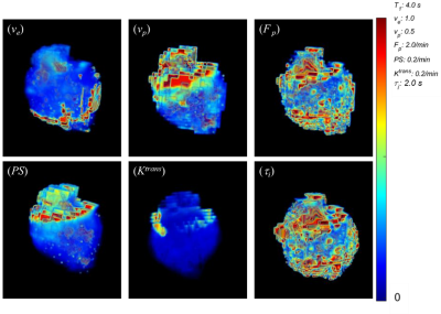 |
Significance of 3D Isotropic Resolution for Image Texture Analysis of Pharmacokinetic Model Parametric Maps
Karl Kiser1, Jin Zhang1, and S. Gene Kim1,2
1Radiology, Weil Cornell Medical College, New York, NY, United States, 2Center for Biomedical Imaging (CBI), Center for Advanced Imaging Innovation and Research (CAI2R), New York University School of Medicine, New York, NY, United States
Quantitative analysis of MRI image features for estimating tumor grading and treatment response is a growing area of research, however a lack of reproducibility and validation present a major challenge in the field. Our study investigates how spatial resolution affects texture features of DCE-MRI images by comparing features generated from 3D isotropic high resolution kinetic parameter maps with typical thick slice maps. We demonstrate that 3D textural features can differ by several orders of magnitude when extracted from isotropic versus thick slice images. These findings have potentially significant implications in the predictive capabilities of texture features.
|
|||
1110.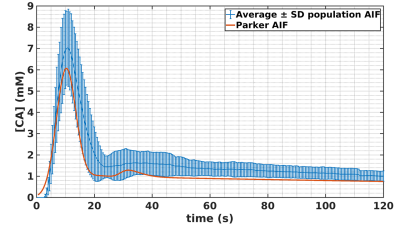 |
Simulation and clinical validation of an algorithm for retrospective Arterial Input Function peak saturation correction
Jean-Sébastien Louis1, Jacques Felblinger1,2, Olivier Huttin3, and Marine Beaumont1,2
1IADI, Inserm U1254, Université de Lorraine, Nancy, France, 2CIC-IT, Inserm 1433, Université de Lorraine and CHRU Nancy, Nancy, France, 3Pôle cardiologie, CHRU Nancy, Nancy, France
Arterial Input Function (AIF) is fundamental for quantitative perfusion analysis. However, AIF peak is underestimated when using common perfusion sequence such SR-turboFLASH due to signal saturation effect. This leads to biased perfusion parameters estimation. Accurate AIF sampling requires specific sequence or protocol imaging not widely available. We proposed a solution to correct AIF peak retrospectively from standard perfusion data. We evaluated the proposed algorithm in simulation on more than 90000 different AIF sampling scenarios. Eventually, we tested our algorithm on clinical data and compared the population AIF estimated from our solution to literature population AIF.
|
|||
1111.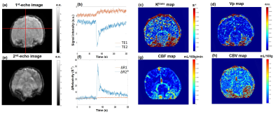 |
High spatial-temporal resolved perfusion imaging: simultaneously DCE- and DSC-MRI acquisition using improved Spiral-Out-In (iSOI) sequence
Yupeng Cao1,2, Jun Zhao1,2, Weinan Tang3, Wentao Liu1, and Dong Han1,2
1National Center for Nanoscience and Technology, Beijing, China, 2University of Chinese Academy of Sciences, Beijing, China, 3Wandong Medical Technology, Beijing, China
The combination of DCE and DSC MRI contribute to the higher accuracy of diagnosis than either alone. However, high spatial-temporal resolution sequences for DCE and DSC are absent in the clinical use. Herein, we propose an improved Spiral-Out-In (iSOI) sequence to simultaneously sample DCE and DSC signal with high spatial resolution. A model-based strategy for reconstruction of 8-fold accelerated MRI were employed to realize high temporal resolution imaging. The in-plane spatial resolution is 0.78 mm and the temporal resolution is 0.52 s. The perfusion parameters were calculated to verify the proposed method which has potential to benefit the accurate diagnosis.
|
|||
1112.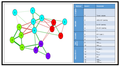 |
Correlation assessment between quantitative multimodality imaging metrics using a community detection algorithm
Ramesh Paudyal1, Milan Grkovski1, Jung Hun Oh1, Heiko Schoder2, David Aramburd Nunez1, Vaios Hatzoglou2, Joseph O Deasy1, John L Hum1, Nancy Lee3, and Amita Shukla-Dave1,2
1Medical Physics, Memorial Sloan Kettering Cancer Center, New York, NY, United States, 2Radiology, Memorial Sloan Kettering Cancer Center, New York, NY, United States, 3Radiation Oncology, Memorial Sloan Kettering Cancer Center, New York, NY, United States
This study aims to assess the correlation between the pre-treatment quantitative imaging metrics obtained from multimodality imaging (MMI) techniques such as 18[F]-FMISO PET/CT, 18[F]-FDG PET/CT, DW- and DCE- MRI describing tumor metabolism, hypoxia, diffusion, perfusion, and cell metabolic activity, using a community detection algorithm. The method partitioned the network into four groups with strong and weak connections. The community connection results show complementary, rather than competitive, information about tumor metabolism, hypoxia, diffusion, and perfusion.
|
The International Society for Magnetic Resonance in Medicine is accredited by the Accreditation Council for Continuing Medical Education to provide continuing medical education for physicians.