Digital Posters
Even More (Artificial) Intelligence in the Body
ISMRM & SMRT Annual Meeting • 15-20 May 2021

Digital Posters
Even More (Artificial) Intelligence in the Body
| Concurrent 5 | 15:00 - 16:00 |
2986.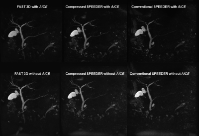 |
FAST 3D vs. Compressed Sensing vs. Parallel Imaging: Image Quality Improvement on MRCP with and without Deep Learning Reconstruction
Takahiro Matsuyama1, Yoshiharu Ohno1,2, Kaori Yamamoto3, Kazuhiro Murayama2, Masato Ikedo3, Masao Yui3, Akiyoshi Iwase4, Takashi Fukuba4, Satomu Hanamatsu1, Yuki Obama1, Takahiro Ueda1, Hirotaka Ikeda1, and Hiroshi Toyama1
1Radiology, Fujita Health University School of Medicine, Toyoake, Japan, 2Joint Research Laboratory of Advanced Medical Imaging, Fujita Health University School of Medicine, Toyoake, Japan, 3Canon Medical Systems Corporation, Otawara, Japan, 4Radiology, Fujita Health University Hospital, Toyoake, Japan
We hypothesize that the newly developed FAST 3D can reduce examination time as well as Compressed SPEEDER and obtain MRCP without any degradation of image quality as compared with conventional parallel imaging technique in patients with hepatobiliary and pancreatic diseases. In addition, deep learning reconstruction (i.e.AiCE) has a potential to improve image quality of MRCP obtained by different protocols in routine clinical practice. The purpose of this study was to compare the capability of image quality improvement on MRCP with and without AiCE among FAST 3D, Compressed SPEEDER and conventional parallel imaging (SPEEDER) in patients with hepatobiliary and pancreatic diseases.
|
|||
2987. |
The value of radiomics-based model on contrast-enhanced MRI for predicting microvascular invasion in HCC before Partial Hepatectomy
Tao Lin1, Ailian Liu1, Lihua Chen1, Qingwei Song1, Renwang Pu1, Ying Zhao1, Xue Ren1, and yan guo2
1Department of Radiology, the First Affiliated Hospital of Dalian Medical University, Dalian, China, 2GE Healthcare, Beijing, China
Hepatocellular carcinoma (HCC), often occurring in patients with chronic viral hepatitis and cirrhosis, was one of the most common malignant tumors. Microvascular invasion (MVI) is one of the well-known potential predictors related to the prognosis of HCC. MVI with higher positive expression indicates more aggressive behaviour of HCC and poorer survival outcomes. Early accurate prediction for MVI status plays an important guiding role in surgical selection and adjuvant therapy for HCC. We proposed a radiomics model based on enhanced MRI to predict MVI in HCC before surgery.
|
|||
2988. |
Deep learning-based detection of liver disease using MRI
Mark A Pinnock1,2, Yipeng Hu1,2, Alan Bainbridge3, David Atkinson4, Rajeshwar P Mookerjee5, Stuart A Taylor4, Dean C Barratt1,2, and Manil D Chouhan4
1Centre for Medical Image Computing, University College London, London, United Kingdom, 2Wellcome/EPSRC Centre for Interventional and Surgical Sciences, University College London, London, United Kingdom, 3Department of Medical Physics and Biomedical Engineering, University College London Hospitals NHS Foundation Trust, London, United Kingdom, 4Centre for Medical Imaging, Division of Medicine, University College London, London, United Kingdom, 5Institute for Liver and Digestive Health, Division of Medicine, University College London, London, United Kingdom
Traditional approaches to MRI detection of liver disease require specialist hardware, sequences and post-processing. Here we propose a deep learning (DL) based model for the detection of liver disease using standard T2-weighted anatomical sequences, as an early feasibility study for the potential of DL-based classification of liver disease severity. Our DL model achieved a diagnostic accuracy of 0.92 on unseen data and achieved a test accuracy of 0.75 when trained with relevant anatomical segmentation masks without images, demonstrating potential scanner/sequence independence. Lastly, we used DL interpretability techniques to analyse failure cases.
|
|||
2989. |
A Two-Stage Deep Learning Model for Accurate Vessel Segmentation and Reconstruction in the MRI of Live
Xu Luo1,2, Ailian Liu3, Yu Yao1,2, Ying Zhao3, Zhebin Chen1,2, Meng Dou1,2, and Han Wen1,2
1Chengdu Institute of Computer Application, Chinese Academy of Sciences, Chengdu, China, 2University of Chinese Academy of Sciences, Beijing, China, 3Department of Radiology, the First Affiliated Hospital of Dalian Medical University, Dalian, China
The deep learning technology was used on the segmentation automatically and reconstruction of the intrahepatic portal vein to get its volume and three-dimensional spatial position information. The results showed that the dice accuracy was 83.32% in the training and 75.89% in the test set among 12 cases. The current study demonstrated the volume of intrahepatic vessels and 3D spatial location information can be obtained by the tow-stage deep learning model and it can be applied in promising clinical effectively.
|
|||
2990. |
Deep Learning 3D Convolutional Neural Network for Noninvasive Evaluation of Pathologic Grade of HCC Using Contrast-enhanced MRI
Ying Zhao1, Han Wen2,3, Ailian Liu1, Yu Yao2,3, Tao Lin1, Qingwei Song1, Xin Li4, Yan Guo4, and Tingfan Wu4
1The First Affiliated Hospital of Dalian Medical University, Dalian, China, 2Chengdu Institute of CoChinese Academy of Sciences, Chengdu, China, 3University of Chinese Academy of Sciences, Beijing, China, 4GE Healthcare (China), Shanghai, China
In recent years, convolutional neural networks (CNNs) have become one of the most advanced deep learning networks. Deep learning with CNNs has reportedly achieved good performance in the pattern recognition of images. In the present study, 3D-CNN based on contrast-enhanced (CE)-MR images was demonstrated to be capable to evaluate pathologic grade of hepatocellular carcinoma (HCC) treated with surgical resection, which will provide more prognostic information and facilitate clinical management.
|
|||
2991. |
T2WI liver MRI with deep learning-based reconstruction: a clinical feasibility study in comparison to conventional T2WI liver MRI
Ruofan Sheng1, Liyun Zheng2, Shu Liao3, Yongming Dai2, and Mengsu Zeng1
1Department of Radiology, Zhongshan Hospital, Shanghai, China, 2United Imaging Healthcare, Shanghai, China, 3Shanghai United Imaging Intelligence, Shanghai, China Liver magnetic resonance imaging (MRI) is limited by several technical challenges, including relatively long acquisition time and respiratory motion artifacts. Recently, deep learning methods have been proposed to reconstruct undersampled k-space data by training deep neural networks. In this study, we raised a U-net convolutional neural network architecture to improve the reconstruction speed and image quality of liver T2-weighted MRI. This technique was able to cover the whole liver during one breath hold and showed promising performance in image quality and lesion detectability. |
|||
2992.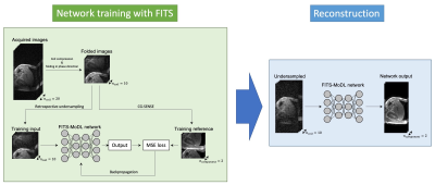 |
Model-based Deep Learning Reconstruction using Folded Image Training Strategy (FITS-MoDL) for Liver MRI Reconstruction
Satoshi Funayama1,2, Utaroh Motosugi3, and Hiroshi Onishi1
1Department of Radiology, University of Yamanashi, Yamanashi, Japan, 2Graduate School of Medicine, University of Yamanashi, Yamanashi, Japan, 3Department of Radiology, Kofu-Kyoritsu Hospital, Yamanashi, Japan
Short acquisition time is one of the key features of liver MRI to acquire images during breath-holding. A combination of undersampling and deep learning-based reconstruction would be a powerful reconstruction method to achieve sufficient speed and SNR. However, it is challenging due to high memory consumption in network training. The folded image training strategy (FITS) is one of the methods to handle this problem. In this study, we demonstrated that the model-based deep learning reconstruction using FITS had better image quality in liver MRI acquired with multiple coils.
|
|||
2993.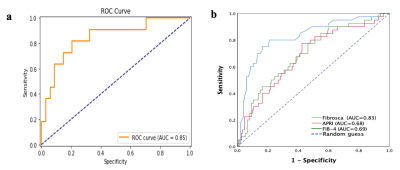 |
The value of Radiomics combined with Machine Learning in the staging of liver Fibrosis
Fengxian Fan1, Weiting Huang2, Yanli Jiang1, Wanjun Hu1, Jing Zhang1, and Jialiang Ren3
1LanZhou University Second Hospital, LanZhou, China, 2LanZhou University, LanZhou, China, 3GE Healthcare, Shanghai, China
The purpose of this study is to develop radiomics models by using T1WI for staging liver fibrosis. Two hundred twenty-four patients had available pathologic reports of liver fibrosis and liver MRI were divided into training (n=179) and testing (n=45) cohorts to develop and validate the radiomics models. The results showed that the T1WI-based radiomics models had a powerful ability to stage liver fibrosis.
|
|||
2994.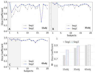 |
Few-shot deep learning for kidney segmentation
Junyu Guo1 and Ivan Pedrosa1
1Radiology, UT southwestern medical center, Dallas, TX, United States
MR kidney image segmentation is an important enabler of radiomics analysis and assessment of kidney size, morphology and renal disease. Deep learning methods are state-of-the-art techniques for segmentation. Training of a robust model with high accuracy requires a large dataset. Manually drawing masks is time-consuming and labor-intensive for a large number of datasets. Furthermore, different masks are required in training for different MR modalities. In this study, we investigated the feasibility of kidney segmentation using deep learning models trained with MR images from only a few subjects. We tested the hypothesis that few-shot deep learning may achieve accurate kidney segmentation.
|
|||
2995.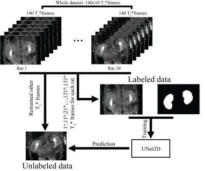 |
Deep learning based kidney segmentation for high temporal resolution tracking renal size changes during sequential gas challenges
Kaixuan Zhao1,2, Joao dos Santos Periquito3, Thomas Gladytz2, Kathleen Cantow3, Luis Hummel3, Jason Millward2, Sonia Waiczies2, Erdmann Seeliger3, Yanqiu Feng1, and Thoralf Niendorf2,4
1School of Biomedical Engineering, Southern Medical University, Guangzhou, China, 2Berlin Ultrahigh Field Facility (B.U.F.F.), Max Delbruck Center for Molecular Medicine in the Helmholtz Association, Berlin, Germany, 3Institute of Physiology, Charité - Universitätsmedizin Berlin, Berlin, Germany, 4Experimental and Clinical Research Center, a joint cooperation between the Charité Medical Faculty and the Max Delbrück Center for Molecular Medicine in the Helmholtz Association, Berlin, Germany
Fast renal volume changes during sequential gas challenges might indicate the dynamic balance between renal filtration and reabsorption. In the present work, a deep learning based semantic segmentation method is employed to monitor renal size changes.
|
|||
2996. |
Is it Feasible? IVIM-DWI and T2WI-based Texture Analysis Predicting Histological Types of Cervical Carcinoma Before Operation
Jiang-Ning Dong1 and Bin Shi1
1The First Affiliated Hospital of USTC, Anhui Provincial Cancer Hospital, Hefei, China The combination of IVIM-DWI biomarkers and T2WI-based texture features had good predictive performance to evaluate three different histological types of cervical carcinoma by synergizing diffusion, perfusion and heterogeneous features, especially for cervical squamous cell carcinoma and adenocarcinoma. A novel quantitative preoperative imaging model composed of IVIM-DWI and TA is a useful supplement for histopathological diagnosis, and might be used in clinical practice to assist in the formulation of treatment strategy. |
|||
2997.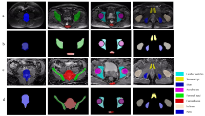 |
Fully Automated Pelvic Bones Segmentation in Multiparameter MRI Using a 3D Convolutional Neural Network
xiang liu1, chao han1, and xiaoying wang1
1department of radiology, peking university first hospital, Beijing, China
This retrospective study aims to perform automated pelvic bones segmentation in multiparametric MRI (mpMRI) using 3D convolutional neural network (CNN). 264 pelvic DWI images and corresponding ADC maps obtained from three MRI vendors from 2018 to 2019 were used for the 3D U-Net CNN development. 60 independent mpMRI data from 2020 were used to externally evaluate the segmentation model using quantitative criteria (Dice similarity coefficient) and qualitative assessment (SCORE system). The results demonstrated that the 3D CNN can achieve fully automated pelvic bone segmentation on multi-vendor DWI and ADC images with good quantitative and qualitative performances.
|
|||
2998.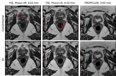 |
Motion Robust High-Resolution Pelvic Imaging using PROPELLER and Deep Learning Reconstruction
Ali Pirasteh1, Lloyd Estkowski2, Daniel Litwiller3, Ersin Bayram4, and Xinzeng Wang5
1Department of Radiology, UW Madison, Madison, WI, United States, 2Global MR Applications & Workflow, GE Healthcare, Madison, WI, United States, 3Global MR Applications & Workflow, GE Healthcare, Denver, CO, United States, 4Global MR Applications & Workflow, GE Healthcare, Houston, TX, United States, 5GE Healthcare, Houston, TX, United States
We evaluated the utility of PROPELLER T2 FSE with deep-learning (DL) reconstruction in the setting of prostate and rectal imaging, with the goal of overcoming respiratory and peristaltic motion, improving image sharpness, and achieving high-resolution imaging within a comparable scan time to the traditional T2 FSE techniques. We demonstrated that in absence of DL reconstruction, the PROPELLER T2 FSE images suffer from excessive noise and lower subjective quality. However, with utilization of DL reconstruction, High-resolution and motion robust images were obtained at clinically acceptable scan times.
|
|||
2999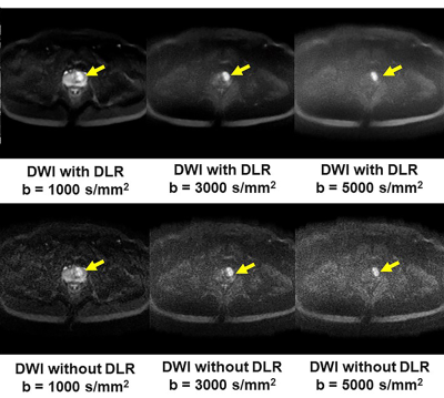 |
Deep Learning Reconstruction for DWI with b Values < 5000s/mm2: Improvement of Image Quality and Diagnostic Performance for Prostatic Cancer Video Permission Withheld
Takahiro Ueda1, Yoshiharu Ohno1,2, Kaori Yamamoto3, Kazuhiro Murayama2, Masato Ikedo3, Masao Yui3, Akiyoshi Iwase4, Takashi Fukuba4, Satomu Hanamatsu1, Yuki Obama1, Hirotaka Ikeda1, and Hiroshi Toyama1
1Radiology, Fujita Health University School of Medicine, Toyoake, Japan, 2Joint Research Laboratory of Advanced Medical Imaging, Fujita Health University School of Medicine, Toyoake, Japan, 3Canon Medical Systems Corporation, Otawara, Japan, 4Radiology, Fujita Health University Hospital, Toyoake, Japan
There have been no reports of major studies to the utility of DLR for DWI with high b values for improving image quality and detection performance for patients with prostatic cancers. We hypothesized that DLR could improve image quality and diagnostic performance of DWI with high values, and that an appropriate b value might be determined for this setting. The purpose of this study was thus to determine the utility of the DLR method for DWI and the appropriate b value for detecting prostatic cancer using a 3T MR system in routine clinical practice.
|
|||
3000.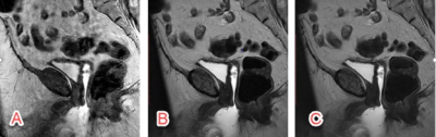 |
T2-weighted Pelvic MR Imaging Using PROPELLER with Deep Learning Reconstruction for Improved Motion Robustness
Mohammed Saleh1, Sanaz Javadi1, Manoj Mathew2, Jong Bum Son3, Jia Sun4, Ersin Bayram5, Xinzeng Wang5, Jingfei Ma3, Janio Szklaruk1, and Priya Bhosale1
1Radiology, MD Anderson Cancer Center, Houston, TX, United States, 2Radiology, Stanford University, Stanford, CA, United States, 3Imaging Physics, MD Anderson Cancer Center, Houston, TX, United States, 4Biostatistics, MD Anderson Cancer Center, Houston, TX, United States, 5Global MR Applications and Workflow, GE Healthcare, Houston, TX, United States
In oncologic MRI, sagittal T2 weighted images are usually acquired to assess gynecologic malignancy. Motion artifacts may render pathology difficult to detect due to patient or bowel motion or the presence of air in the rectum. PROPELLER sequence has shown promising results to reduce motion-related artifacts. Our work shows that DL Recon can be combined with PROPELLER and further help reduce noise and improve the overall image quality for T2-weighted imaging of gynecological malignancies. The combination of PROPELLER (Non-DL) and DL reconstruction could be synergistic in improving image quality.
|
|||
3001.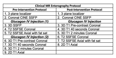 |
Quantifying Efficiency and Variability of Clinical MRI Exams with Advanced Analytics Tools
Sheena Y Chu1, Scott B Reeder1,2,3,4,5, and John W Garrett1,3
1Department of Medical Physics, University of Wisconsin-Madison, Madison, WI, United States, 2Department of Medicine, University of Wisconsin-Madison, Madison, WI, United States, 3Department of Radiology, University of Wisconsin-Madison, Madison, WI, United States, 4Department of Biomedical Engineering, University of Wisconsin-Madison, Madison, WI, United States, 5Department of Emergency Medicine, University of Wisconsin-Madison, Madison, WI, United States
By quantifying the efficiency and variability of clinic MRI exams, sources of potential improvement can be identified. An increase in efficiency and decrease in variability of MRI exams are essential to plan appropriate exam slot lengths, which can contribute to better patient access and reduced cost. Analytics are also essential to measure the impact on interventions to improve workflow. In this work we demonstrate the utility of analytics to identify inefficiencies in clinical MRI protocol in the context of MR enterography, a common abdominal MRI exam.
|
|||
3002.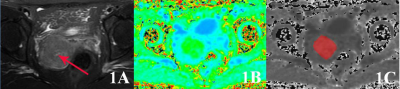 |
Application value of Radiomics Methods Based on DKI Sequence MK Map for Differentiating squamous Cell carcinoma from cervix Adenocarcinoma
Shifeng Tian1, Ailian Liu1, Yan Guo2, and Yuan Wei1
1Department of Radiology, The First Affiliated Hospital of Dalian Medical University, Dalian, China, China, 2GE Healthcare, Dalian City, China, China
Squamous cell carcinoma is the most common pathological type in cervical cancer, followed by cervix adenocarcinoma. There are differences in the evaluation of prognosis between the two. Radiomics can quantitatively analyze a large number of image data, and then quantify tumor heterogeneity and noninvasively evaluate tumor biological behavior. The results suggest that radiomics based on mean kurtosis maps of diffusion kurtosis imaging sequences can identify different types of cervical cancer.
|
|||
3003.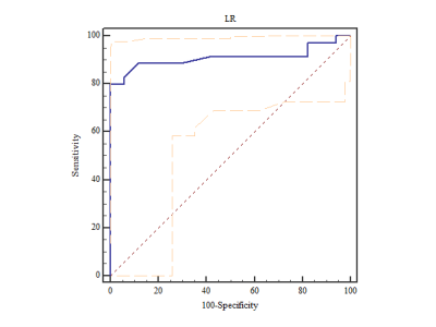 |
Radiomics Based on MR Imaging of Rectal Mucinous Adenocarcinoma: Assess Treatment Response to Neoadjuvant Chemoradiotherapy
Fu Shen1, Minglu Liu1, Zhihui Li1, Xiaolu Ma1, Jianping Lu1, and Yuwei Xia2
1Changhai Hospital, Shanghai, China, 2Huiying Medical Technology Co., Ltd., Beijing, China
The goal of this study was to investigate the value of high resolution T2-weighted–based radiomics in prediction of treatment response to neoadjuvant chemoradiotherapy (nCRT) in patients with rectal mucinous adenocarcinoma (RMAC). The result demonstrated that the MRI based radiomics machine learning model could assess tumoral treatment response to nCRT in patients with RMAC.
|
|||
3004.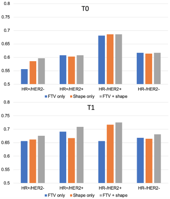 |
Breast MRI radiomic shape features for the prediction of neoadjuvant therapy response
Wen Li1, Rohan Nadkarni1, David C Newitt1, Bo La Yun1,2, Deep Hathi1, Alex Nguyen1, Natsuko Onishi1, Lisa J Wilmes1, Ella F Jones1, Jessica Gibbs1, Teffany Joy Bareng1, Bonnie N Joe1, Elissa Price1, Rita Mukhtar1,
John Kornak1, Efstathios Gennatos1, I-SPY 2 Consortium3, Laura J Esserman1, and Nola M Hylton1
1University of California, San Francisco, San Francisco, CA, United States, 2Seoul National University Bundang Hospital, Seoul, Korea, Republic of, 3Quantum Leap Healthcare Collaborative, San Francisco, CA, United States
A previous study demonstrated that tumor sphericity, measured from breast DCE-MRI during neoadjuvant therapy, is predictive of pathologic complete response and adds value to a predictive model based on functional tumor volume (FTV) alone. This study further explores the additive value of alternative radiomic shape features by breast cancer subtype. A subset of shape features were selected using visually assessed MRI morphological patterns as guidance. The analysis of treatment response prediction was conducted retrospectively using data from the multi-center neoadjuvant I-SPY 2 TRIAL. Improved predictive performance when adding shape features was observed at both pre-treatment and early treatment time points.
|
|||
3005.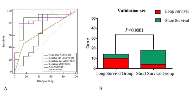 |
The nomogram of MRI-based radiomics with complementary visual features by machine learning improves stratification of glioblastoma patients
ZHENYU SHU1, YUYUN XU1, and YONG ZHANG2
1Zhejiang Provincial People’s Hospital, Hangzhou, China, 2MR Research, GE healthcare (China), SHANG HAI, China
This preliminary study explored the application of radiomics MRI in overall survival(OS) of glioblastoma patients. We found that EPI, age, and radiomic signature are independent predictors of OS for glioblastoma patients. The nomogram was created by integrating the three independent predictors, had the best performance when stratifying glioblastoma patients into long- versus short-term survival, which could help clinicians develop optimal treatment plans.
|
The International Society for Magnetic Resonance in Medicine is accredited by the Accreditation Council for Continuing Medical Education to provide continuing medical education for physicians.