Digital Posters
Perfusion & Permeability: Contrast & Non-Contrast
ISMRM & SMRT Annual Meeting • 15-20 May 2021

Digital Posters
Perfusion & Permeability: Contrast & Non-Contrast
| Concurrent 3 | 13:00 - 14:00 |
1844.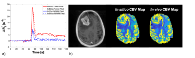 |
How to Benchmark DSC-MRI: the technical development of an anthropomorphic phantom for software validation
Laura C. Bell1, Natenael B Semmineh1, Sudarshan Ragunathan1, and C. Chad Quarles1
1Barrow Neurological Institute, Phoenix, AZ, United States
Clinical adoption of DSC-MRI for brain cancer imaging is plagued by reproducibility concerns. To support and advance reproducible science for DSC-MRI, an anthropomorphic phantom is developed in order to benchmark post-processing pipelines and software platforms. In addition to this anthropomorphic phantom, the technical computation of relative cerebral blood volume (rCBV) is discussed since rCBV is not an absolute measurement in DSC-MRI. In summary, this DRO-based benchmark can then be used to characterize the accuracy of commonly employed DSC-MRI algorithms and clinical software.
|
|||
1845. |
Reproducibility and Validation of Water Permeability in Human Brain using Magnetization Transfer based ASL at 7T
Sultan Zaman Mahmud1,2, Thomas S. Denney1,2, and Adil Bashir1,2
1Department of Electrical and Computer Engineering, Auburn University, Auburn, AL, United States, 2Auburn University MRI Research Center, Auburn University, Auburn, AL, United States
Blood-brain barrier (BBB) is crucial to prevent brain tissue from circulating toxins while allowing the delivery of nutrients from intravascular space to the central nervous system (CNS). Compromised BBB can result in brain dysfunction and degeneration. Development of reproducible non-invasive techniques to assess BBB permeability is of particular interest. Previously we had demonstrated a non-invasive technique to estimate BBB permeability using magnetization transfer (MT) effect on labeled arterial spins at 7T. In this work, we demonstrate the reproducibility of the technique. The feasibility was evaluated in healthy subjects at baseline and after caffeine challenge.
|
|||
1846.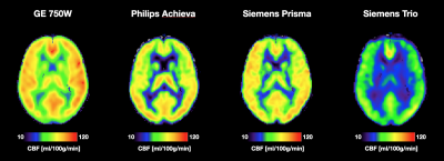 |
Cross-Vendor Test-Retest Analysis of 3D pCASL Cerebral Blood Flow
Kay Jann1, Xingfeng Shao1, Samantha J Ma1,2, Karl G Helmer3, Michael Magaletta3, Mitchell J Horn4, Andrew D Warren4, Vanessa A Gonzalez4, Hanzhang Lu5, Yang Li5, Zixuan Lin5, Kaisha Hazel5, George Pottanat5, and Danny JJ Wang1
1Laboratory of Functional MRI Technology (LOFT), USC Stevens Neuroimaging and Informatics Institute, Keck School of Medicine, University of Southern California (USC), Los Angeles, CA, United States, 2Siemens Medical Solutions USA, Inc., Los Angeles, CA, United States, 3Department of Radiology, Massachusetts General Hospital and Athinoula A Martinos Center for Biomedical Imaging, Harvard Medical School, Charlestown, MA, United States, 4Department of Neurology, Massachusetts General Hospital and Athinoula A Martinos Center for Biomedical Imaging, Harvard Medical School, Charlestown, MA, United States, 5The Russell H. Morgan Department of Radiology & Radiological Science, Johns Hopkins University School of Medicine, Baltimore, MD, United States
Standardized acquisition protocols have been recommended for Arterial Spin Labeling perfusion MRI across major MRI platforms. However, there are still vendor and sequence specific differences for ASL implementations that limit the use of ASL for clinical trials. Here we evaluated the repeatability of CBF measurements across 4 different MR platforms on a traveling cohort of 10 volunteers using standardized 3D background suppressed pCASL sequences. We show that while there are differences in global CBF values, relative CBF values are reproducible in major vascular territories across the 4 MRI platforms.
|
|||
1847.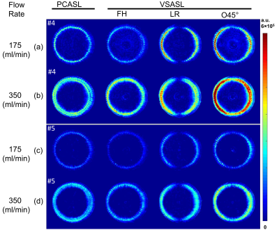 |
Velocity-Selective Inversion prepared Arterial Spin Labeling: Examination in a Commercial Perfusion Phantom
Feng Xu1,2, Dan Zhu3, Hongli Fan2,3, Hanzhang Lu1,2, Dapeng Liu1,2, Wenbo Li1,2, and Qin Qin1,2
11The Russell H. Morgan Department of Radiology and Radiological Science, Johns Hopkins University, Baltimore, MD, United States, 2F.M. Kirby Research Center for Functional Brain Imaging, Kennedy Krieger Institute, Baltimore, MD, United States, 3Biomedical Engineering, Johns Hopkins University, Baltimore, MD, United States
Standardized perfusion phantom allows for controlling reproducibility of arterial spin labeling (ASL) techniques across multiple sites, field strength, and vendors. Here using a commercial perfusion phantom, velocity-selective inversion (VSI) prepared ASL was compared with PCASL with an emphasis on velocity-encoding directions. While this effect is amplified on this phantom with feeding channels having only vertical and transverse flow directions, VSI-ASL with a tilted encoding direction achieved higher labeling efficiency through more uniform labeling of the entire feeding tubes. Careful selection of velocity-encoding directions along the major feeding arteries is recommended for VSASL applications to attain optimal labeling efficiency.
|
|||
1848.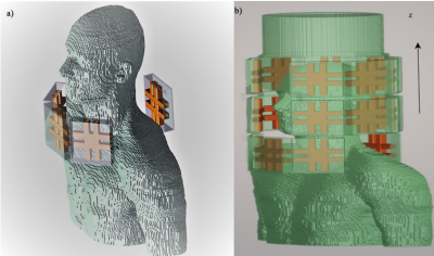 |
A separate RF Neck Coil for Arterial Spin Labeling at 7T MRI
Salem Alkhateeb1, Tales Santini2, Tiago Martins2, nadim farhat2, and Tamer S. Ibrahim2
1Bioengineering, University of Pittsburgh, Pittsburgh, PA, United States, 2University of Pittsburgh, Pittsburgh, PA, United States
A dedicated labeling coil for arterial spin labeling (ASL) technique can alleviate the challenges at 7T MRI, in this work we propose a separate 16-channel RF neck coil for transmit only. Finite-difference time-domain (FDTD) simulations and RF shimming have demonstrated the feasibility of this design to produce a homogeneous B1+ fields in the labeling region (left and right common carotid arteries) while minimizing SAR to within safe limits.
|
|||
1849.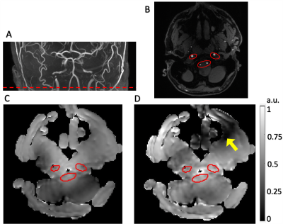 |
Increased labeling efficiency with Maxmin pTx B1+ shimming for pseudo-continuous Arterial Spin Labeling at 7T
Kai Wang1, Samantha J Ma2, Xingfeng Shao1, and Danny JJ Wang1
1University of Southern California, Los Angeles, CA, United States, 2Siemens Medical Solutions USA, Inc, Los Angeles, CA, United States
The transmit magnetic field (B1+) drop and B0 field inhomogeneity at the labeling plane make it challenging to achieve high labeling efficiency at ultrahigh field for pseudo-continuous Arterial Spin Labeling (pCASL). In this study, we designed the pCASL sequence with “Maxmin” shimming for the labeling and “MinCV” shimming for the acquisition utilizing the parallel transmission (pTx). Phantom and in-vivo experiments suggest MinCV shimming can improve the profile homogeneity within ROI and that Maxmin-shimmed labeling is a promising approach to increase the labeling efficiency for pCASL.
|
|||
1850.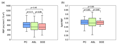 |
Blood flow measurements in diabetic kidney disease: A comparison of phase contrast, arterial spin labelling and dynamic contrast enhanced MRI
Bashair Alhummiany1, David Shelley1,2, Margaret Saysell1,2, Maria-Alexandra Olaru3, Bernd Kühn3, David L. Buckley1, Julie Bailey2, Michael Mansfield2, Steven Sourbron4, and Kanishka Sharma4
1Department of Biomedical Imaging Sciences, University of Leeds, Leeds, United Kingdom, 2Leeds Teaching Hospitals, NHS Trust, Leeds, United Kingdom, 3Siemens Healthcare GmbH, Erlangen, Germany, 4Department of Imaging, Infection, Immunity and Cardiovascular Disease, The University of Sheffield, Sheffield, United Kingdom
Kidney perfusion can be measured using DCE or ASL, but clinical confidence in the assays is undermined by the large variability in published values. In this study, an intra-subject comparison was performed of DCE and ASL against phase-contrast RBF in 25 patients with diabetic kidney disease. Results show that the three techniques agree well on average, but pairwise agreement on the single-subject level remains poor. ASL agreed better with PC than DCE, but the difference was driven by a single DCE outlier. While RBF is a useful biomarker for studies at population level, individual patient management will require further optimization.
|
|||
1851.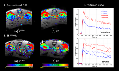 |
High Temporal Resolution Wideband Dynamic Contrast-Enhanced Magnetic resonance imaging : The Mice Renal Function Study
Wei-Hao Huang1, Chia-Ming Shih1, Po-Wei Cheng1, and Jyh-Horng Chen1,2
1Graduate Institute of Biomedical Electronics and Bioinformatics, National Taiwan University, Taipei, Taiwan, Taipei, Taiwan, 2Interdisciplinary MRI/MRS Lab, Department of Electrical Engineering, National Taiwan University, Taipei, Taiwan, Taipei, Taiwan
In this study, we aim to combine dynamic contrast enhanced magnetic resonance imaging (DCE-MRI) and Wideband technique and use this accelerated sequence to assess the renal function of mice. With temporal resolution improvement, we can get more information in the same scan time, helping to perform a more accurate analysis. The quantitative analysis based on the Tofts model was performed and compare the result to the conventional DCE. All in all, we validate the feasibility of high temporal resolution Wideband DCE.
|
|||
1852.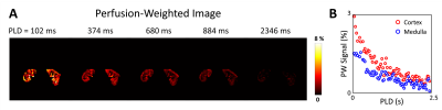 |
Free-breathing Renal Perfusion Imaging with Multi-Delay Arterial Spin Labeling Using Subspace-Based Fast MR
Paul Han1, Thibault Marin1, Yanis Djebra1,2, Georges El Fakhri1, Jinsong Ouyang1, and Chao Ma1
1Gordon Center for Medical Imaging, Department of Radiology, Massachusetts General Hospital and Harvard Medical School, Boston, MA, United States, 2LTCI, Télécom Paris, Institut Polytechnique de Paris, Paris, France
Renal perfusion imaging with multi-delay arterial spin labeling (ASL) can provide multi-parametric information along with more robust quantification of renal blood flow. However, renal perfusion imaging with ASL is challenging especially due to respiratory motion. This work presents a subspace-based fast MR method for free-breathing multi-delay ASL imaging of the kidney. The feasibility of the proposed method is shown using in vivo data obtained from a healthy volunteer on a 3T MR scanner.
|
|||
1853.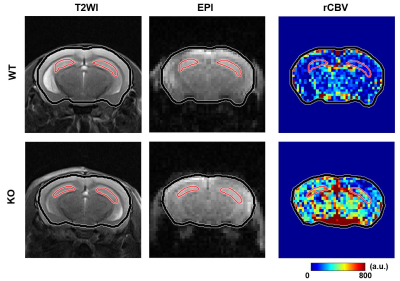 |
Quantification of Relative Cerebral Blood Volume in Aging Collapsin Response Mediator Protein 1 Gene Knockout Mice
Tzu-Ming Hung1, Sheng-Min Huang2, Yun-Chieh Tsai3, Ting-Yu Chin4, and Hsu-Hsia Peng1
1Department of Biomedical Engineering and Environmental Sciences, National Tsing Hua University, Hsinchu, Taiwan, 2Institute of biomedical engineering and nanomedicine, National Health Research Institutes, Miaoli, Taiwan, 3Graduate Institute of Life Sciences, National Defense Medical Center, Taipei, Taiwan, 4Department of Bioscience Technology, Chung Yuan Christian University, Taoyuan, Taiwan
Mice deficient of collapsin response mediator protein 1 (CRMP-1) gene may cause neural disorganization in hippocampus and demonstrate memory and spatial learning dysfunction. Relative cerebral blood volume (rCBV) can reflect the blood volume within the tissue and was served as an index to correlate with psychosis progression. The purpose of this study was to quantify the rCBV of hippocampus and to explore the difference of vascular distribution in wild type (WT) and aging CRMP-1 knockout (KO) mice. KO mice possessed significantly higher rCBV in the hippocampus than WT mice, indicating the increased blood volume in the hippocampus of KO mice.
|
|||
1854.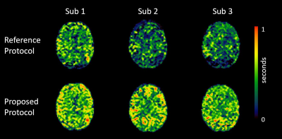 |
Robust blood brain barrier integrity measurements in clinically significant short scan time
Amnah Mahroo1, Nora-Josefin Breutigam1, Jörn Huber1, and Matthias Günther1,2
1MR Physics, Fraunhofer MEVIS, Bremen, Germany, 2MR-Imaging and Spectroscopy, University of Bremen, Bremen, Germany
Improved blood brain barrier integrity measurements in shorter scan times
|
|||
1855.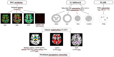 |
An increased Normal Appearing White Matter perfusion: a possible radiological inflammatory marker in relapsing-remitting multiple sclerosis
Caterina Lapucci1,2, Marco Fiorelli3, Annunziata Stefanile4, Silvana Zannino4, Maria Maddalena Filippi5, Antonio Cortese3, Carlo Piantadosi6, Marco Salvetti7, Matilde Inglese1,8, and Tatiana Koudriavtseva4
1DINOGMI, University of Genoa, Genoa, Italy, 2Department of Experimental Neurosciences, Ospedale Policlinico San Martino IRCCS, Genoa, Italy, 3Department of Human Neurosciences, Sapienza University of Rome, Rome, Italy, Rome, Italy, 4Department of Clinical Experimental Oncology, IRCCS Regina Elena National Cancer Institute, IFO, Rome, Italy, Rome, Italy, 5Fatebenefratelli Foundation, Afar Division, Fatebenefratelli Hospital, Isola Tiberina, Rome, Italy, Rome, Italy, 6Neurology Unit, San Giovanni-Addolorata Hospital, Rome, Italy, Rome, Italy, 7Department Of Neuroscience Mental Health And Sensory Organs (NEMOS), Sapienza University, Sant’Andrea Hospital, Rome, Italy, Rome, Italy, 8Department of Neurology, Icahn School of Medicine at Mount Sinai, New York, NY, USA/, New York, NY, United States
Hemodynamic changes by Dynamic Susceptibility Contrast enhanced perfusion (DSC) in Multiple Sclerosis (MS) have been poorly evaluated. The aim of the study was to compare relapsing and remitting (RR) MS patients by DSC. 45 RRMS patients, 22 with (REL) and 23 without (REM) relapse in the previous 2 months were included. A hyperperfusion of the Normal Appearing White Matter (NAWM) compared to FLAIR lesions was noted. The correlations between NAWM perfusion, disease duration and 1-year before Annualized Relapse Rate in REM patients seemed to suggest that an increased NAWM perfusion may be a radiological marker of higher inflammatory activity.
|
|||
1856.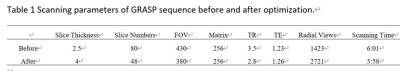 |
Image Quality Optimization: DCE imaging of the Liver at 3T using a Continuously Acquired Radial Golden-angle Compressed Sensing Acquisition
Hui Liu1, Gaofeng Shi1, Qinglei Shi2, Weishuai Wang3, Jiangyang Pan1, and Yang Li1
1Fourth Hospital of Hebei Medical University, shijiazhuang, China, 2MR Scientific Marketing, Siemens Healthcare, beijing, China, 3CS, Services,, Siemens Healthcare, jinan, China
The sequence that continuously acquired Golden-angle RAdial Sparse Parallel acquisition employing compressed sensing reconstruction (“GRASP”) can acquire high spatial and high temporal resolution as well as motion robustness to DCE MRI in liver imaging. However, there are still some artifacts in abdominal imaging, especially in the early arterial phase. In this study, we proposed an optimization scheme which can significantly improve the image quality both in plain and all enhanced phases, which may have important value in the study of abdominal disease using GRASP based DCE in future.
|
|||
1857.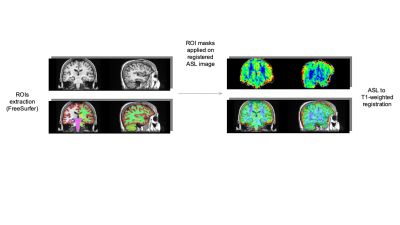 |
Disentangling the heterogeneity of MCI condition by unsupervised clustering of brain measurements on ASL and T1w MR imaging
Paolo Bosco1, Laura Biagi1, Giovanni Cioni2, Michela Matteoli3, Alessandro Sale3, Nicoletta Berardi3, Michela Tosetti1, and the Train the Brain Consortium4
1FiRMLAB, IRCCS Stella Maris Foundation, Pisa, Italy, 2IRCCS Stella Maris Foundation, Pisa, Italy, 3Institute of Neuroscience of the CNR, Pisa, Italy, 4the Train the Brain Consortium, Pisa, Italy
One of the main challenges in identifying people at risk of dementia is their clinical heterogeneity. One hypothesis is that the clinical symptoms may be the result of different biological processes. We applied a data-drive clustering approach on structural and perfusion brain-MR imaging on a cohort of 141 MCI subjects in order to elucidate homogeneous structural and perfusion profiles and we observed the correspondent clinical features. Unsupervised clustering identified 6 different clusters on both ASL and gray matter volume data. Perfusion and atrophy showed to be variable in the different clusters and showed dissimilar patterns at subcortical and cortical levels.
|
|||
1858.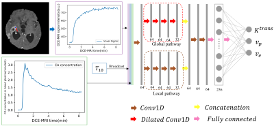 |
A Convolutional Neural Network for Accelerating the Computation of the Extended Tofts Model in DCE-MRI
Ke Fang1, Zejun Wang2,3, Zhaoqing Li2,3, Bao Wang4, Guangxu Han2,3, Zhaowei Cheng1, Zhihong Chen1, Chuanjin Lan5, Yi Zhang6, Peng Zhao7, Xinyu Jin1, Yingchao Liu8, and Ruiliang Bai2,3
1College of Information Science & Electronic Engineering, Zhejiang University, Hangzhou, China, 2Department of Physical Medicine and Rehabilitation of The Affiliated Sir Run Run Shaw Hospital AND Interdisciplinary Institute of Neuroscience and Technology, School of Medicine, Zhejiang University, Hangzhou, China, 3Key Laboratory of Biomedical Engineering of Ministry of Education, College of Biomedical Engineering and Instrument Science, Zhejiang University, Hangzhou, China, 4Department of Radiology, Qilu Hospital of Shandong University, Jinan, China, 5School of Medicine, Shandong University, Jinan, China, 6Shandong Medical Imaging Research Institute, Shandong University, Jinan, China, 7Department of Neurosurgery, Provincial Hospital Affiliated to Shandong First Medical University, Jinan, China, 8Provincial Hospital Affiliated to Shandong First Medical University, Jinan, China
We proposed a customized conventional neural network (CNN) to fasten the computation time of non-linear pharmacokinetic models in DCE-MRI. The results demonstrated that the CNN could shorten the computation time of extended Tofts model of whole-brain data to less than a minute without sacrificing the agreements with conventional non-linear least square (NLLS) fitting. This CNN could serve as an alternative to conventional NLLS fitting for fast assessment of pharmacokinetic parameters in clinical practice.
|
|||
1859.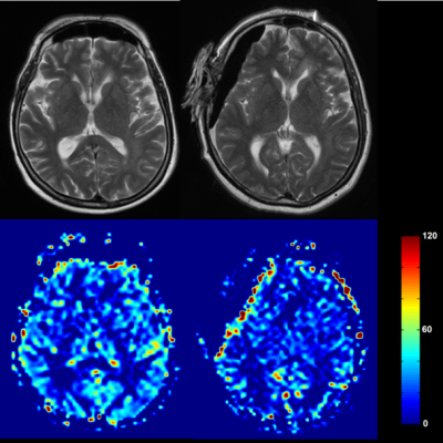 |
Changes of brain perfusion under anesthesia in humans – an explorative Arterial Spin Labeling study
Thomas Lindner1,2, Hajrullah Ahmeti3, Dana Voß3, Monika Huhndorf2, Friederike Austein1,2, Michael Helle4, Olav Jansen2, Michael Synowitz3, and Stephan Ulmer2,5
1Department of Diagnostic and Interventional Neuroradiology, University Hospital Hamburg-Eppendorf, Hamburg, Germany, 2Department of Radiology and Neuroradiology, University Hospital Schleswig-Holstein, Kiel, Germany, 3Neurosurgery, University Hospital Schleswig-Holstein, Kiel, Germany, 4Tomographic Imaging Department, Philips Research Laboratories, Hamburg, Germany, 5Radiology, Kantonsspital Winterthur, Winterthur, Switzerland
Arterial Spin Labeling allows to intraoperatively monitor changes in perfusion induced by anesthesia.
|
|||
1860.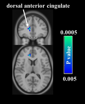 |
Brain response to acupuncture treatment in dysmenorrhea: An arterial spin labeling study
Hui-Chieh Yang1, Cheng-Hao Tu2, and Shin-Lei Peng1
1Department of Biomedical Imaging and Radiological Science, China Medical University, Taichung, Taiwan, 2Graduate Institute of Acupuncture Science, China Medical University, Taichung, Taiwan
Studies on the brain responses to the pain-relieving treatments are relative limited, especially in dysmenorrhea. In this study, we investigated the brain response to acupuncture treatment in dysmenorrhea by using the arterial spin labeling technique. Results showed that after acupuncture treatment, significant decreases in cerebral blood flow were found in the pain-related regions, such as dorsal anterior cingulate cortex in the acupuncture group while orbitofrontal cortex, caudate, and insula in the sham acupuncture group. These findings suggest that acupuncture improves the descending pain modulation system by decreasing the neuronal activity in these brain regions.
|
|||
 |
1861.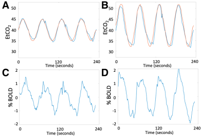 |
Perfusion mapping with sinusoidal CO2 respiratory challenge
Chau Vu1, Jian Shen1, Matthew Borzage2, Soyoung Choi3, and John Wood4
1Biomedical Engineering, University of Southern California, Los Angeles, CA, United States, 2Fetal and Neonatal Institute, Children's Hospital Los Angeles, Los Angeles, CA, United States, 3Neuroscience Graduate Program, University of Southern California, Los Angeles, CA, United States, 4Pediatrics and Radiology, Children's Hospital Los Angeles, Los Angeles, CA, United States
Dynamic susceptibility contrast MRI is a popular perfusion technique that requires the use of intravenous exogenous contrast. To avoid gadolinium injection, this study proposed a perfusion MRI method based on a CO2 respiratory challenge, which delivers a sinusoidally-modulated CO2 stimulus. We evaluated this technique at two different stimulus amplitudes and reported cerebral blood flow (CBF), cerebral blood volume (CBV) and transit time (TT) within the acceptable range of the previous literature.
|
||
1862.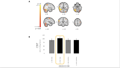 |
Evidence for a sustained cerebrovascular response following motor practice
Eleonora Patitucci1, Michael Germuska1, James Kolasinski1, Valentina Tomassini1,2,3, and Richard G Wise1,2
1CUBRIC, Cardiff University, Cardiff, United Kingdom, 2Institute for Advanced Biomedical Technologies (ITAB), Department of Neurosciences, Imaging and Clinical Sciences, University of Chieti-Pescara, Chieti, Italy, 3MS Centre, Neurology Unit, SS. Annunziata University Hospital, Chieti, Italy
Functional MRI can detect modifications in the brain’s resting state with learning-related behavioural improvements. However, the impact of learning on local cerebral blood flow after task execution remains unclear. Here we investigate changes in CBF after the execution of a motor task and demonstrate a sustained increase in resting CBF that is localised in regions relevant for the learning of the task. Our results show that learning induces sustained changes of local cerebral blood flow (over a timescale of minutes).
|
|||
1863.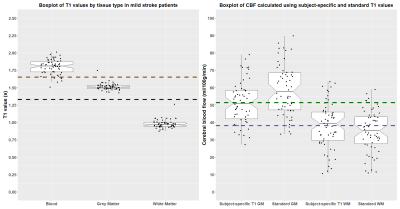 |
Effect of subject-specific T1 values for arterial spin labelling on cerebral blood flow in mild stroke patients
Michael S Stringer1,2, Cameron Manning1,2, Una Clancy1,2, Alasdair Morgan1,2, Zahra Shirzadi3,4, Francesca M Chappell1,2, Dany Jaime Garcia1,2, Angela CC Jochems1,2, Maria Valdes-Hernandez1,2, Stewart Wiseman1,2, Eleni Sakka1,2, Gordon W Blair1,2, Rosalind Brown1,2,
Bradley MacIntosh3,4, Ian Marshall1,2, Fergus Doubal1,2, and Joanna M Wardlaw1,2
1Centre for Clinical Brain Sciences, University of Edinburgh, Edinburgh, United Kingdom, 2UK DRI at the University of Edinburgh, Edinburgh, United Kingdom, 3Hurvitz Brain Sciences Research Program, Sunnybrook Health Sciences Centre, Toronto, ON, Canada, 4Department of Medical Biophysics, University of Toronto, Toronto, ON, Canada
Accurate cerebral blood flow (CBF) quantification using arterial spin labelling (ASL) relies on physiological and MR parameters. Longitudinal relaxation time (T1) of blood, which depends on haematocrit, can be a factor in some patient groups. We determined subject-specific T1 using the DESPOT-1 HIFI method in a mild stroke cohort, calculating CBF using nominal and subject-specific values. CBF calculated with subject-specific T1 values was lower in grey and higher in white matter, though there was not a proportional bias. CBF was lower in patients with higher disease burden. Subject-specific T1 values can reduce variance, potentially improving CBF quantification in clinical ASL.
|
The International Society for Magnetic Resonance in Medicine is accredited by the Accreditation Council for Continuing Medical Education to provide continuing medical education for physicians.