Digital Posters
System & Component Safety
ISMRM & SMRT Annual Meeting • 15-20 May 2021

| Concurrent 4 | 19:00 - 20:00 |
2477.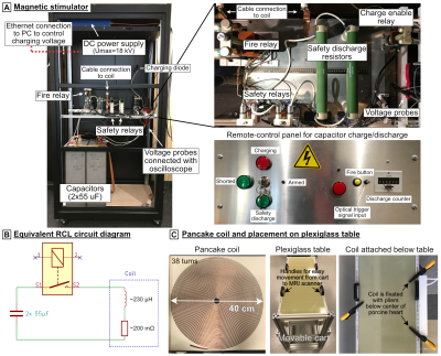 |
Device and simulation workflow for validating cardiac magneto-stimulation thresholds in porcine models
Valerie Klein1,2, Mathias Davids1,2,3, Donald Straney2, Livia Vendramini2, Lothar R. Schad1, Maaike van den Boomen2,3,4, Christopher Nguyen2,3,4, Lawrence L. Wald2,3,5, and Bastien Guerin2,3
1Computer Assisted Clinical Medicine, Medical Faculty Mannheim, Heidelberg University, Mannheim, Germany, 2A. A. Martinos Center for Biomedical Imaging, Department of Radiology, Massachusetts General Hospital, Charlestown, MA, United States, 3Harvard Medical School, Boston, MA, United States, 4Cardiovascular Research Center, Cardiology Division, Massachusetts General Hospital, Charlestown, MA, United States, 5Harvard-MIT Division of Health Sciences and Technology, Cambridge, MA, United States
We developed a magnetic stimulator to measure cardiac stimulation (CS) thresholds of time-varying magnetic fields in pigs. The stimulator consists of a high-power capacitor bank (C=110 µF) discharging into a pancake coil. We will use the measured thresholds to validate a previously developed CS simulation framework that combines electromagnetic field simulations with electrophysiological models of excitable cardiac fibers. We generate whole-body porcine voxel models from multi-bed MRI Dixon and CINE acquisitions to reproduce the anatomy and posture of the animals in our simulations. Carefully validated CS simulations may eventually be useful in setting cardiac safety limits for MRI gradient applications.
|
|||
2478.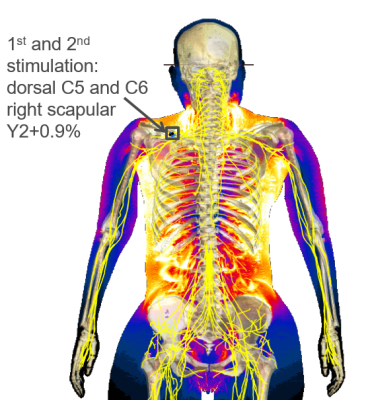 |
Are interventionalists prone to nerve stimulation during MR interventions? PNS simulation study with posable human body models
Feng Jia1, Sebastian Littin1, Philipp Amrein1, Maximilian Frederik Russe1, and Maxim Zaitsev1,2
1Department of Radiology, Medical Physics, University of Freiburg, Faculty of Medicine, Freiburg, Germany, 2High Field MR Center Center for Medical Physics and Biomedical Engineering, Medical University of Vienna, Vienna, Austria
TODO Simulation of peripheral nerve stimulation of MRI gradient coils for an interventional radiologist.
|
|||
2479.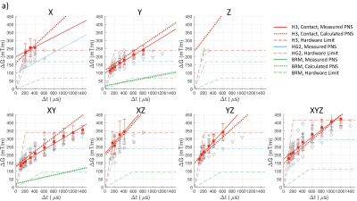 |
Electric Field Calculation and PNS Prediction for Head and Body Gradient Coils
Koray Ertan1,2, Trevor Wade3,4, Andrew Alejski4, Charles A McKenzie3, Paolo Decuzzi2, Brian Rutt1, and Peter B Roemer5
1Department of Radiology, Stanford University, Stanford, CA, United States, 2Laboratory of Nanotechnology for Precision Medicine, Italian Institute of Technology, Genoa, Italy, 3Department of Medical Biophysics, Western University, London, ON, Canada, 4Robarts Research Institute, Western University, London, ON, Canada, 5Roemer Consulting, Lutz, FL, United States
Computationally efficient methods are presented which allow calculation of switched-gradient-induced electric field distributions on realistically sized body model with uniform interior properties (compatible with regulatory IEC standards). Electric fields were calculated for three classes of gradient coils (asymmetric head, symmetric head and body gradients) using 100-member male and female body model populations; these were then used to estimate population-mean PNS parameters which were validated against experimental PNS measurements, showing high accuracy. Our computationally efficient methods can calculate whole-body E-field distributions in seconds with updates for different gradient designs in tens of milliseconds, providing an important tool for PNS-constrained gradient design.
|
|||
2480.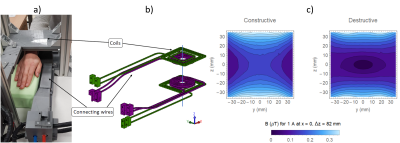 |
Peripheral Nerve Stimulation limits with fast narrow and broad-band pulses
Joseba Alonso1,2, Daniel Grau2,3, Juan Pablo Rigla3, Eduardo Pallás1,2, José Miguel Algarín1,2, José Borreguero1,2, Rubén Bosch1,2, Guillermo Comazzi1,2, Elena Díaz3, Fernando Galve1,2, José Manuel González3, Carlos Gramage1,2, Rubén Pellicer1,2,
Alfonso Ríos3, and José María Benlloch1,2
1Spanish National Research Council (CSIC), Valencia, Spain, 2Universitat Politècnica de València, Valencia, Spain, 3Tesoro Imaging S.L., Valencia, Spain
We present an apparatus for PNS threshold determination on a subject's limb, capable of narrow and broad-band magnetic excitation with pulse characteristic times down to 40 us. From measurements on 51 volunteers, we observe that PNS limits coincide for sinusoidal and triangular excitations, and are slightly lower for trapezoidal pulses. We measure significant correlations of PNS sensitivity with arm size and body weight, and none with height or gender. We confirm thresholds increase significantly for short trains also in these fast timescales. Finally, we propose our versatile low-cost system for fast offline determination of a subject's limits prior to scanning.
|
|||
2481.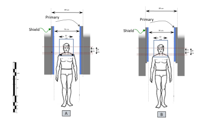 |
Design and Electric Field Analyses of a Shorter and Wider Asymmetric Gradient Coil for Whole Body MRI Scanners: A Comparison with a Symmetric Design
Afis Ajala1, Yihe Hua1, Desmond Teck Beng Yeo1, Mark Vermilyea1, and Thomas Foo1
1GE Global Research, Niskayuna, NY, United States A unique approach to providing a more open, less claustrophobic whole-body MRI is presented. A design for an asymmetric gradient coil moves the region of interest (ROI) towards one end of the gradient coil, shortening the gradient coil length from the isocenter to the patient opening. This results in a stepped configuration where the patient-bore diameter outside of the imaging ROI is substantially wider compared to using a conventional symmetric gradient coil design. The stepped patient bore has previously been used in head-only gradient systems where the greater openness has resulted in increased patient comfort and reduced claustrophobia. |
|||
2482.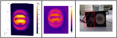 |
Thermal simulations for a high power diffusion weighting MR gradient coil
Philipp Amrein1, Feng Jia1, Sebastian Littin1, and Maxim Zaitsev1
1Dept.of Radiology, Medical Physics, University Medical Center Freiburg, Freiburg, Germany
In the context of the development of a high power MR diffusion probe for the human breast, a computer model for thermal simulation was created that allows evaluation of different duty cycles and current strengths. The proposed method includes simulating constant effective currents rather than time resolved pulse sequences. This allows reasonable computational effort to analyze the heating effects for a given diffusion weighting sequence on a given gradient system. The simulated results show reasonable agreement with experimental data.
|
|||
2483.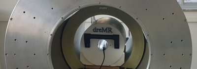 |
Induced heating of compact cryogen-free superconducting magnet during field-cycling insert coil operation
Matthew A. McCready1, William B. Handler1, and Blaine A. Chronik1
1Physics and Astronomy, Western University, London, ON, Canada
Delta relaxation enhanced magnetic resonance (dreMR) is a field-shifting quantitative molecular imaging method. The dreMR method is expected to produce higher contrast images at low-fields. Our low-field magnet has a small bore and may interact more strongly with the dreMR system causing a quench. No investigation of induced heating within the bore has been carried out for dreMR. Here, we investigate this interaction with our 0.5T superconducting magnet and find that dreMR pulse sequences do not cause significant heating in the magnet. We therefore state that dreMR is safe to carry out in such systems without quenching the magnet.
|
|||
2484.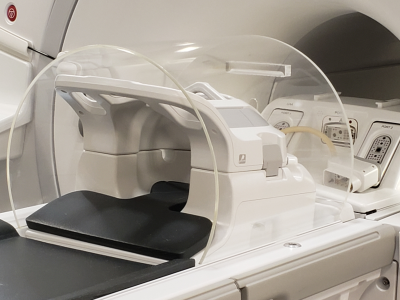 |
Evaluation of a Novel Acoustic Noise Shield for PET-MR
Chen Lin1, LeRoy H Stecker1, Brittany L Benson1, Craig A Hildestad2, Shengzhen Tao1, and Robert A Pooley1
1Radiology, Mayo Clinic, Jacksonville, FL, United States, 2Mayo Clinic, Jacksonville, FL, United States
A novel acoustic noise shield constructed of clear acrylic has been developed and evaluated. The results suggest that: 1) it is effective for MR acoustic noise reduction, 2) PET image quality exceeds ACR accreditation testing standards with the proposed acoustic shield in use, showing its compatibility with PET imaging, 3) there is a small impact on PET SUV which can be compensated with attenuation correction, and 4) it does not cause claustrophobia.
|
|||
2485.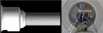 |
Acoustic evaluation of carbon fiber RF shield structure for a 3T head-only imaging system
Matthew Tarasek1, Tom Foo1, Mark Vermilyea1, Desmond Yeo1, Isabelle Jansen1, Eric Budesheim 1, and Keith Park1
1GE Global Research, Niskayuna, NY, United States
In this work we evaluate the MRI acoustic changes brought about by modifying the RF shield structure in a head-only (MAGNUS) 3T imaging system. Specifically, we use a standardized acoustic measurement protocol to compare sound pressure levels (SPL) in a fiberglass patient-bore structure to a similar newly developed carbon fiber reinforced polymer (CFRP) structure. Results indicate that a modification of the structural properties of the patient-bore structure properties can provide SPL reduction for MR imaging applications, and is likely due to the different modulus of elasticity between the two materials.
|
|||
2486.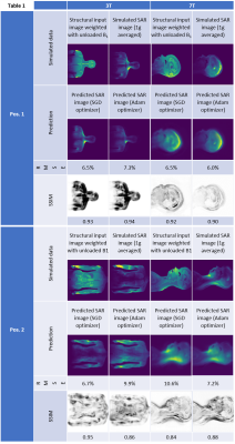 |
MRSaiFE: towards the real-time prediction of tissue heating in MRI - a feasibility study
Simone Angela Winkler1, Elizaveta Motovilova1, Sayim Gökyar1, Isabelle Saniour1, Fraser Robb2, and Akshay Chaudhari3
1Department of Radiology, Weill Cornell Medicine, New York, NY, United States, 2GE Healthcare, Aurora, OH, United States, 3Stanford University, Stanford, CA, United States
A crucial safety concern for UHF MRI is the significant RF power deposition in the body in the form of local specific absorption rate (SAR) hotspots, leading to dangerous tissue heating/damage. This work is a proof-of-concept demonstration of an artificial intelligence (AI) based real-time MRI safety prediction software (MRSaiFE) facilitating safe generation of 3T and 7T images by means of accurate local SAR-monitoring at sub-W/kg levels. This feasibility study demonstrates that SAR patterns can be predicted with a root-mean-square error (RMSE) of <11% along with a structural similarity (SSIM) level of >84% for both field strengths.
|
|||
2487.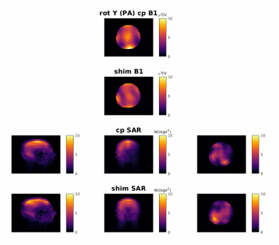 |
Motion and Pose Variability of SAR Estimation with Parallel Transmission at 7T
Amer Ajanovic1, Joseph V Hajnal1,2, Raphael Tomi-Tricot1,3, and Shaihan Malik1
1Biomedical Engineering Department, School of Biomedical Engineering and Imaging Sciences, King's College London, London, United Kingdom, 2Centre for the Developing Brain, School of Biomedical Engineering and Imaging Sciences, King's College London, London, United Kingdom, 3MR Research Collaborations, Siemens Healthcare Limited, Frimley, United Kingdom
Contemporary safety assessment for parallel transmit (pTx) MRI relies on computationally expensive electromagnetic (EM) simulations; hence, safety evaluations rely on the use of small numbers of models. The effect of patient movement inside the coil on patient safety is, therefore, difficult to capture. In this work we compare 'within-scan' variability with the alternative scenario in which a subject’s head is positioned arbitrarily within the coil, but that this position is different to the one used for the SAR estimation model; we refer to this as ‘pose’ variability.
|
|||
2488.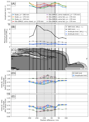 |
Robustness of pTx safety concepts to varying subjects and subject positions
Johannes Petzold1, Bernd Ittermann1, and Frank Seifert1
1Physikalisch-Technische Bundesanstalt (PTB), Braunschweig and Berlin, Germany
This simulation study compares two concepts ensuring IEC-compliant SAR values for parallel transmission (pTx) in respect to their robustness against subject- and position-changes: 1) direct SAR assessment (SL) and 2) an amplitude limit for all pTx channels (AL). For the example case of a 3T pTx body coil, SL resulted in ten-fold the allowed SAR in one model for the same excitation vector that obeys IEC limits in another model. For AL, less than twice the SAR was found in this case. AL allows for a lower safety factor, therefore, resulting in a higher mean(B1+) compared to SL.
|
|||
2489.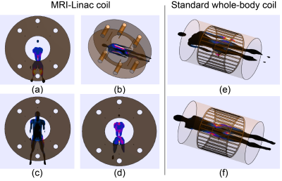 |
Radiofrequency safety evaluation and management of the Australian MRI-Linac system
Mingyan Li1, Ewald Weber1, Aurelien Destruel2, Feng Liu1, and Stuart Crozier1
1School of Information Technology and Electrical Engineering, The University of Queensland, Australia, Australia, 2Center for Magnetic Resonance in Biology and Medicine, Aix-Marseille University, MARSEILLE, France
The Australian MRI-Linac system has a split magnet with wide opening, which offers treatment flexibility with different patient positioning. However, the versatile patient setup and individual physique differences also lead to uncertainty for specific absorption rate (SAR) management. In particular, a standing patient may have body regions close to radiofrequency (RF) coil rungs, which may lead to high local SAR. In this work, numerical simulations were employed to investigate the SAR distribution while a human model was in different treatment positions and thus give safety management suggestions. A standard whole-body RF coil was also simulated as a reference.
|
|||
2490.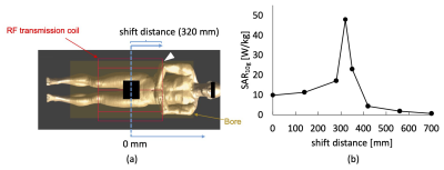 |
Electromagnetic simulation analysis of an RF burn injury case occurred at the elbow-bore wall contact point
Minghui Tang1, Kiyoi Okamoto2, and Toru Yamamoto1
1Faculty of Health Sciences, Hokkaido University, Sapporo, Japan, 2Graduate School of Health Sciences, Hokkaido University, Sapporo, Japan
RF burn injuries occasionally occur at the contact points of skin and the bore wall, albeit out of common belief of hazardous loop forming. To investigate the mechanism of the RF burn injury occurred by the bore wall contact, we modeled an elbow-bore wall contact case on an electromagnetic analysis software by using the computable human phantom. The simulated SAR at the contact point varied up to 480% depending on the patient position in a scanner. This tendency was in accordance with the simulated electric field distribution in the scanner which is dominantly determined by the RF transmission coil.
|
|||
2491.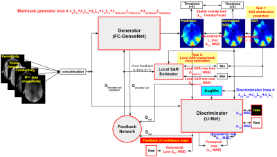 |
A Novel Specific Absorption Rate Prediction Framework Using Multi-Task Feedback Generative Adversarial Learning: Application to 10.5 T Head MRI
Jinyoung Kim1, Alireza Sadeghi-Tarakameh1, Angel Torrado-Carvajal2,3, and Yigitcan Eryaman1
1Center for Magnetic Resonance Research, Department of Radiology, University of Minnesota, Minneapolis, MN, United States, 2Athinoula A. Martinos Center for Biomedical Imaging, Department of Radiology, Massachusetts General Hospital and Harvard Medical School, Boston, MA, United States, 3Medical Image Analysis and Biometry Laboratory, Universidad Rey Juan Carlos, Madrid, Spain
In this study, we propose a novel local SAR prediction framework based on deep learning. To this end, we introduce multi-task feedback adversarial learning to simultaneously predict local SAR distribution and its peak SAR value. The proposed model learns a mapping between simulated B1+ magnitude/tissue property maps and local SAR provided by EM simulations. Given query inputs with the properly trained model, the generator produces the local SAR distribution slice by slice, and the local SAR peak estimator predicts the upper bound of local SAR values. Validation results show that the proposed model may allow online subject-specific local SAR prediction.
|
|||
2492. |
Realistic model of the 3T Siemens Connectom birdcage coil and its validation
Mikhail Kozlov1, Kerrin J. Pine1, and Nikolaus Weiskopf1,2
1Max Planck Institute for Human Cognitive and Brain Sciences, Leipzig, Germany, 2Felix Bloch Institute for Solid State Physics, Leipzig, Germany
We devised a numerical model of a 123.2MHz 32-rung high-pass whole-body birdcage coil of the Siemens 3T Connectom scanner. The model was developed based on realistic geometrical data for most of coil elements and a reverse engineering approach. Our simulations suggest that RF exposure at level of 3.2W/kg head SAR may result in higher than 20W/kg of SAR10g in the Connectom scanner; (ii) B1+ mapping alone cannot be used for validation of numerical model of whole-body birdcage coil, (iii) developed RF-circuit and 3D-EM co-simulation approach with reverse engineering resulted in a reasonable agreement between simulations and measurements in human subject.
|
|||
2493.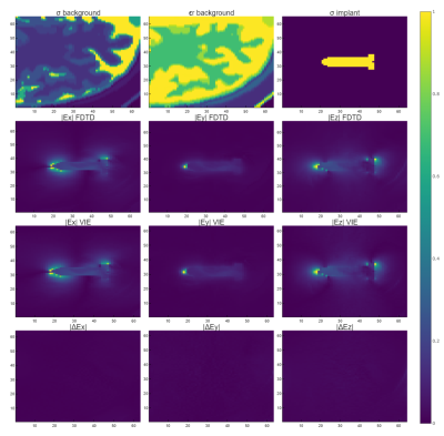 |
GPU accelerated calculations of the scattered RF-field due to a dielectric update without extensive pre-calculated data.
Peter Stijnman1, Bart Steensma1, Cornelis van den Berg1, and Alexander Raaijmakers1,2
1Computational Imaging, UMC Utrecht, Utrecht, Netherlands, 2Biomedical Engineering, Eindhoven University of Technology, Eindhoven, Netherlands
Previous work showed that the scattered RF-fields created by dielectric pads and medical implants can quickly be calculated. The method requires an extensive offline calculation stage and a lot of memory. We propose an extension to the method that removes these two problems by using a volume integral equation solver. This allows us to do the entire calculation on a GPU for more acceleration. The resulting scattered electric fields have a maximum difference of 3.2% with respect to FDTD, while currently being a 106 times faster. Eventually, we want to use this method for patient-specific implant RF-safety assessment.
|
|||
2494.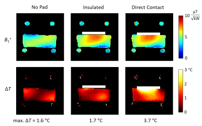 |
RF Safety Assessment of High Permittivity Dielectric Pads - Impact of Material Insulation
Wyger Brink1, Rob Remis2, and Andrew Webb3
1Leiden University Medical Center, Leiden, Netherlands, 2Circuits and Systems, dept. of Microelectronics, Delft University of Technology, Delft, Netherlands, 3C.J. Gorter Center, dept. of Radiology, Leiden University Medical Center, Leiden, Netherlands
High permittivity dielectric pads are known to be effective for tailoring the RF field and improving image quality in high field MRI systems. Despite the number of studies reporting benign SAR effects, their safety remains a concern and should be evaluated on an application-specific basis using RF simulations. In this work we demonstrate the impact of insulation material on the RF safety of high permittivity pads, using both RF simulations as well as phantom experiments.
|
|||
2495.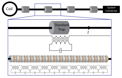 |
Advances in Caterpillar Traps: A Highly Flexible, Distributed System of Toroid Cable Traps
Ekin Karasan1, Alison Hammerschmidt1, Victor Taracila2, Fraser Robb2, and Michael Lustig1
1University of California, Berkeley, CA, United States, 2GE Healthcare, Coils, Aurora, OH, United States
Conducting wires within the scanner may interact with the transmit field causing high shield currents and posing safety hazards. Closely spaced RF traps are used to mitigate common-mode currents. However, these bulky and rigid traps hinder the flexibility of the cable. We previously proposed caterpillar traps: a distributed system of smaller, flexible toroids covering the full length of the cable. Their potential to attenuate shield currents while being flexed was demonstrated. This work makes several improvements to the manufacturing process to reduce time and cost and improve usability. Furthermore, we present results from experiments to evaluate their performance and robustness.
|
|||
2496.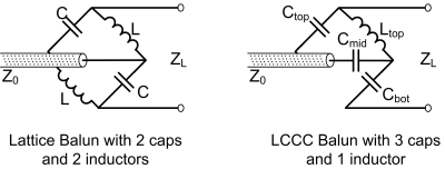 |
LCCC Balun for RF Coil
Xinqiang Yan1,2 and John C. Gore1,2
1Vanderbilt University Institute of Imaging Science, Nashville, TN, United States, 2Department of Radiology and Radilogical Science, Vanderbilt University Medical Center, Nashville, TN, United States
We introduced a novel LCCC lumped-element balun to suppress the unwanted common-mode current in RF coils and RF systems. Unlike the 4-element Lattice balun consisting of two capacitors and two inductors (LCCL), this design has 3 capacitors and only 1 inductor. With fewer inductors (previous lumped-element baluns need at least 2 inductors), this LCCC balun can further be miniaturized and has lower insertion loss.
|
The International Society for Magnetic Resonance in Medicine is accredited by the Accreditation Council for Continuing Medical Education to provide continuing medical education for physicians.