Digital Posters
Hot-Wired Systems: Gradients & Magnets
ISMRM & SMRT Annual Meeting • 15-20 May 2021

Digital Posters
Hot-Wired Systems: Gradients & Magnets
| Concurrent 2 | 17:00 - 18:00 |
3085.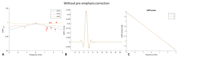 |
Gradient Characterization for High Performance Gradients
Nastaren Abad1, Afis Ajala1, Yihe Hua1, and Tom K.F Foo1
1General Electric Global Research, Niskayuna, NY, United States
Impulse response function (IRF) harvesting is a powerful technique for gradient characterization. In this study a field monitoring system was utilized to characterize a head-only, high-performance gradient coil (MAGNUS), enabling expansion of the spatial response pattern into higher order spherical harmonics. The predictive power of IRF-estimated waveforms and field evolution is demonstrated by comparing to nominal and active field monitored approaches for arbitrary gradient waveforms. The calculated modulation function indicated good correspondence for arbitrary waveforms and k-space trajectories varying with slew rate, amplitudes and number of interleaves.
|
|||
3086.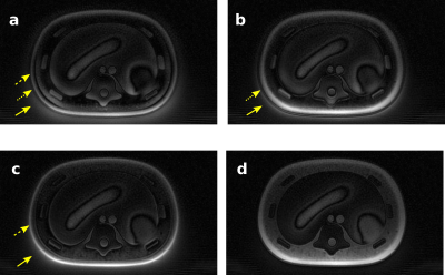 |
Measuring the Gradient Impulse Response Function (GIRF) for UTE Imaging on an MR-Linac
Rosie Goodburn1, Tom Bruijnen2, Wajiha Bano1, Uwe Oelfke1, and Andreas Wetscherek1
1Radiotherapy and Imaging, The Institute of Cancer Research, London, United Kingdom, 2Department of Radiotherapy, University Medical Center Utrecht, Utrecht, Netherlands
Synthetic CT (sCT) is a key component of adaptive MR-guided radiotherapy. Generation of thoracic sCT is challenging and benefits from ultrashort echo-time (UTE) imaging that provides contrast in short-T2* tissues such as bone and lung. However, UTE imaging is susceptible to artifacts caused by eddy currents and gradient delays. In this work, we measured the gradient impulse response function (GIRF) of an MR-Linac with standard scanner hardware and used it to retrospectively correct k-space trajectories. Images reconstructed with GIRF and gradient-delay corrected trajectories exhibited higher image quality, including reduced halo artifacts, blurring, destructive phase artifacts, and improved uniformity and contrast.
|
|||
3087.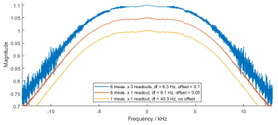 |
Phantom-based high-resolution measurement of the gradient system transfer function
Hannah Scholten1, Manuel Stich2, and Herbert Köstler1
1Department of Diagnostic and Interventional Radiology, University of Würzburg, Würzburg, Germany, 2Siemens Healthcare, Erlangen, Germany
The gradient system transfer function (GSTF) constitutes a comprehensive tool for correcting k-space trajectory distortions due to gradient infidelities. We developed a new phantom-based measurement approach that allows determining the GSTF with a high frequency resolution without relying on long readout durations. Our first results include self- and B0 cross-terms with high SNR and resolutions below 10 Hz, resolving the true width of several resonances. The high resolution enables us to capture long-lasting eddy current effects, which is especially promising for the application of GSTF-based correction methods in, for example, diffusion-weighted imaging.
|
|||
3088.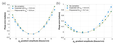 |
Calibration of Concomitant Field Compensation using Phase Contrast MRI
Thomas K.F. Foo1, Louis Frigo2, Myung-Ho In3, Nastaren Abad1, Vincent B Ho4, and Matt A Bernstein3
1GE Research, Niskayuna, NY, United States, 2GE Healthcare, Waukesha, WI, United States, 3Mayo Clinic, Rochester, MN, United States, 4Uniformed Services University of the Health Sciences, Bethesda, MD, United States
A phase contrast pulse sequence was modified to use a single-sided bipolar encoding to calibrate the transverse gradient offsets for asymmetric gradient coils. The phase difference approach cancels the concomitant gradient field effects from all gradient waveforms except for the bipolar gradient waveforms. By fitting the measured phase offset in the phase difference images to the applied bipolar gradient amplitudes, the gradient offset value can be calculated. This was used for pre-emphasis compensation for the zeroth and first order concomitant gradient fields.
|
|||
3089.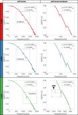 |
Direct Comparison of Gradient Modulation Transfer Functions and Acoustic Noise Spectra of the same MRI at High- (3T) and Lower-Field (0.75T)
Hannes Dillinger1, Sebastian Kozerke1, and Christian Guenthner1
1Institute for Biomedical Engineering, University and ETH Zurich, Zurich, Switzerland
In this work, gradient fidelity is compared between high-field (3T) and lower-field (0.75T) configurations on the same MRI system by measuring the Gradient Modulation Transfer Function (GMTF) and audio noise spectra. Results demonstrate that at lower field mechanical resonances are reduced, leading to improved gradient fidelity and reduction of audio noise levels of up to 70%. Furthermore, it is shown that spectral peaks in the audio spectrum can be related to resonances in the GMTF, which suggests mechanical resonances can be identified using the audio spectrum.
|
|||
3090.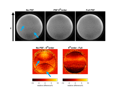 |
PSF-based reconstruction for removal of artifacts caused by misalignment between a silent gradient insert and the body gradients
Edwin Versteeg1, Dennis W.J. Klomp1, and Jeroen C.W. Siero1,2
1Radiology, University Medical Center Utrecht, Utrecht, Netherlands, 2Spinoza centre for neuroimaging Amsterdam, Amsterdam, Netherlands
A silent gradient axis driven at 20 kHz can reduce sound in MR-sequences. This silent gradient axis consists of a separate coil that is positioned on the patient table and operated in synergy with the whole-body gradients. The positioning of this coil with respect to the whole-body gradients is prone to operator errors which leads to image artifacts. In this work, we show that this misalignment can be characterized and corrected by measuring the point spread function (PSF) of the silent gradient axis. Using this method, a significant reduction in ghosting artifacts was observed in phantom experiments.
|
|||
3091.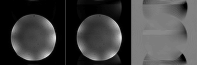 |
Eddy current correction for field probes mounted in a head coil
Jennifer Nussbaum1, Maria Engel1, and Klaas Paul Pruessmann1
1Institute for Biomedical Engineering, ETH Zurich and University of Zurich, Zurich, Switzerland
Concurrent acquisition of dynamic fields has become a prominent way to correct for field imperfections. However, when using field probes in close proximity to a head coil, the field measurement can be corrupted by eddy currents on the head coil that are induced by the gradient fields. In this work, we provide a one-time calibration solution to correct for this issue.
|
|||
3092.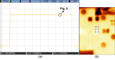 |
Design and Implementation of High Switching Frequency Gradient Power Amplifier Using eGaN Devices
Soheil Taraghinia1, Volkan Acikel2, Reza Babaloo1,3, and Ergin Atalar1,3
1UMRAM, Bilkent University, Ankara, Turkey, 2Aselsan A.S., Ankara, Turkey, 3Electrical and Electronics Engineering, Bilkent University, Ankara, Turkey
In this work, a switching H-bridge gradient power amplifier utilizing eGaN devices is implemented. A single-stage 150 V/ 50 A full-bridge GPA for an insert gradient array system at 1 MHz effective switching frequency is fabricated and tested. Pulse width modulation signals are generated digitally using a Virtex 7 family FPGA board. A single-stage LC low pass filter is designed to attenuate the ripple current.
|
|||
3093.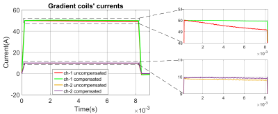 |
Droop compensation of gradient current waveforms in gradient array systems
Reza Babaloo1,2, Soheil Taraghinia2, and Ergin Atalar1,2
1Department of Electrical and Electronics Engineering, Bilkent University, Ankara, Turkey, 2National Magnetic Resonance Research Center (UMRAM), Ankara, Turkey
Providing accurate gradient currents is challenging due to the nonlinearity of the gradient system arising from gradient power amplifiers and power supply stages which causes droop in the output currents. This work introduces a nonlinear model for the gradient array system by using the state-space averaging technique and the nonlinear inversion of the model compensates for droop. The feasibility of the method is depicted by simulation and experimental results. The proposed method can provide desired currents by lower voltages which results in minimizing the needed power. It is also possible to use small capacitors to lower the cost of system.
|
|||
3094.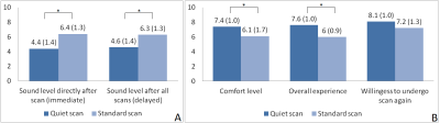 |
Fast and quiet MPRAGE using a silent gradient axis at 7T – subject experience and qualitative image assessment
Sarah M Jacobs1, Edwin Versteeg1, Leonie NC Visser2, Anja G van der Kolk1,3, Dennis WJ Klomp1, and Jeroen CW Siero1,4
1Department of Radiology and Nuclear Medicine, University Medical Center Utrecht, Utrecht, Netherlands, 2Alzheimer Center Amsterdam, Department of Neurology, Amsterdam Neuroscience, Vrije Universiteit Amsterdam, Amsterdam UMC, Amsterdam, Netherlands, 3Department of Radiology, the Netherlands Cancer Institute, Amsterdam, Netherlands, 4Spinoza Centre for Neuroimaging Amsterdam, Amsterdam, Netherlands
Acoustic noise can negatively impact patients from anxiety and communication problems to transient and permanent hearing loss. In this pilot study we used a silent gradient axis that is switched at the inaudible frequency of 20 kHz with a silent readout module implemented in an MPRAGE sequence, and investigated subject experience and image quality of fast and quiet anatomical brain imaging at 7T. Here we show preliminary evidence that our silent gradient axis with silent readout module incorporated into a T1-weighted MPRAGE sequence is perceived more quiet and positive and delivers images of largely acceptable quality.
|
|||
3095.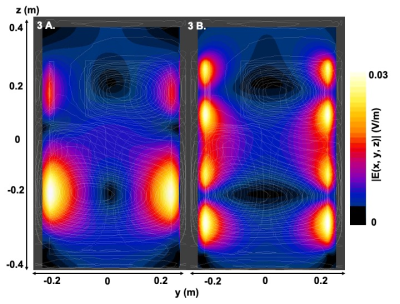 |
Gradient induced electric field within a shoulder cut-out gradient coil built for head and neck imaging.
Arjama A Halder1,2, Eric J Lessard1,2, William B Handler1, and Blaine A Chronik1,2
1xMR Labs, Physics and Astronomy, London, ON, Canada, 2Medical Biophysics, Western University, London, ON, Canada
This abstract aims to investigate the electromagnetic environment within a high-performance shoulder cut-out gradient coil designed to be used as a part of an all-in-one platform for head and neck imaging applications. The results achieved in this abstract and future measurements that will be performed with this coil will allow the quantification for gradient induced electric field effects experienced by patients and ensure that the coil is within the expected PNS limits.
|
|||
3096.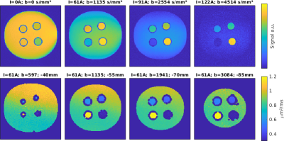 |
Approaching order of magnitude increase of gradient strength: Non-linear breast gradient coil for diffusion encoding
Sebastian Littin1, Tristan A. Kuder2, Feng Jia1, Arthur Magill2, Philipp Amrein1, Frederik B. Laun3, Sebastian Bicklehaupt4, Mathias Davids5,6,7, Valerie Klein5,7, Mark E. Ladd2, and Maxim Zaitsev1,8
1Department of Radiology, Medical Physics, University Freiburg, Faculty of Medicin, Freiburg, Germany, 2Medical Physics in Radiology, German Cancer Research Center, Heidelberg, Germany, 3Department of Radiology, MR Physics, University Medical Center Erlangen, Erlangen, Germany, 4Department of Radiology, University Medical Center Erlangen, Erlangen, Germany, 5Department of Radiology, A. A. Martinos Center for Biomedical Imaging, Charlestown, MA, United States, 6Harvard Medical School, Boston, MA, United States, 7Computer Assisted Clinical Medicine, Heidelberg University, Medical Faculty Mannheim, Heidelberg, Germany, 8High Field MR Center Center for Medical Physics and Biomedical Engineering, Medical University of Vienna, Vienna, Austria
Local gradient coils allow for enhanced performance in terms of both gradient amplitude and switching rate. We recently presented the concept of a single channel non-linear breast gradient coil. Here we show results of the characterization, safety assessment and initial diffusion encoded images from phantom experiments of the prototype coil. Is an order of magnitude increase of gradient strength for diffusion weighting feasible using local gradient coils?
|
|||
3097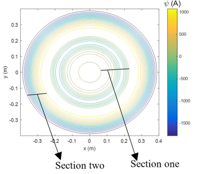 |
Towards a More Power Efficient Two-Channel Biplanar Z-Gradient Coil Design Using Target Field Method Video Permission Withheld
Haile Baye Kassahun1, Sadeq S Alsharafi1, Ahmed M Badawi1, and AbdEl-Monem M El-Sharkawy1
1Systems and Biomedical Engineering, Cairo University, Cairo, Egypt
A two-channel biplanar Z-gradient coil design is introduced by dividing the conventional coil surface into two sections radially. The corresponding lower and upper sections are driven by the same current strength but in opposite directions. Coils were designed using the Fourier series expansion (target field) method. For the same target field, the dissipated power of the two-channel biplanar Z-gradient coil was lower than that of the conventional coil (with corresponding dimensions) by at least 25% thereby reducing the ohmic losses. In the future, the benefit of using multi-channel bi-planar gradient coils may be further investigated.
|
|||
3098.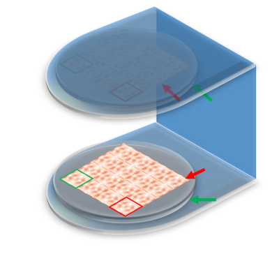 |
Design of Biplanar Matrix Z2 Nonlinear Gradient Coil with An Open Structure for O-Space imaging
Congcong Liu1,2, Shi Su1, Ye Li1,2, Xin Liu1,2, Hairong Zheng1,2, Dong Liang1,2,3, and Haifeng Wang1,2
1Paul C. Lauterbur Research Center for Biomedical Imaging, Shenzhen Institutes of Advanced Technology, Chinese Academy of Sciences, Shenzhen, China, 2Shenzhen College of Advanced Technology, University of Chinese Academy of Sciences, Shenzhen, China, 3Research Center for Medical AI, Shenzhen Institute of Advanced Technology,Chinese Academy of Sciences, Shenzhen, China
In this work, a novel biplanar matrix Z2 nonlinear gradient coil with an open structure has been designed, which could be employed in open spatial low-field MRI system, and an efficient encoding strategy for O-Space imaging was proposed in combination with the proposed biplanar matrix nonlinear gradient coil, which has the potential to shift the center placements (CP’s) in nonlinear gradient imaging without an external linear gradient field. The biplanar Z2 encoding field generated by a nonlinear gradient element consisting of curve segments was optimized and implemented to show the feasibility of this proposed biplanar matrix coil and encoding approach.
|
|||
3099.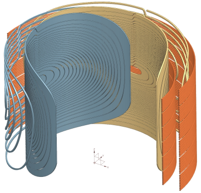 |
MRI Hybrid Gradient Coil Equipped with a Programmable Z-Array and Conventional X- and Y- Elements
Manouchehr Takrimi1 and Ergin Atalar1,2
1UMRAM, Bilkent University, Ankara, Turkey, 2Department of Electrical and Electronics Engineering, Bilkent University, Ankara, Turkey
A novel hybrid gradient coil consisting of two conventional actively shielded x/y gradient coils and a programmable active-shield z-gradient array coil is introduced and simulated. The proposed hybrid gradient coil can dynamically provide the required magnetic profile based on diverse applications in MRI. This is achieved by using a set of independent power amplifiers that feed the conventional coils and the array elements. To show the flexibility, three magnetic profiles are simulated: (a) a conventional profile; (b) a profile with twice z-gradient intensity and a shifted FOV; (c) a profile with quadruple z-gradient intensity when the active-shield is reverse fed.
|
|||
3100.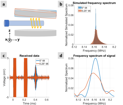 |
Mechanical Tilt-Induced Gradient Fields for Low-Field Spokes-and-Hub Permanent Magnet MR Imagers
Irene Kuang1, Jason Stockmann2,3, Elfar Adalsteinsson1,4, and Jacob White1
1Electrical Engineering and Computer Science, Massachusetts Institute of Technology, Cambridge, MA, United States, 2A. A. Martinos Center for Biomedical Imaging, Massachusetts General Hospital, Charlestown, MA, United States, 3Harvard Medical School, Boston, MA, United States, 4Institute for Medical Engineering and Science, Massachusetts Institute of Technology, Cambridge, MA, United States
As recently demonstrated by Cooley et al., for Halbach-array based permanent-magnet MR imagers, time-varying encoding fields can be generated mechanically, by rotating the entire array. In this paper we show that in spokes-and-hub based permanent magnet imagers, time-varying encoding fields can also be generated mechanically, but with much smaller motions. In particular, we show that a quarter-degree magnet tilt, along an easily precessed axis, can produce a spatially-discriminating field gradient. We demonstrate the spatial discrimation using both simulation and measurements from our recently-presented prototype imager.
|
|||
| 3101. | Systematic, Linear Algebra-based Dimensional Analysis of Gradient Inductance Scaling with Coil Radius
Matt A Bernstein1 and Seung-Kyun Lee2
1Radiology, Mayo Clinic, Rochester, MN, United States, 2Biomedical Engineering, Sungkyunkwan University, Suwon, Korea, Republic of
Applying systematic dimensional analysis based linear algebraic methods, we derive the important scaling relation for MR gradient design: gradient coil inductance L scales as the square gradient efficiency (measured in T/m/A) and the fifth power of radius. The inclusion of "turns" as a unit in the dimensional matrix produces the result uniquely, greatly simplifying the analysis.
|
|||
3102.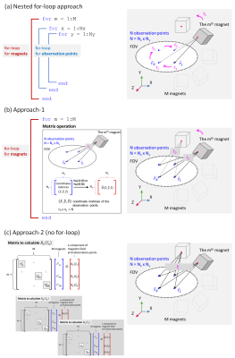 |
Acceleration of magnetic field calculation of permanent magnet arrays for their optimizations
Ting-Ou Liang1, Yi-Dan Chen2, Shao Ying Huang2, and Erping Li1
1Zhejiang University, Hangzhou, China, 2Singapore University of Technology and Design, Singapore, Singapore
Two current-model-based approaches are presented to accelerate the calculation of the magnetic field of permanent-magnet-array(PMA) that consists of magnet blocks. It enables a speedy convergence of optimization of a PMA design that is a key component of the popular body-part-dedicated portable MRI in the recent years. This method can be extended to magnet of other shapes. Both Halbach(60blocks) and sparse Inward-Outward ring-pair-PMA(1,200 blocks) are used as examples to demonstrate the speed and accuracy of the algorithm. The best acceleration is 78.92% and 77.94%calculation-time reduction compared to the conventional nested for-loops, and 99.81% and 99.91%reduction compared to FEM-based commercial software.
|
|||
3103.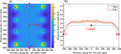 |
Design and Development of a Hybrid Helmholtz Coil System for Production of Low Magnetic Field System (up to 7 mT)
Yenal Gokpek1 and Ozkan Doganay1
1Institute of Health Sciences, Ege University, IZMIR, Turkey
In this study, the design and construction of a hybrid Helmholtz coil system and numerical modelling were investigated for production of homogeneous magnetic field over a volume of interest that consist of cylindrical geometry (h=700mm, r=90mm). The 4‑coil system was constructed and tested by two commercially available low power DC power supplies. The magnetic field created by the 4‑coil system was found to be B=2.5182±0.0035mT over the volume of interest. We found that the simulated and measured magnetic fields are in good agreement (R=0.9759, p<0.05).
|
|||
3104.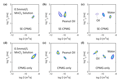 |
D-T2 Distribution Obtained Using CPMG-only Sequence Compared with Traditional SE-CPMG Sequence on the Single-sided NMR Device
Ziyi Pan1, Jieying Zhang1, Hai Luo2, Weiqian Wang2, Xiao Chen2, Ziyue Wu2, and Hua Guo1
1Center for Biomedical Imaging Research, Department of Biomedical Engineering, School of Medicine, Tsinghua University, Beijing, China, 2Marvel Stone Healthcare, Wuxi, Jiangsu, China
Two-dimension (2D) diffusion-relaxation (D-T2) correlation measured by a low-cost, portable single-sided NMR system can separate fat and water in biological tissues (for example for liver steatosis detection). Diffusion-editing CPMG sequence is commonly used for such measurement. Recently, a novel approach is proposed to measure the D-T2 correlation using the simplest CPMG-only sequence. The CPMG-only sequence is less sensitive to motion, and can extend to systems with more imperfect magnetic and radiofrequency fields. This work compares the proposed CPMG-only sequence with the traditional diffusion-editing SE-CPMG sequence. Results show the SE-CPMG and CPMG-only sequences can achieve comparable performance on water-fat separation.
|
The International Society for Magnetic Resonance in Medicine is accredited by the Accreditation Council for Continuing Medical Education to provide continuing medical education for physicians.