Digital Posters
MR-Guided Interventions: Methods
ISMRM & SMRT Annual Meeting • 15-20 May 2021

Digital Posters
MR-Guided Interventions: Methods
| Concurrent 3 | 19:00 - 20:00 |
4242.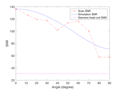 |
Exploration of the Surgical Placement of the Local Pituitary Coil for Microadenomas
Jiahao Lin1,2, Siyuan Liu2, Rock Hadley3, Marvin Bergsneider4, Giyarpuram N Prashant4, Sophie Peeters4, Robert Candler2,5, and Kyunghyun Sung1
1Department of Radiological Sciences, David Geffen School of Medicine, University of California, Los Angeles, LOS ANGELES, CA, United States, 2Department of Electrical and Computer Engineering, University of California, Los Angeles, LOS ANGELES, CA, United States, 3Department of Radiology, University of Utah, Salt Lake City, UT, United States, 4Department of Neurosurgery, University of California, Los Angeles, LOS ANGELES, CA, United States, 5California NanoSystems Institute, Los Angeles, CA, United States
We construct an agar phantom to examine the SNR variation as the coil plane rotates from 0° to 90°, respect to B0. Our local pituitary coil has improved SNR with a factor, ranged from 3.3 to 4.2, compared to the commercial Siemens head coil as long as the surgical positioning of the coil to be within 0° and 70° with respect to the B0 field.
|
|||
4243.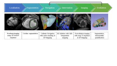 |
Toward automatic lesion transmurality assessment using machine learning: a proof of concept in preclinical EP studies under MRI-guidance
Valéry Ozenne1,2,3,4, Pierre Bour2,3,4, Marylène Delcey2,3,4, Nicolas Cedilnik5, Maxime Sermesant5, and Bruno Quesson2,3,4
1Centre de Résonance Magnétique des Systèmes Biologiques, UMR 5536, CNRS, Bordeaux, France, 2IHU Liryc, Electrophysiology and Heart Modeling Institute, Fondation Bordeaux Université, Bordeaux, France, 3Univ. Bordeaux, Centre de recherche Cardio-Thoracique de Bordeaux, U1045, Bordeaux, France, 4INSERM, Centre de recherche Cardio-Thoracique de Bordeaux, U1045, Bordeaux, France, 5Université Côte d’Azur, Inria, Epione, Sophia Antipolis, France MR-guidance of electrophysiological (EP) procedures requires manual segmentation of the cardiac cavities either at the beginning of the procedure to produce the roadmap volume or after radiofrequency ablation (RFA) to assess the lesion transmurality in post ablation images. The purpose of this work is to evaluate the feasibility of automatic in-line segmentation in the context of routine preclinical EP studies. |
|||
4244.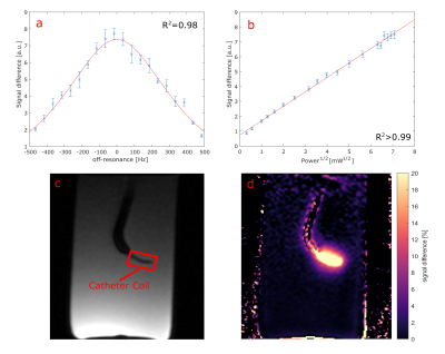 |
Towards Catheter-based Intra-Arterial Spin Labeling for Perfusion Measurements
Kevin Waescher1, Simon Reiss1, Ali Caglar Özen1,2, Thomas Lottner1, and Michael Bock1
1Dept. of Radiology, Medical Physics, Medical Center University of Freiburg, Faculty of Medicine, University of Freiburg, Freiburg, Germany, 2German Consortium for Translational Cancer Research Partner Site Freiburg, German Cancer Research Center (DKFZ), Heidelberg, Germany
MR-guided catheterization with injection of Gd-based contrast agent allows for quantitative perfusion measurement of the myocardium in coronary interventions. However, Gd is contraindicated in patients with impaired renal function, and it changes the relaxation times in the target organ after injection, so that a characterization of the target tissue becomes difficult. In this study, we provide a different solution to quantify perfusion using arterial spin labelling with a catheter based coil. The results show that ASL using a local catheter based labelling coil provides an alternative technique for perfusion measurements during cardiovascular interventions without exogenous contrast agents.
|
|||
4245.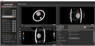 |
Prototype Platform for Real-time MR-guided Brain Clot Evacuation
Robert Moskwa1, Azam Ahmed2, and Walter Block1
1Medical Physics, University of Wisconsin-Madison, Madison, WI, United States, 2Neurological Surgery, University of Wisconsin-Madison, Madison, WI, United States
The international Phase III MISTIE Trial, which used CT-image guidance to position a catheter at the site of intracerebral hemorrhage (ICH) failed to meet its clinical endpoint for all patients. The Trial concluded that improved guidance was needed to help a wider set of neurosurgeons. MR-guidance of ICH evacuation could meet these needs. We present an MR-guidance prototype to provide neurosurgeons with a real-time interface with similar viewpoints as commonly used in stereotactic operating room settings. The MR-guidance prototype shows potential to be a reliable tool to assist as monitoring and guiding tools and lysing drugs during clot-evacuation procedures.
|
|||
4246.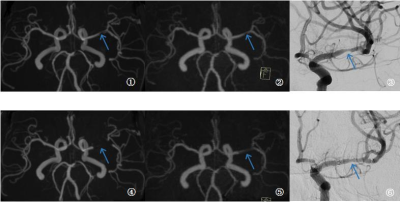 |
PETRA subtraction-based MRA to assess middle-cerebral-artery stenosis before and after treatment with angioplasty
Feifei Zhang1, Yuncai Ran1, Shujian Li1, Jinxia Zhu2, Xuemei Gao1, Jingliang Cheng1, and Chengcheng Zhu3
1The first Affiliated Hospital of Zhengzhou University, Zhengzhou, China, Zhengzhou, China, 2MR Collaboration, Siemens Healthcare Ltd., Beijing, China, Beijing, China, 3Department of Radiology, University of Washington, Seattle, Seattle, WA, United States
This study investigated the feasibility of PETRA-MRA to evaluate stent angioplasty of middle-cerebral-artery stenosis before and after treatment. PETRA-MRA was shown to significantly reduce susceptibility artifacts induced by metallic devices, decrease the phase dispersion of labeled flow, and increase the signal intensity within the stent. Compared to TOF-MRA, PETRA-MRA has better consistency with DSA in image quality and stenosis measurement, especially for follow-up assessments after stent angioplasty.
|
|||
4247.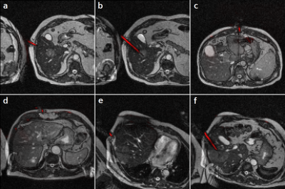 |
Deep Learning-Based Needle Tracking Trained on Bloch-Simulated Data and Evaluated on Clinical Real-Time bSSFP Images
Ralf Vogel1,2, Dieter Ritter2, Jonathan Weine2,3, Jonas Faust2,4, Elodie Breton5, Julien Garnon5,6, Afshin Gangi5,6, Andreas Maier1, and Florian Maier2
1Pattern Recognition Lab, Friedrich-Alexander-Universität Erlangen-Nürnberg, Erlangen, Germany, 2Siemens Healthcare, Erlangen, Germany, 3TU Dortmund, Dortmund, Germany, 4Universität Heidelberg, Heidelberg, Germany, 5ICube UMR7357, University of Strasbourg, CNRS, FMTS, Strasbourg, France, 6Imagerie Interventionnelle, Hôpitaux Universitaires de Strasbourg, Strasbourg, France
Recently, Deep Learning-based methods were used to track the position and orientation of needles in MR images in real-time. Synthetic training data can be generated in large amounts, without data privacy restrictions, and without the need of animal experiments. Therefore, we have simulated the image acquisition using virtual human phantoms containing randomly placed metallic needles in a Bloch simulator. The synthetic images were used to train a U-net to predict the position and orientation of the needle within the susceptibility artifacts of clinical images in less than $$$90\,\text{ms}$$$.
|
|||
4248.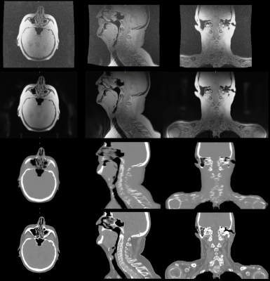 |
Deep Learning-based MR-only Radiation Therapy Planning for Head&Neck and Pelvis
Florian Wiesinger1, Sandeep Kaushik1, Mathias Engström2, Mika Vogel1, Graeme McKinnon3, Maelene Lohezic1, Vanda Czipczer4, Bernadett Kolozsvári4, Borbála Deák-Karancsi4, Renáta Czabány4, Bence Gyalai4, Dorottya Hajnal4, Zsófia Karancsi4,
Steven F. Petit5, Juan A. Hernandez Tamames5, Marta E. Capala5, Gerda M. Verduijn5, Jean-Paul Kleijnen5, Hazel Mccallum6, Ross Maxwell6, Jonathan J. Wyatt6, Rachel Pearson6, Katalin Hideghéty7, Emőke Borzasi7, Zsófia Együd7, Renáta Kószó7,
Viktor Paczona7, Zoltán Végváry7, Suryanarayanan Kaushik3, Xinzeng Wang3, Cristina Cozzini1, and László Ruskó4
1GE Healthcare, Munich, Germany, 2GE Healthcare, Stockholm, Sweden, 3GE Healthcare, Waukesha, WI, United States, 4GE Healthcare, Budapest, Hungary, 5Erasmus MC, Rotterdam, Netherlands, 6Newcastle University, Newcastle, United Kingdom, 7University of Szeged, Szeged, Hungary
MR imaging offers unique advantages for Radiation Therapy Planning (RTP) via excellent soft-tissue contrast for the delineation of the tumor target volume and surrounding organs-at-risk (OARs). Remaining challenges include absent CT information (required for accurate dose calculation) and time-consuming manual tumor and OAR contouring. Here we describe the application of Deep Learning for MR-only RTP in terms of synthetic CT conversion and automated OAR delineation. Exemplary results are illustrated from an ongoing MR-only RTP study in head&neck and pelvis.
|
|||
4249.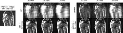 |
Neural network based denoising of high temporal resolution cine images for tumor tracking in MR-guided radiotherapy
Florian Friedrich1,2, Juliane Hörner-Rieber3, Peter Bachert1,2, Mark E. Ladd1,2,4, and Benjamin R. Knowles1
1Medical Physics in Radiology, German Cancer Research Center (DKFZ), Heidelberg, Germany, 2Department of Physics and Astronomy, Heidelberg University, Heidelberg, Germany, 3Department of Radiation Oncology, University Hospital of Heidelberg, Heidelberg, Germany, 4Faculty of Medicine, Heidelberg University, Heidelberg, Germany MR-linac systems allow for real-time tumor position updates. Higher temporal resolution imaging through k-space undersampling allows for an increased number of position updates, however iterative reconstructions may negate the decrease in acquisition time and undersampling artifacts may impact tracking stability. In this study a fast method to denoise and suppress image artifact using a U-net is presented. Undersampled Cartesian and radial cine images were acquired from a patient with a liver tumor on an MR-linac. Tumor tracking stability was assessed. Denoising was found to improve tracking stability and has potential in high temporal resolution cine imaging on MR-linac systems. |
|||
4250.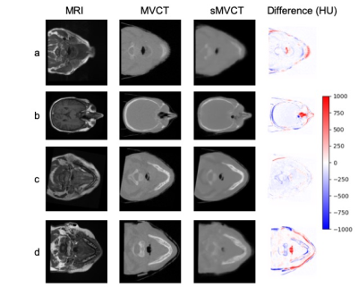 |
Towards higher accuracy mapping of MRI to electron density using a 3D deep CNN for MRI-only radiotherapy treatment planning
Jessica E Scholey1, Abhejit Rajagopal2, Elena Grace Vasquez3, Atchar Sudhyadhom4, and Peder Eric Zufall Larson2
1Department of Radiation Oncology, University of California, San Francisco, San Francisco, CA, United States, 2Department of Radiology, University of California, San Francisco, San Francisco, CA, United States, 3Department of Physics, University of California, Berkeley, Berkeley, CA, United States, 4Department of Radiation Oncology, Harvard Medical School, Boston, MA, United States
We implemented a novel approach of using MRI to synthesize CT datasets acquired at MV photon energies for more accurate electron density mapping in radiotherapy treatment planning. We used a 3D deep convolutional neural network and demonstrated clinical proof-of-concept by evaluating the dosimetric impact of using synthetic datasets in a test radiotherapy treatment plan. The proposed method produced mean MAE of 72.8±17.3 HU and SSIM of 0.82 in the test dataset. The dose distributions computed on the test case produced 100% gamma passing rate (computed at 3%/3mm) indicating that synthetic MV images may be used for clinical treatment planning.
|
|||
4251.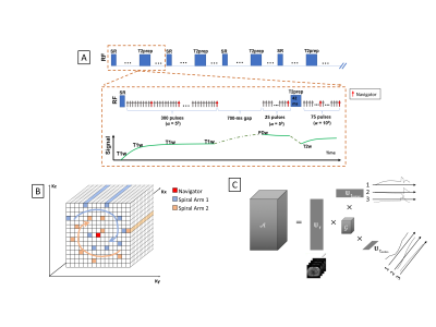 |
Multi-Task MR Simulation for Abdominal Radiation Treatment Planning: Technical Development
Junzhou Chen1,2, Pei Han1,2, Fei Han3, Zhehao Hu1,2, Nan Wang1,2, Wensha Yang4, Anthony G Christodoulou1,2, Debiao Li1,2, and Zhaoyang Fan1,2,5
1Biomedical Imaging Research Institute, Cedars-Sinai Medical Center, Los Angeles, CA, United States, 2Department of Bioengineering, University of California, Los Angeles, Los Angeles, CA, United States, 3Siemens Medical Solutions USA, Inc., Los Angeles, CA, United States, 4Department of Radiation Oncology, University of Southern California, Los Angeles, CA, United States, 5Department of Radiology, University of Southern California, Los Angeles, CA, United States
MRI can provide superior soft tissue contrasts for radiation therapy planning. However, radiation planning in the abdomen is especially difficult because respiratory motion can cause misregistration across separate scans with different contrast weightings due to inconsistent breath holds or poor patient compliance. While some studies have produced volumetric and motion-resolved images, they are all limited to a single contrast which is suboptimal for radiation planning. To address these issues, we present a free-breathing MR imaging platform that produces multi-contrast and motion-resolved volumetric images using MR-multitasking, dedicated for radiation therapy planning, and under a 9:30 mins scan time.
|
|||
4252.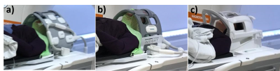 |
Image quality comparisons of novel and commercial coil setups in MRI for head and neck radiotherapy simulation
Evangelia Kaza1, Jeffrey P Guenette2, Christian V Guthier1, Steven Hatch3, Alexander Marques3, Lisa Singer1, and Jonathan D Schoenfeld1
1Radiation Oncology, Brigham and Women’s Hospital, Dana-Farber Cancer Institute, Harvard Medical School, Boston, MA, United States, 2Division of Neuroradiology, Brigham and Women’s Hospital, Dana-Farber Cancer Institute, Harvard Medical School, Boston, MA, United States, 3Radiation Oncology, Brigham and Women’s Hospital, Dana-Farber Cancer Institute, Boston, MA, United States
Quality characteristics of healthy volunteer head images (SNR, CNR, artifact size) were compared between two flexible coil arrangements encompassing radiotherapy immobilization masks relative to a gold standard diagnostic Head/Neck20 coil that cannot accommodate the masks. The novel arrangement of two UltraFlexLarge18 coils provided higher SNR ratios and was more spacious than the commercially recommended arrangement of two FlexLarge4 coils. Artifact size and CNR ratios were similar for the two coil setups. Clinical application of the UltraFlexLarge18 coil arrangement would be advantageous for head and neck radiotherapy MRI simulations due to higher SNR and increased patient comfort.
|
|||
4253.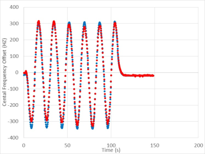 |
Real-Time B0 Correction in a MRI Guided Radiotherapy System
Austen Curcuru1, Deshan Yang1,2, and Michael Gach1,2,3
1Biomedical Engineering, Washinton University in Saint Louis, Saint Louis, MO, United States, 2Radiation Oncology, Washinton University in Saint Louis, Saint Louis, MO, United States, 3Radiology, Washinton University in Saint Louis, Saint Louis, MO, United States
MRI linear accelerator hybrid systems use balanced steady state free precession sequences (bSSFP) during radiotherapy due to the sequence’s high signal to noise ratio and rapid acquisition times. However, bSSFP sequences are highly sensitive to off-resonance effects.1 As a result, the position and velocity of the radiotherapy gantry can cause imaging artifacts that can impact tumor tracking and cause fluctuations in the main magnetic field that can result in radiotherapy/MRI isocenter misalignments. Sequence parameters were adjusted during the sequence run time to reduce these issues by integrating a navigator into the bSSFP sequence and measuring B0 variations in real-time.
|
|||
4254.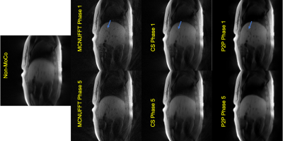 |
Respiratory Motion Detection and Reconstruction Using CAPTURE and Deep Learning for a 0.35T MRI-LINAC System: An Initial Study
Sihao Chen1, Cihat Eldeniz1, Weijie Gan1, Ulugbek Kamilov1, Deshan Yang1, Michael Gach1, and Hongyu An1
1Washington University in St. Louis, Saint Louis, MO, United States
Magnetic Resonance Imaging Guided Linear Accelerator (MRI-LINAC) combines an MRI system with a linear accelerator radiotherapy system to treat patients. The MRI-LINAC uses MR to track organ or lesion motion and gates the radiation beam accordingly. In this study, we demonstrate the combination of a T1-weighted self-navigated respiratory motion detection method (CAPTURE) with a deep learning 4D reconstruction (Phase2Phase, P2P) method to derive the 3D deformable respiratory motion field from data acquired on a 0.35 T ViewRay MRI-LINAC system. This initial study demonstrated promising results despite the low SNR at this field strength.
|
|||
4255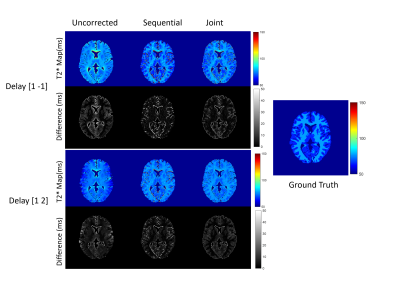 |
Joint Radial Trajectory Correction for Fast T2* Mapping on an MR-Linac Video Permission Withheld
Wajiha Bano1,2, Will Holmes1,2, Mohammad Golbabaee3, Alison Tree1,2, Uwe Oelfke1,2, and Andreas Wetscherek1,2
1Joint Department of Physics, The Institute of Cancer Research, London, United Kingdom, 2The Royal Marsden NHS Foundation Trust, London, United Kingdom, 3Computer Science Department, The University of Bath, Bath, United Kingdom
Measuring T2* relaxation during the course of MR-guided radiotherapy can characterize tumour hypoxia, which is associated with treatment resistance. T2* mapping with radial trajectories allows for efficient coverage of k-space but is susceptible to errors arising from gradient delays. We propose a method that jointly estimates gradient delays and T2* using model-based reconstruction. Using the numerical phantom and the in-vivo prostate data we demonstrated that the proposed approach performs better for different noise levels for both fully sampled and undersampled datasets. This will allow better integration of T2* mapping for hypoxia imaging into an MR-linac treatment work flow.
|
|||
4256.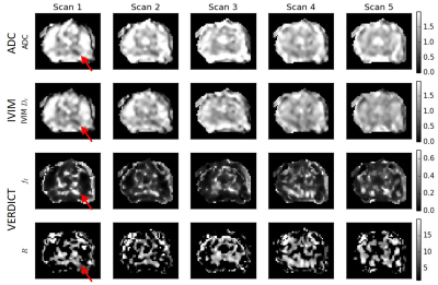 |
Diffusion changes in prostate cancer patients undergoing radiation treatment on an MR-Linac system: Preliminary findings
Colleen Bailey1,2, Rachel W Chan1, Jay Detsky3,4, Danny Vesprini3,4, and Angus Z Lau1,2
1Odette Cancer Centre, Sunnybrook Research Institute, Toronto, ON, Canada, 2Medical Biophysics, University of Toronto, Toronto, ON, Canada, 3Department of Radiation Oncology, Sunnybrook Health Sciences Centre, Toronto, ON, Canada, 4Department of Radiation Oncology, University of Toronto, Toronto, ON, Canada
Eight prostate cancer patients undergoing radiation treatment were scanned on an MR-Linac with conventional ADC and high-b diffusion protocols at each of five treatment fractions. With the system’s weaker gradients and longer echo times, 6/8 patients had visible lesions at the maximum b-value of 2000 s/mm2. Fits to the VERDICT model demonstrated higher intracellular fraction in the lesion, inversely correlated with ADC. In three patients, ADC increased starting at the third treatment time point; a fourth patient exhibited a transient ADC increase. Future work will correlate these changes with biochemical recurrence to test their relevance as a biomarker.
|
|||
4257.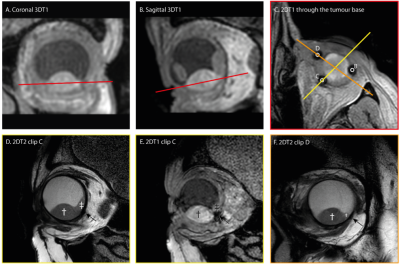 |
MRI-based tumor localisation after tantalum clip placement for proton beam therapy planning of uveal melanoma
Myriam Jaarsma-Coes1,2, Teresa Ferreira1, Marina Marinkovic2, Khanh Vu2, Gre Luyten2, Coen Rasch3, Berit Verbist1, and Jan-Willem Beenakker1,2
1Radiology, Leiden University Medical Center, Leiden, Netherlands, 2Ophthalmology, Leiden University Medical Center, Leiden, Netherlands, 3Radiotherapy, Leiden University Medical Center, Leiden, Netherlands
For proton beam therapy (PBRT) of Uveal Melanoma, tantalum clips are surgically sutured on the outside of the eye to mark the tumour edge. To aid in the 3D clip localisation and modelling of the tumor, a dedicated MRI protocol was developed and evaluated. In 51% of the clips, the MRI and per-operative measurements differed less than 1.0mm. In 28% of the clips the discrepancy between the MRI and peroperative measurement was attributed to the complex tumor geometry or anterior tumor localisation. For the majority of the patients MRI added valuable information to the peroperative measurements, improving PBRT planning.
|
|||
4258.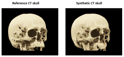 |
Deep learning based synthetic CT skull for transcranial MRgFUS interventions using 3D V-net–Transfer learning implications
Pan Su1,2, Sijia Guo2,3, Steven Roys2,3, Florian Maier4, Thomas Benkert4, Himanshu Bhat1, Elias R. Melhem2, Dheeraj Gandhi2, Rao P. Gullapalli2,3, and Jiachen Zhuo2,3
1Siemens Medical Solutions USA, Inc., Malvern, PA, United States, 2Department of Diagnostic Radiology and Nuclear Medicine, University of Maryland, School of Medicine, Baltimore, MD, United States, 3Center for Metabolic Imaging and Therapeutics (CMIT), University of Maryland Medical Center, Baltimore, MD, United States, 4Siemens Healthcare GmbH, Erlangen, Germany
Transcranial MRI-guided focused ultrasound (tcMRgFUS) is a promising technique for treating multiple diseases. It is desirable to simplify the clinical workflow of tcMRgFUS treatment planning. Previously, feasibility of leveraging deep learning to generate synthetic CT skull from ultra-short echo time (UTE) MRI has been demonstrated for tcMRgFUS planning. In this study, 3D V-Net was used for skull estimation, by taking advantage of 3D volumetric images. Furthermore, feasibility of applying pre-trained model in new dataset was studied, demonstrating the possibility of generalization across various sequences/protocols and scanners.
|
|||
4259.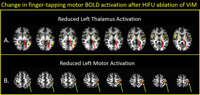 |
FMRI of Post High-Frequency Focused Ultrasound Ablation of ViM Shows Reduced Ipsilateral Thalamic and Cortical Motor Activation
Anna Crawford1, Mark Lowe1, Sean Nagel2, Daniel Lockwood1, Emmanuel Obusez1, Andre Machado2, and Stephen Jones1
1Imaging Institute, Cleveland Clinic Foundation, Cleveland, OH, United States, 2Neurological Institute, Cleveland Clinic Foundation, Cleveland, OH, United States
High Intensity Focused Ultrasound (HIFU) in now entering clinical practice, for example to treat essential tremor (ET) by creating small lesions in the thalamus. Due to small size of treatment lesions, treatment success depends critically on targeting, which is classically done using measurements and landmarks. We explore an alternative method using functional imaging to guide targeting and assess efficacy, specifically using 7T task-related fMRI. We present preliminary data of the patterns of BOLD activation in a set of patients before and after HIFU.
|
|||
4260.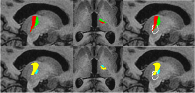 |
Comparison of Automated Thalamic Segmentation Techniques: Applications in MRgFUS Planning
Kain Kyle1, Jerome Maller2, Yael Barnett3, Stephen Tisch3, Benjamin Jonker3, Michael Barnett1, Arkiev D'Souza1, and Chenyu Wang1
1University of Sydney, Sydney, Australia, 2GE Healthcare, Sydney, Australia, 3St Vincent's Hospital Sydney, Sydney, Australia
Investigation of thalamic segmentation tools FreeSurfer and THOMAS for use in MRgFUS planning.
|
|||
4261.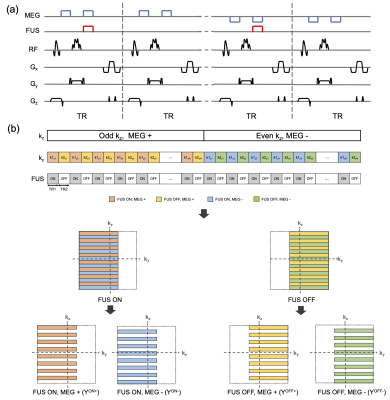 |
Reduced-FOV 3D MR-ARFI with a joint reconstruction for localizing the focused ultrasound beam in neuromodulation
Huiwen Luo1,2, Michelle K. Sigona1,2, Li Min Chen2,3, Charles F. Caskey2,3, and William A. Grissom1,2,3
1Biomedical Engineering, Vanderbilt University, Nashville, TN, United States, 2Vanderbilt University Institute of Imaging Science, Nashville, TN, United States, 3Radiology and Radiological Sciences, Vanderbilt University Medical Center, Nashville, TN, United States
Magnetic resonance acoustic radiation force imaging (MR-ARFI) can be used to localize the focal spot in MR-guided focused ultrasound (FUS) by encoding the ultrasound-induced displacements into the phase of an MR image. A two-minute reduced-FOV 3D MR-ARFI scan with a joint image reconstruction method at 3 Tesla is proposed to image and localize the entire focus in FUS neuromodulation with a low FUS duty-cycle of 0.85%. In this work the reduced FOV MR-ARFI pulse sequence is demonstrated and the proposed k-space undersampling with joint sparse difference image reconstruction to achieve a two-minute scan time is validated.
|
The International Society for Magnetic Resonance in Medicine is accredited by the Accreditation Council for Continuing Medical Education to provide continuing medical education for physicians.