Digital Posters
Engineering & Applying Low-Field MRI
ISMRM & SMRT Annual Meeting • 15-20 May 2021

Digital Posters
Engineering & Applying Low-Field MRI
| Concurrent 3 | 17:00 - 18:00 |
4025.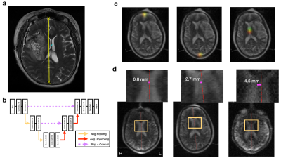 |
Low-Field Point-of-Care MRI: Automated Estimates of Brain Midline Shift Correlate With Clinical Outcomes in Stroke
Prantik Kundu1, Sadegh M. Salehi1, Bradley A. Cahn2, Mercy H. Mazurek2, Matthew M. Yuen2, Jo Schlemper1, Barbara Gordon-Kundu2, Rafael O'Halloran1, Michal Sofka1, and Kevin N. Sheth2
1Hyperfine Research Inc., Guilford, CT, United States, 2Yale University School of Medicine, New Haven, CT, United States
Low-field (64 mT) point-of-care (POC)-MRI was acquired at the bedside from patients with ischemic and hemorrhagic stroke in the neurointensive care unit at a major academic medical center (n=128). An AI system was trained to quantify brain midline shift (MLS), a standard neuroradiological marker of brain injury from POC-MRI, using anatomical annotations from independent neuroradiologists. A cross-validation experiment showed that AI estimates of MLS from POC-MRI were associated with stroke severity and disability at subsequent discharge. AI estimates of MLS greater than 1.5 mm were positively predictive of poor discharge outcome in the full sample and in ischemic stroke.
|
|||
4026.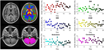 |
Reliability of brain volumetrics in low-field portable MRI
Thomas Campbell Arnold1, Ramya Muthukrishnan2, Steven N Baldassano1, Samantha By3, Brian Welch3, Brian Litt1,4, and Joel M. Stein5
1Bioengineering, University of Pennsylvania, Philadelphia, PA, United States, 2Computer Science, University of Pennsylvania, Philadelphia, PA, United States, 3Hyperfine Research, Guilford, CT, United States, 4Neurology, Perelman School of Medicine, Philadelphia, PA, United States, 5Radiology, Perelman School of Medicine, Philadelphia, PA, United States
A growing body of literature demonstrates the value of MRI-based volumetric measures in diagnosing and treating neurodegenerative disorders. However, clinicians cannot offer biomarker screening for at-risk patients due to MRI systems’ high cost and limited access. Low-field MRI scanners offer a potential method for collecting low-cost images that could be analyzed for longitudinal biomarker changes or used in population-level studies. Here we examine the reliability of volumetric measurements made on a low-field MRI system. We compare the variability of 6 tissue volumes collected over 40 scans for 3T and 64mT systems, as well as the overlap between volume segmentations.
|
|||
4027.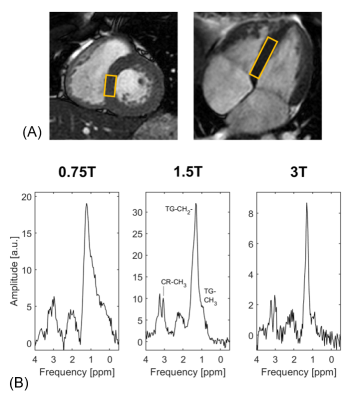 |
Feasibility of human in vivo skeletal muscle and cardiac proton spectroscopy at 0.75T
Sophie M. Peereboom1, Christian Guenthner1, Mohammed M. Albannay1, and Sebastian Kozerke1
1Institute for Biomedical Engineering, University and ETH Zurich, Zurich, Switzerland
Proton MR spectroscopy can assess cardiac triglyceride levels. Although spectral separation and thermal polarization decrease at lower field strength, increased T2* and T2 values, decreased T1 values, decreased echo times and the possibility to decrease receiver bandwidth are clear advantages compared to higher fields. In this work the feasibility of in vivo skeletal muscle and cardiac proton spectroscopy at 0.75T is demonstrated. For this purpose, a clinical 3T Philips Achieva scanner was ramped down to a field strength of 0.75T. Results are compared to spectra acquired at 1.5T and 3T and simulations confirm experimental findings.
|
|||
4028.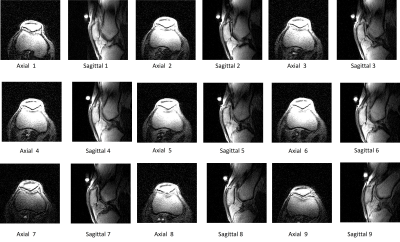 |
Novel Technique for Assessment of Patellar Tracking During Weight-Bearing Knee Flexion Using Dynamic Upright MRI
Neeraj Kulkarni1, John Greenhalgh1, Robert Wolf1, David Chu1, Rachel Caffrey2, Brianna Elizabeth Damadian1, and Raymond Damadian1
1FONAR Corporation, Melville, NY, United States, 2Lehigh University, Bethlehem, PA, United States
Despite decades of imaging research focused on the knee, little attention has been paid to dynamic, weight-bearing knee MRI emulating the physiologic condition of the joint. Here, we utilize dynamic imaging in alternating axial and sagittal planes to capture the patellofemoral joint of one volunteer performing one cycle of knee flexion/extension. Our findings indicate that it is possible to characterize patellar tracking in both axial and sagittal planes utilizing fast imaging sequences (<2 minutes) on mid-field upright MRI. Additionally, it is possible to quantify the patellofemoral kinematics on these images using patellar flexion angle and anteroposterior patellar translation.
|
|||
4029.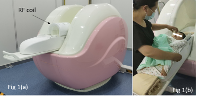 |
A Novel Dedicated 0.35T Open Neonatal-Infant Brain MRI system
Mao Sheng1, ZhongChang Miao2, Jian Bao3, Renjie Zong3, SiSeung Kim3, Huiyao Zhang3, and Bing Keong Li3
1Department of Radiology, Children’s Hospital of Soochow University, Suzhou, China, 2Department of Radiology, The First People’s Hospital of Lianyungang, Jiangsu Province, China, 3Jiangsu LiCi Medical Device Co., Ltd, Lianyungang, China
A dedicated 0.35T Neonatal-Infant Brain MRI system is developed to investigate the effectiveness and safeness for neonatal and infant patients. 66 volunteers from 0-12 months are recruited to undergo a clinical trial and it is found that low field brain images displayed high grey-white tissue contrast, which is notably helpful for clinical diagnosis. The newly developed system also has very low acoustic noise and that most of the volunteer showed no sign of temperature raise. Low field MRI system therefore has the potential to be a effective and safer alternative MRI system for neonatal and infant patients.
|
|||
4030.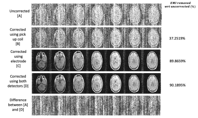 |
In vivo human imaging on a 47.5mT open MRI system with active Electromagnetic Interference (EMI) mitigation using an electrode
Sai Abitha Srinivas1,2, Stephen Cauley3,4, Jason P Stockmann3,4, Charlotte R Sappo1,2, Christopher E Vaughn1,2, Lawrence L Wald3,4,5, William A Grissom1,2,6,7, and Clarissa Zimmermann Cooley3,4
1Vanderbilt University Institute of imaging science, Nashville, TN, United States, 2Department of Biomedical Engineering, Vanderbilt University, Nashville, TN, United States, 3Harvard Medical School, Boston, MA, United States, 4Dept. of Radiology, Massachusetts General Hospital, Athinoula A Martinos Center for Biomedical Imaging, Boston, MA, United States, 5Harvard-MIT Division of Health Sciences and Technology, Cambridge, MA, United States, 6Department of Electrical Engineering, Vanderbilt University, Nashville, TN, United States, 7Department of Radiology, Vanderbilt University, Nashville, TN, United States
Low-field MRI scanners can operate outside MR safe rooms, but their image quality is adversely affected due to the presence of electromagnetic interference signals which can severely obscure the images. We demonstrate a generalized dynamic model that can handle time-varying external interference sources by using simultaneously acquired data from multiple EMI detectors - specifically from an electrode, traditional RF pick up coils and the primary MR coil - in-vivo, in real world EMI settings on a 47.5mT permanent magnet open MRI system.
|
|||
4031.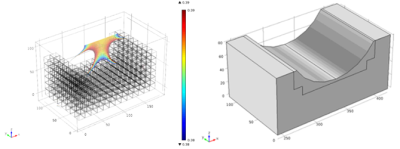 |
Design of a portable, low-field magnetic resonance sensor for clinical measurement of volemic status
Sydney Sherman1,2 and Michael Cima2,3
1Health Science and Technology, Massachusetts Institute of Technology, Cambridge, MA, United States, 2Koch Institute for Integrative Cancer Research, Cambridge, MA, United States, 3Massachusetts Institute of Technology, Cambridge, MA, United States
A low-field portable MR-based sensor was designed for the acquisition of clinical T2 relaxometry measurements in skeletal muscle. A range of low-field permanent magnet array configurations were modeled with varying sensitive region depths and homogeneous volumes; an optimization score for each design was calculated. The optimal design has a sensitive region 15-20mm from the surface of the magnet, making it capable of acquiring measurements deep into the leg such that the measurement is fully localized to skeletal muscle. The exclusion of subcutaneous fat tissue in the sensitive region will improve sensitivity to fluid shifts within the skeletal muscle.
|
|||
4032. |
In vivo hypoxia monitoring using a novel single sided NMR device
Dion G Thomas1, Freya G Harrison2, Paul D Teal3, Petrik Galvosas1,4, Mary J Berry2,5, Sergei Obruchkov6, and Yu-Chieh Tzeng2
1School of Chemical and Physical Sciences, Victoria University of Wellington, Wellington, New Zealand, 2Centre for Translational Physiology, University of Otago, Wellington, New Zealand, 3School of Engineering and Computer Science, Victoria University of Wellington, Wellington, New Zealand, 4MacDiarmid Institute for Advanced Materials and Nanotechnology, Wellington, New Zealand, 5Department of Paediatrics and Child Health, University of Otago, Wellington, New Zealand, 6Robinson Research Institute, Victoria University of Wellington, Wellington, New Zealand
In this study, we investigate how a low field single-sided NMR device can be used to monitor brain tissue properties in-vivo. This device produces a B0 field with a sweet spot in the brain, which defines the region of tissue that is measured. An ovine model of brain hypoxia was developed, to allow tissue oxygenation to be controlled. We observed that the T2 decreased during hypoxia, recovering once normal oxygenation levels were re-established. These results shows that single-sided NMR devices have the potential to be used for real-time monitoring applications.
|
|||
4033. |
Zero-Dead-Time Earth’s Field NMR Using Two-Photon Excitation
Victor Han1 and Chunlei Liu1,2
1Electrical Engineering and Computer Sciences, University of California, Berkeley, Berkeley, CA, United States, 2Helen Wills Neuroscience Institute, University of California, Berkeley, Berkeley, CA, United States
Two-photon excitation allows for excitation at half of the Larmor frequency. In Earth’s magnetic field, this corresponds to a Larmor frequency of about 2 kHz and an excitation frequency of about 1 kHz. By transmitting at 1 kHz and receiving at 2 kHz, we could transmit and receive at the same time by using filters to protect the receiver. While some harmonic distortion prevented us from fully separating the transmitted from the received signal during excitation, the always connected and unsaturated receive circuitry allowed us to eliminate receiver dead time, which can be quite long at low frequencies.
|
|||
4034.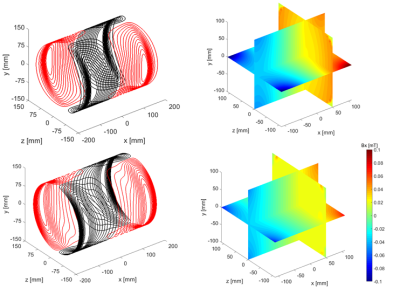 |
An axial gradient coil with improved linearity for Halbach-based MR systems
Bart de Vos1, Thomas O'Reilly1, Wouter Teeuwisse1, Rob Remis2, and Andrew Webb1
1C.J. Gorter Center for High Field MRI, Leiden University Medical Center, Leiden, Netherlands, 2Circuits and Systems, Delft University of Technology, Delft, Netherlands
We reduced image artifacts and increased the attainable axial field-of-view by designing a highly linear x-gradient coil for Halbach array-based MR systems. A truncated sum of sinusoidal basis function is used for the current density. Higher order modes combined with specifying the target field inside a volume lead to a stable solution of the inverse source problem. The coil was installed on our 50 mT system and resulted in a 150% increase in linear DSV with respect to our previously designed gradient coil. Whole-brain three-dimensional images have been acquired using turbo spin echo sequences in less than 10 minutes.
|
|||
4035.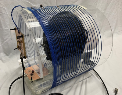 |
A Linear Gradient Solenoid for Slice-Selective Brain Imaging using B1+-Selective RF Pulses
Matthew Wilcox1,2, Sai Abitha Srinivas1,2, Christopher E Vaughn1,2, Charlotte R. Sappo1,2, and William A. Grissom1,2,3,4
1Biomedical Engineering, Vanderbilt University, Nashville, TN, United States, 2Institute of Imaging Science, Vanderbilt University, Nashville, TN, United States, 3Radiology, Vanderbilt University, Nashville, TN, United States, 4Electrical Engineering, Vanderbilt University, Nashville, TN, United States
Despite drawbacks including production of acoustic noise and peripheral nerve simulation, long switching times, bulkiness, and high costs, use of B0 gradient coils is ubiquitous in conventional MR imaging. Replacement of B0 encoding with B1+ encoding alleviates these concerns and allows the use of hardware which is lower-cost, more compact, and silent. As a first step towards realizing a full B1+ encoding coil system, this work demonstrates an RF z-gradient solenoid coil developed for brain imaging on a 47.5 mT system. The coil performance is evaluated in simulation, bench, and scanner experiments and slice-selection using B1+-selective pulses is demonstrated.
|
|||
4036. |
Target-field optimized quadrature RF coils for wrist imaging on a 76 mT Halbach-based MR system
Bart de Vos1, Thomas O'Reilly1, Wouter Teeuwisse1, Rob Remis2, and Andrew Webb1
1C.J. Gorter Center for High Field MRI, Leiden University Medical Center, Leiden, Netherlands, 2Circuits and Systems, Delft University of Technology, Delft, Netherlands
We describe a design method for quadrature RF coils for Halbach based magnets using a target field approach. The resulting current densities corresponding to these coils make them inherently decoupled. We constructed a coil pair for a newly built 76 mT system and imaged the wrist of a volunteer. The images showed a 33% SNR improvement over a linear coil, in close agreement with simulations.
|
|||
4037.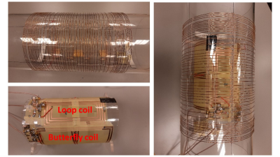 |
Design and testing of a four-channel receive array coil on a 50 mT permanent magnet system.
Javad Parsa1, Thomas O'Reilly1, Bart de Vos1, and Andrew Webb1
1Leiden University Medical Center, Leiden, Netherlands
An integrated transmit coil and four-element receive array has been simulated, constructed, characterized and tested on a low field MRI system operating at 2.15 MHz. Using a combination of loops and butterfly coils neighbouring inter-element coupling is below -18 dB, with directly opposite coils ~-9 dB, and <-17 dB coupling for all receive coils to the transmit coil. Images of a phantom have been acquired with a simple sum-of-squares reconstruction.
|
|||
4038.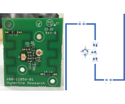 |
Dipole position sensors integrated in receiver array for motion detection and correction
Ed Boskamp1, Mike Twieg1, and Rafael O'Halloran1
1Hyperfine, Guilford, CT, United States
MRI is sensitive to patient motion. There are a number of pulse sequence techniques like navigators and propeller to prevent motion artifacts. We are introducing a sensitive sensor based technique to detect motion and pose without touching the patient. The sensors are a number of miniaturized dipole resonators integrated into the receiver array. Motion corrupted lines in k space are flagged and rescanned, or when motion is between a limited number of poses, the data can be binned and combined before the recon process.
|
|||
4039.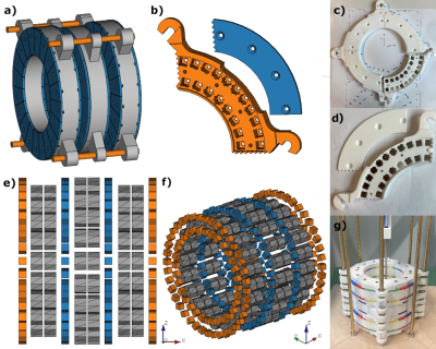 |
B0-shimming methodology for affordable, compact, and homogeneous low-field MR magnets
Konstantin Wenzel1, Hazem Alhamwey1, Tom O'Reilly2, Layla Riemann1, and Lukas Winter1
1Physikalisch-Technische Bundesanstalt (PTB), Braunschweig and Berlin, Germany, 2Leiden University Medical Center (LUMC), Leiden, Netherlands
In this work, B0-shimming techniques are investigated allowing the construction of simple, low-cost, and homogeneous Halbach-based low-field MR magnets. The techniques are applied to build a desktop MR magnet at B0=0.1T and can be easily scaled to magnet designs of larger diameter. The presented shimming approach improved B0 homogeneity by a factor of ~8 from 5448ppm to 682ppm in a 2D target region. All design files and code concerning this work will be made available open source on www.opensourceimaging.org.
|
|||
4040.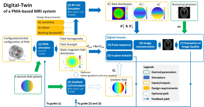 |
The Development of a Digital-twin of a Permanent-Magnet-Array (PMA)-Based Portable MRI System
Shao Ying Huang1, Yi-Dan Chen1, Ting-Ou Liang2, Yan Hao Koh1, and Wenwei Yu3
1Singapore University of Technology and Design, Singapore, Singapore, 2Zhejiang University, Hangzhou, China, 3Chiba University, Chiba, Japan
This abstract presents the development of a digital-twin of a permanent-magnet-array-based portable MRI system. It is to guide the design, optimization, hardware debug and calibration, and evaluation of such a system at both a sub-system level and a system level. Meanwhile, it facilitates the development of a sub-system without building the whole system. It consists of the simulators for magnet array, RF coil, gradient coils, pulse sequence, k-space analysis, image reconstruction, and image quality evaluation.
|
|||
4041.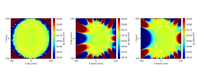 |
The design of a low-weight homogenous Halbach helmet for imaging the adult brain.
Thomas O'Reilly1 and Andrew Webb1
1C.J. Gorter Center for High Field MRI, Leiden University Medical Center, Leiden, Netherlands
Arrays of permanent magnets have shown promise as a relatively light weight, low cost, sustainable way of generating magnetic fields suitable for MRI. In this work we show the design of a new homogenous helmet shaped, Halbach-array based magnet optimized for imaging the adult head. The magnet has a mean magnetic field strength of 59.7 mT and a homogeneity of 1313 ppm over a brain sized 25x20x15 cm3 ellipsoid. By truncating one side of the magnet the weight of the system has been reduced compared to symmetric designs while increasing the obtained field strength.
|
|||
4042.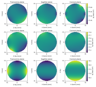 |
The role of non-random magnet rotations on main field homogeneity of permanent magnet assemblies
Thomas O'Reilly1 and Andrew Webb1
1C.J. Gorter Center for High Field MRI, Leiden University Medical Center, Leiden, Netherlands Arrays of permanent magnets are an attractive approach to designing magnets for low field MRI systems as they are affordable, easy to handle and offer great flexibility in magnet designs. However, translating designs from simulations to realised systems is challenging. The difference between the two is often attributed to imperfections in the individual magnets that make up the permanent magnet array. Here we show that non-random errors in the magnet rotations due to imperfect magnet holders can be the dominant cause of this discrepancy and if the imperfections are known they can easily be corrected for during manufacturing. |
|||
4043.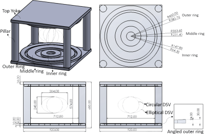 |
Triple-ring permanent magnet design for ultra-low-field MRI systems
Yang Gao1,2, Alex T. L. Leong1,2, Yilong Liu1,2, and Ed X. Wu1,2
1Laboratory of Biomedical Imaging and Signal Processing, The University of Hong Kong, Hong Kong SAR, China, 2Department of Electrical and Electronic Engineering, The University of Hong Kong, Hong Kong SAR, China
MRI has impacted modern healthcare tremendously. However, MRI accessibility is low and extremely inhomogeneous globally. The recent shift in prioritizing MRI’s value has seen the emergence in developing point-of-care systems at ultra-low-field (<0.1T), particularly the use of permanent magnets for its light-weight, low power and low cost benefits. However, fundamentally, a relatively strong and uniform magnetic field is still desired for ultra-low-field MRI systems. Here, we propose a triple-ring permanent magnet design that aims to preserve and improve upon the fundamental field homogeneity requirements of MR imaging at ultra-low-field, without sacrificing the flexibility to perform both body and head imaging.
|
|||
4044.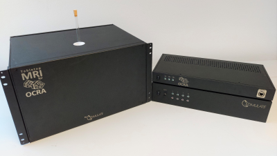 |
Educational Tabletop MRI system using the Open-Source Console for Real-time Acquisition (OCRA)
Marcus Prier1,2, David Schote1,2, Ivan Fomin1,2, Thomas Witzel3, Georg Rose1,2, and Oliver Speck1,2
1Otto-von-Guericke University, Magdeburg, Germany, 2Research Campus STIMULATE, Magdeburg, Germany, 3Q Bio Inc, San Carlos, CA, United States
A Tabletop MRI system was developed that merges the Marinos Center Tabletop and OCRA projects, and is improved in many aspects compared to the original system. The proposed system includes a fully functional MRI console with a customizable GUI and microcontroller server. The hardware was extended with an optimized magnet design with passive shimming, active TR switches with reverse bias and RFPA noise blanking, and a low cost and fast response 4 channel gradient amplifier. It can be used for educational purposes and student courses in medical engineering.
|
The International Society for Magnetic Resonance in Medicine is accredited by the Accreditation Council for Continuing Medical Education to provide continuing medical education for physicians.