Digital Posters
Renal Functional Imaging
ISMRM & SMRT Annual Meeting • 15-20 May 2021

| Concurrent 5 | 19:00 - 20:00 |
2517.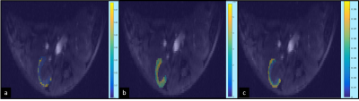 |
Comparison of pharmacokinetic models for assessing murine renal function by DCE-MRI
Soham Mukherjee1, Mahon L Maguire1, Jack Sharkey1, Sourav Bhaduri1, Patricia Murray2, Rachel Bearon3, Bettina Wilm2, and Harish Poptani1
1Centre for Preclinical Imaging, University of Liverpool, Liverpool, United Kingdom, 2Department of Cellular and Molecular Physiology, University of Liverpool, Liverpool, United Kingdom, 3Department of Mathematical Sciences, University of Liverpool, Liverpool, United Kingdom
Quantitative analysis of kidney function is essential part of monitoring disease progression. The most common parameter of evaluating kidney functioning is by measurement of glomerular filtration rate. Here, we describe that dynamic contrast enhanced magnetic resonance imaging can be used to determine the permeability parameter Ktrans, which can be used to assess renal activity. We used three different pharmacokinetic models and arterial input functions to get the Ktrans value. It was seen that the raw arterial input function used along with shutter speed model has strong correlation with findings obtained from transcutaneous GFR measurement device.
|
|||
2518.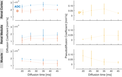 |
Diffusion Time Dependence of Apparent Diffusion Coefficient and Intravoxel Incoherent Motion Diffusion Parameters in the Human Kidney
Julia Stabinska1, Hans-Joerg Wittsack1, and Alexandra Ljimani1
1Department of Diagnostic and Interventional Radiology, Heinrich Heine University Dusseldorf, Dusseldorf, Germany
Due to non-Gaussian nature of water diffusion in tissues, the measured diffusion-related parameters depend on b-values and diffusion time. In fact, our study shows that the apparent diffusion coefficient (ADC) and IVIM-related diffusion coefficient D obtained in the human kidney slightly increase with the diffusion time in the range between 20 ms and 45 ms, as opposed to ADC and D of skeletal muscle. Among all investigated diffusion parameters, the IVIM-based pseudodiffusion coefficient D* exhibits the strongest dependence on diffusion time and could potentially provide valuable information regarding the blood vessels and urine flow in the kidney.
|
|||
2519.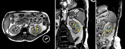 |
Phosphorus Magnetic Resonance Spectroscopy of Healthy Human Kidney in-situ at 3T
Maysam Jafar1 and Jan Weis2
1Clinical Science, Philips Healthcare, Stockholm, Sweden, 2Department of Medical Physics, Uppsala University Hospital, Uppsala, Sweden
Phosphorous (31P) spectra of healthy human kidney are invaluable in studies of kidney physiology, disease and in the evaluation of allograft viability pre and post renal transplantation. However, 31P-spectroscopy of the kidney in-situ is challenging due to the relatively large distance between the kidney and the surface coil. The aim of this study was to investigate whether it is possible to acquire 31P spectra of normal kidney in a clinically acceptable measurement time using a 3T MR scanner. We demonstrate that localized phosphorous spectroscopy of normal kidney in-situ is feasible in today’s 3T clinical MR systems.
|
|||
2520.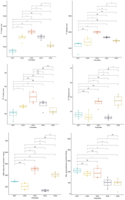 |
Repeatability of multi-parametric renal MRI biomarkers in healthy subjects: An iBEAt pilot study
Kanishka Sharma1, Bashair Alhummiany2, David Shelley2,3, Margaret Saysell2,3, Maria-Alexandra Olaru4, Bernd Kühn4, Julie Bailey3, Kelly Wroe3, Cherry Coupland3, Michael Mansfield3, and Steven Sourbron1
1Department of Imaging, Infection, Immunity and Cardiovascular Disease, University of Sheffield, Sheffield, United Kingdom, 2Department of Biomedical Imaging Sciences, University of Leeds, Leeds, United Kingdom, 3Leeds Teaching Hospitals, Leeds, United Kingdom, 4Siemens Healthcare GmbH, Erlangen, Germany
The iBEAt study MRI biomarker panel has been developed to determine if imaging biomarkers can improve predictions of renal function decline in diabetic kidney disease. The aim of this pilot study was to inform the approach to quality control and image processing of iBEAt data. Repeatability in 5 healthy volunteers (4 scans per subject) was analysed with a prototype image-processing pipeline. The results indicated that repeatability of renal T1, T2* and phase-contrast renal blood flow was comparable to literature, and measurement precision was sufficient to detect differences between volunteers. ASL had poor repeatability presumably from B0 inhomogeneities in labelling plane.
|
|||
2521.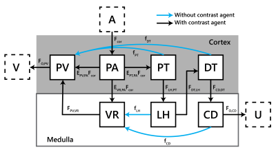 |
Measurement of renal medullary perfusion using a 7-compartment model for MR Renography
Anneloes de Boer1, Bashair Al Hummiany2, Kanishka Sharma3, and Steven Sourbron3
1University Medical Center Utrecht, Utrecht, Netherlands, 2University of Leeds, Leeds, United Kingdom, 3University of Sheffield, Sheffield, United Kingdom
Medullary perfusion potentially is an early marker of renal damage, but cannot be measured using existing models in MR renography. In this study a novel 7-compartment, 10-parameter model is proposed and evaluated in synthetic and patient data. Robustness of an iterative fitting approach was assessed by the coefficient of variation over multiple fits, resulting in median values <2.5% both in synthetic and patient data. According to simulations, medullary perfusion was underestimated by 5.3%, but in diabetic patients medullary perfusion was relatively high (81 mL/100mL/min). Future studies will be needed to determine this model’s sensitivity to pathophysiological changes in medullary perfusion.
|
|||
2522.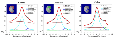 |
Improved Accuracy of Ratiometric CEST pH Mapping using Two Iodinated Agents with Nonequivalent Amide Protons and a Single Low Saturation
Quan Tao1, Peiwei Yi1, Zimeng Cai1, Yingjie Mei2, Ruiyuan Liu1, and Yanqiu Feng1
1School of Biomedical Engineering, Southern Medical University, Guangzhou, China, 2Philips healthcare, Guangzhou, China
Various Iodinated agents were supposed to be pH sensors in kidney or tumor based on chemical exchange saturation transfer (CEST) mechanism. We compared different contrast agents (CAs) and their combination, established a ratiometric CEST pH imaging method to improve the accuracy of pH measurement in vivo, by combined the molar concentration ratio of two nonequivalent amide protons resonated at 4.3 ppm and 5.6 ppm.
|
|||
2523.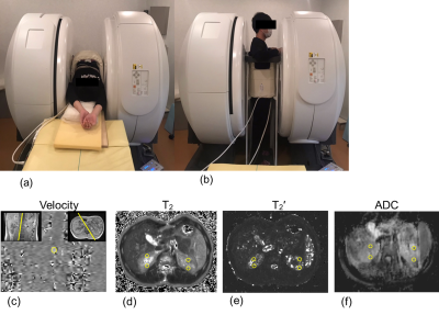 |
Effect of gravity on kidney function: evaluation using multiposture MRI
Yuki Oda1, Tosiaki Miyati1, Naoki Ohno1, Seiya Nakagawa1, and Satoshi Kobayashi1
1Division of Health Sciences, Kanazawa University, Kanazawa, Japan
We assessed the effect of gravity on regional kidney function in the supine and upright positions using an original MRI that can obtain functional information in any posture (multiposture MRI). We compared the mean blood flow, T2, and T2’, apparent diffusion coefficient of the kidney between the supine and upright positions. Gravity reduced the blood flow and T2 of the kidney, and these differences between postures potentially provide new diagnostic information.
|
|||
2524.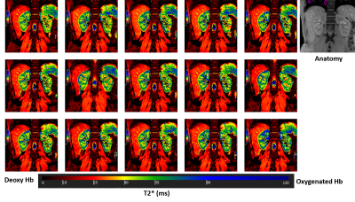 |
BOLD MRI for evaluating intra-renal oxygenation level during acute saline loading
El-Sayed H Ibrahim1, Abdul Parchur1, Srividya Kidambi1, Allen Cowley1, and Mingyu Liang1
1Medical College of Wisconsin, Milwaukee, WI, United States
Enhanced sensitivity of blood pressure to salt intake is observed in many hypertensive patients. The kidney plays a key role in the development of hypertension including salt-sensitive hypertension. In this study, we used BOLD MRI to examine the temporal response of renal medullary and cortical oxygenation levels during saline infusion in a small set of young subjects. The results showed that during the period of fast saline infusion, tissue oxygenation levels remained almost constant in the cortex but showed 10%-15% average reduction in the medulla. The responses, however, varied between subjects, which may reflect differences in salt-sensitive versus salt-insensitive individuals.
|
|||
2525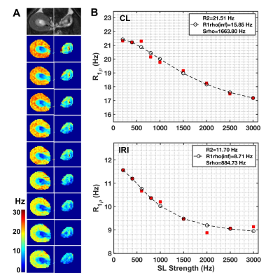 |
Severity of Tubular Atrophy and Fibrosis in Acute Kidney Injury Revealed by Multi-parametric MRI Video Permission Withheld
Feng Wang1,2, Tadashi Otsuka3, Zhongliang Zu1,2, Mark P de Caestecker3, Raymond C Harris3, Takamune Takahashi3, and John C Gore1,2
1Vanderbilt University Institute of Imaging Science, Vanderbilt University Medical Center, Nashville, TN, United States, 2Department of Radiology and Radiological Sciences, Vanderbilt University Medical Center, Nashville, TN, United States, 3Division of Nephrology and Hypertension, Vanderbilt University Medical Center, Nashville, TN, United States
Acute kidney injury (AKI) is a predictor of mortality, often resulting in incomplete recovery, giving rise to chronic kidney disease (CKD). AKI is defined in clinical practice as a rapid decline in kidney function that occurs over a 7-day period or less, but concerns about the risks of renal biopsies in patients with AKI have limited our ability to identify diagnostic features that are predictive of CKD progression. Multi-parametric MRI, including quantitative spin-lock imaging, relaxometry, and magnetization transfer, can be used to identify features of experimental AKI that are predictive of CKD progression without need for renal biopsy.
|
|||
2526.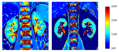 |
Detection of fibrosis in patients with moderate renal impairment with multiparametric MRI
Pete Thelwall1,2, Jehill Parikh1, Benjamin Pippard1, Caroline Wroe3, Rob Janiczek4, Steven Sourbron5, and Neil Sheerin1
1Translational and Clinical Research Institute, Newcastle University, Newcastle upon Tyne, United Kingdom, 2Centre for In Vivo Imaging, Newcastle University, Newcastle upon Tyne, United Kingdom, 3South Tees Hospitals NHS Foundation Trust, Middlesborough, United Kingdom, 4GlaxoSmithKline, Philadelphia, PA, United States, 5University of Sheffield, Sheffield, United Kingdom
A multiparametric renal MRI scan protocol was employed to characterise the impact of renal fibrosis on tissue relaxation properties. The study recruited patients with moderate renal dysfunction (CKD stage 3, eGFR between 30 and 60 ml/min/1.73 m2) with biopsy-proven fibrosis and age-matched healthy volunteers. Statistically significant differences in kidney cortical native T1 and T2 were observed between patients and controls, ascribed to tissue microstructural changes associated with the mild fibrosis observed in the patient group.
|
|||
2527. |
Renal lipid content based on PDFF Imaging is a new potential biomarker for assessing early renal injury in patients with metabolic syndrome
Shisi Li1, Yanjun Chen1, Yingjie Mei2, Xianfu Mo1, Jialing Chen1, Yongqiang Li3, and Xiaodong Zhang1
1Department of Medical Imaging, The Third Affiliated Hospital of Southern Medical University, Guangzhou, China, 2Philips Healthcare, Guangzhou, China, 3Department of Nephrology, The Third Affiliated Hospital of Southern Medical University (Academy of Orthopedics· Guangdong Province), Guangzhou, China
Renal fat content has been known as correlated with renal injury in type 2 diabetes. However, it is not clear the change of renal fat content in patients with metabolic syndrome (MS), which is a more popular disease threatened human health. In the present study, we assess the feasibility and reproducibility of renal fat fraction (FF) using PDFF imaging with MR mDixon-Quant sequence. And we aim to investigate the changes of renal FF in patients with MS, whose estimated glomerular filtration rate (eGFR) grade were G1(normal or elevated) and G2(mild decline) described in KDIGO (Kidney Disease: Improving Global Outcomes). In addition, we evaluate the correlation of renal FF and eGFR and the major factors of eGFR. The results show that with eGFR decreasing, renal FF in patients with MS-G2 group increased significantly compared with control and MS-G1 group. And the renal FF is an important affection factor of eGFR with a significant negative correlation. The noninvasive quantitative Dixon-based MRI may be a new biomarker for the evaluation of early renal impairment.
|
|||
2528. |
Explore the performance of FF and R* value measured by mDIXON-quant for heathy controls, mild and acute CKD patients.
Haoyang Jiang1, Ailian Liu2, Ye Ju2, Jiazheng Wang3, Changyu Du1, Lingli Qi1, Xinmiao Bu2, Wenjun Hu2, Nan Wang2, and Liangjie Lin3
1Dalian Medical University, Dalian, China, 2The First Affiliated Hospital Of Dalian Medical University, Dalian, China, 3Philips Healthcare, Beijing, China
Early diagnosis and treatment of chronic kidney disease (CKD) can significantly improve the prognosis and reduce the occurrence of end-stage renal disease. This study aims to explore the performance of fat-fraction and R* value measured by mDIXON-quant for differential diagnosis between heathy controls, mild and acute CKD patients. The renal cortex R2* values of CKD patients (mild or severe) were significantly higher than those of heathy controls. The fat fraction in renal cortex and medulla of acute CKD patients was significantly higher than those mild CKD patients and healthy controls.
|
|||
2529. |
Value of quantitative susceptibility mapping for detecting renal fibrosis of early diabetic nephropathy in type 2 diabetes
Jiayuan Shan1, Jinggang Zhang1, Jie Chen1, Wei Xing1, and Jilei Zhang2
1Radiology, Third Affiliated Hospital of Soochow University, Changzhou, China, 2Philips Healthcare, Shanghai, China
The purpose was to explore if quantitative susceptibility mapping (QSM) can assess renal fibrosis about early diabetic nephropathy (DN) in type 2 diabetes (T2D). 32 patients with early DN in T2D were included in the study to evaluate the potential clinical relevance of QSM. We found that susceptibility values of the medulla were statistically significant among different fibrosis. Susceptibility value of the medulla was highly correlated with estimated glomerular filtration rate (eGFR). QSM could serve as a quantitative biomarker to assess the renal fibrosis and monitor the treatment in patients with DN.
|
|||
2530.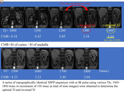 |
Multiparametric MR imaging in diabetic nephropathy: New insights to evaluate early diabetic nephropathy noninvasively
Akira Yamamoto1, Tsutomu Tamada2, Yu Ueda3, Takeshi Fukunaga2, and Atsushi Higaki2
1Radiology, Kawasaki Medical School, Kurashiki, Japan, 2Kawasaki Medical School, Kurashiki, Japan, 3Phillips Japan, Tokyo, Japan
The purpose of this study was to identify the changes in multiparametric magnetic resonance imaging (MRI) findings in early diabetic nephropathy. Measurements were made of the renal cortex and renal medulla T2 values, T2* values and R2* values, as well as optimal TI, inverted TI value and cortico-medullary difference on optimal TI in SSFP with ssIR pulse with multi TI. Also, renal cortical thickness and renal length were measured, representing morphological changes. Significant differences were seen between the healthy and early diabetic nephropathy (grade1 and grade2) in T2 values of cortex and T2* values of medulla. This study suggests the possibility that MRI using the values of T2 of cortex and T2* of medulla can be used to evaluate early diabetic nephropathy non-invasively and in a short period of time.
|
|||
2531.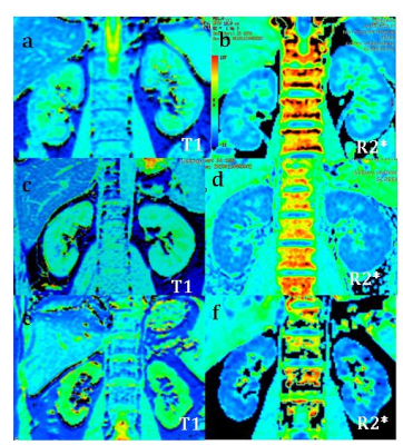 |
Quantitative T1 and R2* mapping in the evaluation of renal function in chronic kidney disease
Jiaxin Yan1, Weiqiang Dou2, Hongmei Gu1, Xinquan Wang1, Weiyin Vivian Liu2, Huijian Lu1, Ying Zhou1, Xuejun Zhou1, and LI Yuan1
1Affiliated Hospital of Nantong University, Nantong, China, 2GE Healthcare, MR Research China, Beijing, China
In this study, we explore the quantitative T1 and R2* mapping in theclassification on renal function in chronic kidney disease (CKD) . 146 CKD patients and 26 healthy volunteers(HVs)underwent T1 mapping and renal BOLD-MRI for R2* mapping. All CKD cases were divided into two groups according to the estimated glomerular filtration (eGFR):mild renal impairment group and moderate to severe group.Clincal information were collected.Compared with HVs,the T1 and R2* values of renal cortex were significantly higher and were well correlated with renal function.Therefore,T1 mapping and BOLD-MRI derived R2* mapping might provide an effective method in assessing renal function.
|
|||
2532.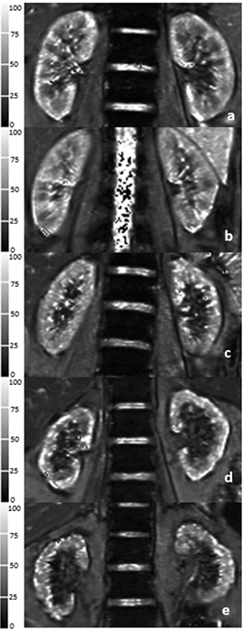 |
Evaluation of renal oxygenation and hemodynamics in patients with chronic kidney disease by BOLD-MRI and intrarenal Doppler ultrasonography
Jing Yang 1, Shuohui Yang 2, Zheng He3, Mengxiao Liu4, and Caixia Fu5
1Nephrology, Shuguang Hospital Affiliated to Shanghai University of Traditional Chinese Medicine, Shanghai, China, 2Radiology, Shuguang Hospital Affiliated to Shanghai University of Traditional Chinese Medicine, Shanghai, China, 3Ultrasonography, Shuguang Hospital Affiliated to Shanghai University of Traditional Chinese Medicine, Shanghai, China, 4MR Scientific Marketing, Siemens Healthcare, Shanghai, China, 5MR Applications Development, Siemens Shenzhen Magnetic Resonance Ltd, Shenzhen, China
The purpose of this study was to observe the alteration of renal oxygenation and renal hemodynamics in CKD patients using blood oxygenation level-dependent (BOLD) magnetic resonance imaging (MRI) and intrarenal Doppler ultrasonography (IDU). The result showed that renal oxygenation and blood flow velocities declined as the CKD stage progressed. The BOLD-MRI and IDU techniques were able to dynamically evaluate intrarenal oxygenation and hemodynamics changes, respectively, in CKD patients. Combination these two techniques can detect the abnormalities associated with CKD stage sensitively.
|
|||
2533.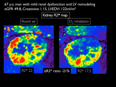 |
Renal hypoxia estimated by O2-inhalation T2* BOLD MRI: association with renal dysfunction and left ventricular remodeling in cardiomyopathy
Michinobu Nagao1, Kiyoe Ando1, Yasuhiro Goto1, Isao Shiina1, Kazuo Kodaira1, Masami Yoneyama2, Takashi Namiki2, Atsushi Yamamoto1, Eri Watanabe1, Akiko Sakai1, Risako Nakao1, and Shuji Sakai1
1Tokyo Women's Medical University, Tokyo, Japan, 2Philips Japan, Tokyo, Japan
O2-inhalation T2*-BOLD MRI is a non-invasive imaging techniqueto evaluate renal oxygenation. This analysis revealed that renal hypoxia is strongly associated with renal dysfunction, and that early left ventricular remodeling in cardiomyopathy leads to renal hypoxia. This renal hypoxia image helps clarify chronic cardiorenal interaction and the effects of drugs for cardiorenal disease.
|
|||
2534.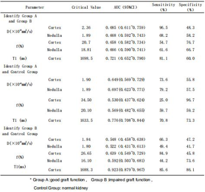 |
The value of intravoxel incoherent motion diffusion-weighted imaging and T1-Mapping in the evaluation of renal transplantation function
Dejuan Shan1,2, Xianquan Cui3, Xiangtao Lin1,2, Ruiyuan Diao2, Peng Zhao2, Mengxiao Liu4, Shuai Zhang2, Xiaoli Li2, Nan Lin2, Zhongyu Hou2, and Bing Liu5
1Department of Radiology, Shandong Provincial Hospital Affiliated to Shandong University, Jinan, China, Jinan, China, 2Department of Radiology,Shandong Provincial Hospital Affiliated to Shandong First Medical Uiversity, Jinan, China, 3Qilu Hospital of Shandong University, Jinan, China, 4MR Scientific Marketing, Diagnostic Imaging, Siemens Healthcare Ltd., Shanghai 201318, China, Shanghai, China, 5Department of Radiology, China-Japan Friendship Hospital, Beijing, China, Beijing, China
The objective of this study was to evaluate renal function of renal transplantation using intravoxel incoherent movement(IVIM) and T1-Mapping. The results showed that the T1-Mapping was significantly superior than IVIM in distinguishing between the groups with good graft function (Group A), impaired graft function (Group B) and normal kidney (Control Group).
|
|||
2535. |
Native T1 mapping in assessment of kidney fibrosis in patients with chronic glomerulonephritis
Zhaoyu Shi1, Fangfang Shang1, Xinquan Wang1, Hongmei Gu1, Xiaoyan Liu1, Weiqiang Dou2, Weiyin Vivian Liu2, Yuan Zhang1, Jianhua Wu1, and Li Yuan1
1Affiliated Hospital of Nantong University, Nantong, China, 2GE Healthcare, MR Research China, Beijing, China
119 patients with chronic glomerulonephritis (CGN) and 19 healthy controls (HCs) were recruited in this study. Among these patients, 43 patients had kidney biopsy. Patients underwent a native renal T1 map examination within 1 week before kidney biopsy. Clinical information and biopsy pathological scores were collected. Compared to HCs, the T1 values of renal cortex in CGN patients were significantly higher and well correlated with renal function, chronic kidney disease (CKD) stage and renal fibrosis. Therefore, native T1 mapping has demonstrated good diagnostic performance in non-invasive detection of CGN fibrosis.
|
|||
2536. |
Evaluation of renal function in healthy volunteers and patients with chronic kidney disease by using APT weighted imaging and R2* mapping
Ye Ju1, Ailian Liu1, Jiazheng Wang2, Wenjun Hu1, Changyu Du3, Lingli Qi3, Haoyang Jiang3, Xinmiao Bu1, Nan Wang1, and Peng Sun2
1First Affiliated Hospital of Dalian Medical University, Dalian, China, 2Philips Healthcare, Beijing, China, 3Dalian Medical University, Dalian, China
Amide proton transfer (APT) and mDixon - Quant techniques have been applied in clinical work, However, there was no study on the degree of renal damage in chronic kidney disease (CKD). We discussed the value of APT combined with R2* to assess the degree of renal damage.
|
The International Society for Magnetic Resonance in Medicine is accredited by the Accreditation Council for Continuing Medical Education to provide continuing medical education for physicians.