Digital Posters
Nobody's Perfect: Hardware & Patient Corrections
ISMRM & SMRT Annual Meeting • 15-20 May 2021

Digital Posters
Nobody's Perfect: Hardware & Patient Corrections
| Concurrent 2 | 19:00 - 20:00 |
3325.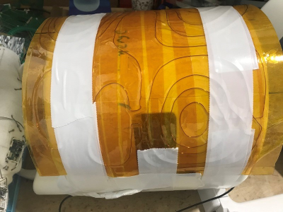 |
Single Half-Cylinder B0 Shim Coil for the Knee
Mingdong Fan1, Sugandima Nishadi Weragoda1, Benjamin Cheung1, Michael Martens1, Labros Petropoulos2, Xiaoyu Yang2, Shinya Handa2, Noah Deetz2, Hiroyuki Fujita2, and Robert Brown1
1Case Western Reserve University, Cleveland, OH, United States, 2Quality Electrodynamics, Mayfield, OH, United States
The void under the knee of a supine patient leads to localized B0 magnetic field distortions due to air-tissue susceptibility differences. To cancel this local artifact, low-order harmonic shims are inadequate, but a single local shim coil can produce higher harmonic suppression. A half-cylinder version wrapped on the outside of the RF coil has been constructed and used to obtain in vivo data consistent with the magnetic field simulations. The detailed results support the possibility of an effective knee shimming approach.
|
|||
3326.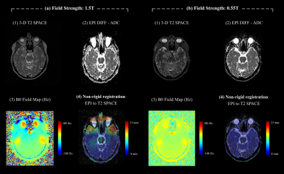 |
B0 Susceptibility-Induced Geometric Image Distortion at Low-Field for Magnetic Resonance Imaging in Radiation Therapy
Manuel Schneider1, Sylvain Doussin1, Dieter Ritter1, and Martin Requardt1
1Siemens Healthcare GmbH, Erlangen, Germany
Our main purpose was to quantify geometric image distortion due to reduced susceptibility at 0.55T compared to 1.5T. Therefore, Bloch simulations of a digital phantom and a 2D GRE sequence were performed. The simulations at both field strengths were calculated twice: once without considering B0 susceptibility, and once considering B0 susceptibility artefacts. Additionally, 3D T2 SPACE images with high readout bandwidth as well as echo-planar diffusion-weighted images were obtained and compared in n=3 volunteers. Less geometric distortion due to B0 susceptibility artefacts was observed at 0.55T compared to higher field strengths.
|
|||
3327.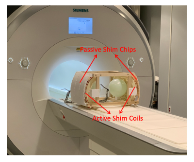 |
Hybrid Active and Passive Local Shimming (HAPLS) for Two-region Magnetic Resonance Imaging (MRI)
Zhi Hua Ren1, Jason P. Stockmann2,3, Andrew Dewdney4, and Ray F. Lee1
1Zuckerman Institute, Columbia University, New York, NY, United States, 2Harvard Medical School, Boston, MA, United States, 3Athinoula A. Martinos Center for Biomedical Imaging, Massachusetts General Hospital, Boston, MA, United States, 4Siemens Healthcare GmbH, Erlangen, Germany
When the bilateral regions of intersest are far off isocenter like in case of shoulders or for brains of two people to be scanned simultaneously, the image quality often suffers from non-optimal spherical harmonic based shimming routine and higher field inhomogeneity in large off-isocenter regions. To address these challenges, a hybrid active and passive local shimming (HAPLS) technique is proposed to shim two isolated areas in one field of view (FOV) simultaneously. Both the simulation and experimental results validated that HAPLS can complementarily address the bifocal and high-order inhomogeneities in the two-region MRI.
|
|||
3328. |
Characterization of 3rd order shim system of human 7T MRI system demonstrates the need of real shim field calibration
Mahrshi Jani1, Ivan Dimitrov1, Bei Zhang1, Binu Thomas1, and Anke henning1
1Advance Imaging Research Center, UT Southwestern Medical Center, Dallas, TX, United States
To perform magnetic resonance imaging (MRI) and magnetic resonance spectroscopy (MRS)in high and ultra-high field homogeneity of field (B0) is required to improve image quality and obtainable metabolic information form MRI scan. In this work we present, real shim field calibration to improve homogeneity of B0 field. The vendor implemented B0 shim system consists of one 0th order, three 1st order, five 2nd order and seven 3rd order shim coils. The proposed calibration to be implemented in existing stand-alone Philips system and further application software shall be developed to integrate latest algorithms and methods to B0 shimming.
|
|||
3329.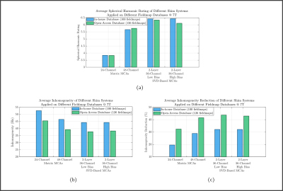 |
The Spherical Harmonic Rating: A Metric for B0 Shim System Performance Assessment
Bruno Pinho-Meneses1 and Alexis Amadon1
1Université Paris-Saclay, CEA, CNRS, BAOBAB, NeuroSpin, Gif-sur-Yvette, France
Different B0 shim system designs are tested on two distinct fieldmap databases for assessment of commonly employed metrics for evaluation of B0 inhomogeneity mitigation. We verify that those metrics might not always be the best choice for performance analysis, as, for a same shim system, they may provide hardly comparable values across sites. We then propose a novel, simple and robust metric for shim system analysis, the Spherical Harmonic Rating, which is shown to provide consistent performance across sites, providing an equivalency of the shim system to a Spherical Harmonics basis.
|
|||
3330.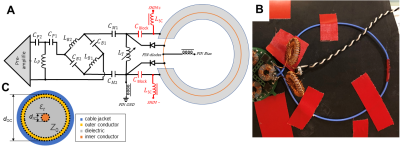 |
On-coil B0 shimming with a flexible coaxial coil element at 3 T
Bernhard Gruber1,2, Maxim Zaitsev1, and Elmar Laistler1
1High Field MR Center, Center for Medical Physics and Biomedical Engineering, Medical University of Vienna, Vienna, Austria, 2Department of Radiology, A.A. Martinos Center for Biomedical Imaging, Massachusetts General Hospital, Harvard Medical School, Charlestown, MA, United States
The combination of highly flexible RF coils and on-coil shimming is an interesting concept, in particular for applications in body parts that have strong inter-patient variability, such as breast, thorax or abdomen. The well-known AC/DC approach on rigid standard loop coils involves many bulky RF chokes and would therefore not be suitable for flexible coil elements. In the presented approach coaxial coils are used instead, where no segmenting capacitors and correspondingly no RF chokes are required. We demonstrate that this conceptual design shows good RF performance and the created magnetic fields for shimming are in good agreement with analytical calculations.
|
|||
3331.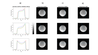 |
Open-Source Gradient Impulse Response Function (GIRF) calibration measurements and their impact in Spiral acquisition
Tiago Timoteo Fernandes1, Andreia S Gaspar1, Andreia C. Freitas1, Nuno A. da Silva2, and Rita G Nunes1
1Institute for Systems and Robotics - Lisboa and Department of Bioengineering, IST-UL, Lisbon, Portugal, 2Hospital da Luz Learning Health, Lisbon, Portugal
A major challenge in MRI, particularly when aiming to reduce equipment costs, is to be able to cope with imperfect hardware performance. Even when using current clinical systems, calibration is often required to reduce image artifacts; an example is the impact of eddy-currents in spiral readouts. Here we controlled for this effect by determining the gradient impulse response function (GIRF) using an open source calibration pulse sequence, demonstrating the impact of the GIRF and its correction both through simulation and in real phantom data. This implementation will contribute towards the development of open source tools for spiral imaging.
|
|||
3332.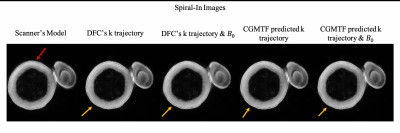 |
Correct K-space coordinates and gradient coupled $$$B_0$$$ variation for Spiral imaging using the current monitor
Tzu-Cheng Chao1, Jürgen Rahmer2, Sandeep Ganji3, Guruprasad Krishnamoorthy3, Peter Börnert2, and James G. Pipe1
1Department of Radiology, Mayo Clinic, Rochester, MN, United States, 2Philips Research, Hamburg, Germany, 3Philips Healthcare, Gainesville, FL, United States
Error from inaccurate eddy current assessment has been a critical issue in spiral imaging. A model is established to estimate both the gradient field and its induced B0 variation from the measured current of the gradient amplifier. The correction improves the geometric consistency between both spiral-in and spiral-out images. The results suggest that the present work have potential to improve quality of spiral imaging.
|
|||
3333.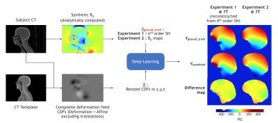 |
On the predictability of B0 maps at ultra-high fields using deep learning
Vidya Prasad1, Sharun S Thazhackal1, Ashvin Srinivasan1, Suja Saraswathy1, Jaladhar Neelavalli2, and Umesh Rudrapatna2
1Philips Research, Philips, Bangalore, India, 2Philips Healthcare, Philips, Bangalore, India
UHF-MRI offers increased scanning sensitivity, but suffers from pronounced B0 inhomogeneities. B0 maps are essential for shimming and reconstruction at UHFs, but suffer from scanning overhead and low measurement confidence. Predicting B0 maps from survey scans can effectively address these issues. We assessed predictability of brain B0 maps at 7T and 11.7T using deep learning on a large synthetic B0 map dataset created from 540 CT images. We found that deep learning can predict 1) spherical harmonics for shimming and 2) the complete B0 maps. Our results indicate that B0 predictions work as effectively as acquiring B0 maps for shimming.
|
|||
3334.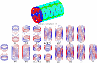 |
A new route of ultrahigh field MRI shimming: hybrid superconducting matrix coil and multi-order spherical harmonics room-temperature shim coils
Yaohui Wang1,2, Qiuliang Wang1,2, Yang Liu1, Jigang Zhao1, Hui Wang1, Junsheng Cheng1, and Feng Liu3
1Institute of Electrical Engineering, Chinese Academy of Sciences, Beijing, China, 2University of Chinese Academy of Sciences, Beijing, China, 3School of Information Technology and Electrical Engineering, The University of Queensland, Brisbane, Australia
A new route for ultrahigh field MRI shimming was proposed which utilized hybrid superconducting matrix coil and multi-order room-temperature shim coils. The proposed scheme was aimed to create a stable and accurate shimming solution for high homogeneous magnetic field magnet. A real 9.4T whole-body MRI magnetic field profile was used to validate the shimming strategy and a remarkable effect was achieved. Therefore, the new shimming method deserves to generalize to ultrahigh field MRI system.
|
|||
3335.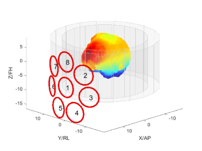 |
Eight channel B0 shim array as add-on for ultra-high field systems
Jan Ole Pedersen1, Esben Thade Petersen2,3, Jason Stockmann4, and Vincent O. Boer2
1Philips Healthcare, Copenhagen, Denmark, 2Danish Research Centre for Magnetic Resonance, Centre for Functional and Diagnostic Imaging and Research, Copenhagen University Hospital, Hvidovre, Denmark, 3Section for Magnetic Resonance, DTU Health Tech, Technical University of Denmark, Kgs Lyngby, Denmark, 4Athinoula A Martinos Center for Biomedical Imaging, Department of Radiology, Massachusetts General Hospital, Charlestown, MA, United States
We have simulated and manufactured a shim coil array specifically designed to address fronto-temporal B0 inhomogeneities at 7T. The shim array is driven by an open-source amplifier, and facilitates slice-wise dynamic shimming. Complexity and costs are purposely kept low to allow manufacturing without prior expertise in electronics design and without access to extensive electronics manufacturing facilities.
|
|||
3336.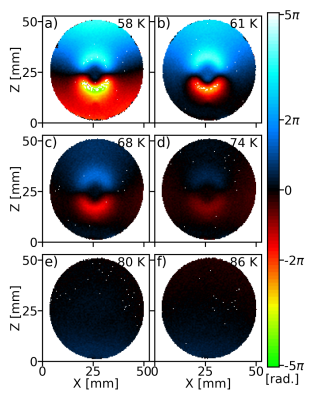 |
Studies of the static field homogeneity artefacts induced by the diamagnetism of HTS coils and solution to avoid them
Aimé Labbé1, Rose-Marie Dubuisson1, Jean-Christophe Ginefri1, Cornelis J van des Beek2, Luc Darrasse1, and Marie Poirier-Quinot1
1Université Paris-Saclay, CEA, CNRS, Inserm, BioMaps, Orsay, France, 2Université Paris-Saclay, CNRS Center for Nanoscience and Nanotechnology, Palaiseau, France
High-Temperature Superconducting (HTS) radiofrequency coils can improve sensitivity by more than an order of magnitude, but their propensity to warp the magnetic field as a result superconducting diamagnetism could cause image degradation. We report on the observation of these B0 artefacts. They are shown to be predominant at lower working temperatures of the HTS coil (60 K) and negligible at 80 K and can be avoided altogether by cooling the HTS coil in B0. We also propose a phenomenological model of these field perturbations. This work studies and solves an important issue in the integration of HTS coil in MRI.
|
|||
3337.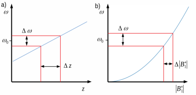 |
Bloch-Siegert |B1+|-Selective Excitation Pulses
Jonathan B Martin1, Christopher E Vaughn1, Mark A Griswold2, and William A Grissom1
1Biomedical Engineering, Vanderbilt University, Nashville, TN, United States, 2Radiology, Case Western Reserve University, Cleveland, OH, United States
We present a new class of pulses which utilize the Bloch-Siegert shift to localize a |B1+| selective excitation. The summation of a far-off-resonant pulse inducing a |B1+|-dependent Bloch-Siegert shift and a frequency-selective excitation pulse excites spins depending on the strength of transmit field that they experience. The passband of the pulse can be shifted by adjusting the frequency modulation of each of the component pulses. We verify the ability of the new pulse to produce a selective excitation in simulation and on a scanner.
|
|||
3338.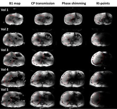 |
B1+ Homogenization in 3D Liver MRI at 7 Tesla Using Eight-Channel Parallel Transmission: Kt-points vs. Phase Shimming
Bobby Runderkamp1, Thomas Roos2, Wietske van der Zwaag2, Matthan Caan3, Gustav Strijkers3, and Aart Nederveen1
1Radiology and Nuclear Medicine, Amsterdam UMC, Amsterdam, Netherlands, 2Spinoza Center for Neuroimaging, Amsterdam, Netherlands, 3Department of Biomedical Engineering and Physics, Amsterdam UMC, Amsterdam, Netherlands
Abdominal MRI at ultra-high field benefits from increased SNR but is challenged by high B1+-inhomogeneity. We use eight-channel parallel transmission to homogenize B1+ signal in 7T liver MRI and compare CP-transmission against phase shimming and a kt-points pulse. Increased signal homogeneity was obtained with the kt-points pulse and phase shimming compared to CP-transmission for small volunteers. Combining the kt-points pulse with a compressed sensing reconstruction facilitated single breath-hold high-resolution liver MRI. With improved B1-mapping and kt-point optimization, kt-points is expected to also improve homogeneity in larger volunteers, and to outperform phase shimming in achieving homogeneous signal in 7T liver MRI.
|
|||
3339.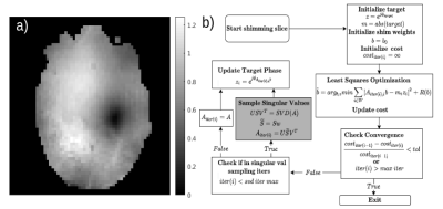 |
Magnitude Least Squares RF Shimming with Singular Vector Minibatching
Jonathan B Martin1, Benjamin M Hardy2, and William A Grissom1
1Biomedical Engineering, Vanderbilt University, Nashville, TN, United States, 2Physics and Astronomy, Vanderbilt University, Nashville, TN, United States
Magnitude-least squares optimization is widely used to design RF shims that produce homogeneous B1+ fields at high field strengths, but the MLS optimization problem is non-convex and prone to becoming stuck in local minima corresponding to unacceptable voids in the shimmed field. We describe a simple, improved Gerchberg-Saxton algorithm for MLS RF shimming in which randomly selected subsets of the B1+ map matrix's singular vectors are used in each shim update. Shims are then refined using conventional GS. Simulations of 8- and 30-channel head coils at 7T verify the method's robustness, and demonstrate advantages over conventional RF shim design techniques.
|
|||
3340.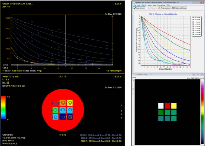 |
A universal framework to build digital reference objects for evaluation of quantitative MRI analysis with multiple measurements
Henry Szu-Meng Chen1, Brian A Taylor1, Joshua P Yung1, Ping Hou1, R. Jason Stafford1, and Ho-Ling A Liu1
1Imaging Physics, MD Anderson Cancer Center, Houston, TX, United States
We propose a framework to easily design and generate digital reference object (DRO) s that conforms with DICOM standard for testing quantitative MRI analysis. The method focuses on using existing phantom scan as the basis and allows user to customize the phantom’s geometry and signal model. We generated a number of DRO for T2* and DSC quantitation and analyzed them with two different software as proof-of-concept.
|
|||
 |
3341.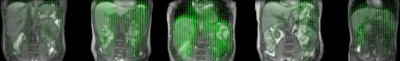 |
Robust real-time 3D motion estimation with MR-MOTUS on an MR-LINAC: a multi-subject validation study
Niek RF Huttinga1, Tom Bruijnen1, Cornelis AT van den Berg1, and Alessandro Sbrizzi1
1Department of Radiotherapy, Computational Imaging Group for MR therapy & Diagnostics, University Medical Center Utrecht, Utrecht, Netherlands
In this work we tighten the gap towards clinical application of MR-MOTUS for real-time tracking of non-rigid 3D respiratory motion-fields during MR-guided radiotherapy. The framework is extended from an experimental setting on a conventional 1.5T MR-scanner, to a practical setting on an MR-LINAC: multi-coil and free-breathing data acquisitions, and the possibility for validation at high temporal resolution. 3D motion-field reconstructions are performed in 160ms/dynamic, for a total of five volunteers. Validations show that spatially realistic motion-fields are reconstructed that have 0.94$$$\pm$$$0.018 temporal correlation with a conventional respiratory-motion surrogate, while satisfying the clinical latency requirement of 5Hz for MR-guided radiotherapy.
|
||
3342.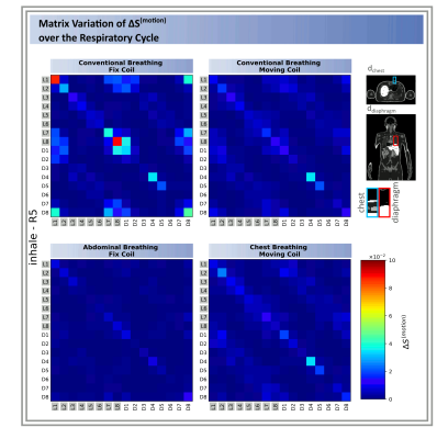 |
Investigation of respiration-induced changes of the scattering matrix by EM simulations and a breathing body model
Natalie Schön1, Frank Seifert1, Gregory J. Metzger2, Bernd Ittermann1, and Sebastian Schmitter1,2
1Physikalisch-Technische Bundesanstalt (PTB), Braunschweig and Berlin, Germany, Berlin, Germany, 2Center for Magnetic Resonance Research, University of Minnesota, Minneapolis, MN, United States
Respiratory motion is a fundamental challenge for thorax MR imaging, particular at ultra-high fields. Recent in-vivo studies have shown that the coil's time-dependent scattering matrix (S-Matrix) can be used for respiration gating to minimize those artefacts. Here, we present electromagnetic simulations of the respiration-induced changes of the transmit RF coil’s S-matrix. Analysing the impact of breathing patterns (conventional, chest, abdominal) illustrates the combined effects of chest/diaphragm motion. The type of respiratory motion and RF coil geometry, position and element type are shown to impact the S-Matrix with implications on coil performance, respiratory triggering, power monitoring and RF pulse design.
|
|||
3343. |
Using gradient-readout, fast spectroscopic imaging and a 3D multi-echo GRE acquisition for scanner Quality Assurance (QA)
Claudiu Schirda1
1Radiology, University of Pittsburgh School of Medicine, Pittsburgh, PA, United States
We propose using a gradient readout, fast MRSI acquisition and a support or stand-alone 3D multi-echo, 3-point Dixon GRE scan for MRI scanner and coil quality assurance (QA). The fast MRSI acquisition with the support 3D GRE scan uses the BRAINO phantom, takes less than 10 minutes and could be used for 1 to 3 months QA. A modified (with an increased FOV), stand-alone 3D GRE scan, takes 3-minutes, could be used either with a spherical or cylindrical phantom and is proposed for daily QA, in addition to the manufacturer EPI-based stability scan. Fully automated reconstruction programs provided at github.com/FastMRSI.
|
|||
3344.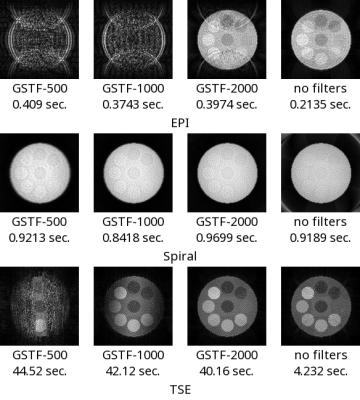 |
A New Representation of the Gradient System Transfer Function Using a Practical Algorithm of Finite Impulse Response Filters
Hidenori Takeshima1
1Advanced Technology Research Department, Research and Development Center, Canon Medical Systems Corporation, Kanagawa, Japan
This work proposes a new representation of the gradient system transfer function (GSTF) based on finite impulse response (FIR) filters. FIR filters can represent arbitrary gradient functions but are not used because of their computational complexity. The proposed method represents the GSTF as a set of average coefficients and their durations. Input signals are represented as sums of signals for the same durations. On an update step, only two signals are accessed for each duration. Therefore, as confirmed experimentally, the proposed method can process arbitrary gradient functions in acceptable computational time, without using infinite impulse response (IIR) filters.
|
The International Society for Magnetic Resonance in Medicine is accredited by the Accreditation Council for Continuing Medical Education to provide continuing medical education for physicians.