Digital Posters
Novel RF Coils & Components
ISMRM & SMRT Annual Meeting • 15-20 May 2021

| Concurrent 2 | 17:00 - 18:00 |
1411.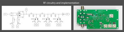 |
Rapid high power transmit-receive switching using a timed cascade of PIN diodes
Christoph Michael Schildknecht1, Markus Weiger1, Romain Froidevaux1, and Klaas Paul Pruessmann1
1Institute for Biomedical Engineering, ETH Zurich and University of Zurich, Zürich, Switzerland
For imaging of short-T2 samples often very short excitation pulses are desired, which requires high peak RF power. In this work, a transmit-receive switch is presented that can handle 18kW peak RF power and switches its state in less than 1µs. The novel design uses a timed cascade of anti-parallel PIN diode pairs to achieve this performance.
|
|||
1412.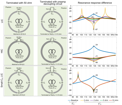 |
Analysis of preamplifier decoupling effect in MRI coil array with electromagnetic field and RF circuit co-simulation
Ming lu1,2, Bei Zhang3, John C. Gore1,2, and Xinqiang Yan1,2
1Vanderbilt University Institute of Imaging Science, Vanderbilt University Medical Center, Nashville, TN, United States, 2Department of Radiology and Radiological Sciences, Vanderbilt University Medical Center, Nashville, TN, United States, 3Advanced Imaging Research Center, University of Texas Southwestern Medical Center, Dallas, TX, United States
Preamplifier decoupling is a standard way of suppressing inter-element inductive coupling in MRI receive arrays. A well-known method to evaluate and optimize RF coils before practical fabrication is to use circuit-level and electromagnetic (EM) field-level simulations. However, to be best of our knowledge, currently there is not a solid method to evaluate the preamp decoupling effect with circuitry and EM field analysis. In this work, we used RF circuit and EM co-simulation4 to evaluate the preamplifier decoupling effect and tested it on the conventional coil (LIC) and the recently introduced high impedance coil (HIC).
|
|||
1413.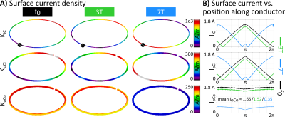 |
Should coaxial coils be operated at their self-resonance? A simulation study
Sigrun Roat1, Andre Kuehne2, Lena Nohava1,3, and Elmar Laistler1
1High Field MR Center, Center for Medical Physics and Biomedical Engineering, Medical University of Vienna, Vienna, Austria, 2MRI.TOOLS GmbH, Berlin, Germany, 3CEA, CNRS, Inserm, BioMaps (Laboratoire d'Imagerie Biomédicale Multimodale Paris Saclay), Université Paris-Saclay, Orsay, France
Flexible RF coils gained large interest in recent years. One possibility to achieve flexibility is using transmission line resonator-type coils made from coaxial cable. These self-resonant structures can be constructed without lumped elements along the loop enhancing flexibility. No consensus has been reached yet whether these coils should be driven at their respective self-resonance. To investigate this question, we have performed a simulation study investigating 11 different setups at 3 frequencies each
|
|||
1414.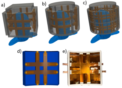 |
Conformal design of radio-frequency head coil for ultra-high field MRI
Tiago Martins1, Tales Santini1, Jacob Berardinelli1, Anthony DeFranco1, and Tamer S Ibrahim1
1University of Pittsburgh, Pittsburgh, PA, United States
In this work, we explore a conformal Tic Tac Toe (TTT) head coil design for ultra-high field MRI. By conforming the antennas to the human head, we can extract better homogeneity allowing for better radiofrequency shimming. The comparison between the traditional TTT design and the conformal TTT design shows expanded coverage, reduction of the coefficient of variation by 0.4% and same intensity for the 16-channel version. The conformal designs are extremely flexible as its advantages can be explored for different configurations and sizes.
|
|||
1415.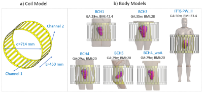 |
Safety and imaging performance of 2-channel RF shimming for fetal MRI at 3T
Filiz Yetisir1, Esra Abaci Turk1,2, P. Ellen Grant1,2,3, Elfar Adalsteinsson4,5, and Lawrence L. Wald3,5,6
1Fetal-Neonatal Neuroimaging and Developmental Science Center, Boston Children's Hospital, Boston, MA, United States, 2Department of Pediatrics, Harvard Medical School, Boston, MA, United States, 3Department of Radiology, Harvard Medical School, Boston, MA, United States, 4Department of Electrical Engineering and Computer Science, Massachusetts Institute of Technology, Cambridge, MA, United States, 5Harvard-MIT Division of Health Science and Technology, Massachusetts Institute of Technology, Cambridge, MA, United States, 6Athinoula A. Martinos Center for Biomedical Imaging, Massachusetts General Hospital, Charlestown, MA, United States
3T MRI provides increased SNR but poses technical challenges for fetal imaging such as increased field inhomogeneity and SAR. RF shimming can address some of these challenges but also adds safety considerations. Using a diverse set of 5 numerical pregnant body models generated from patient MRI datasets, we found that 2-channel RF shimming can improve transmit field amplitude and uniformity by up to 19% and 36% respectively without increasing maternal or fetal SAR. The biggest difference in SAR and transmit field patterns was observed between the supine models and the left lateral model.
|
|||
1416.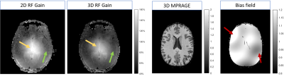 |
Stabilization of bias field on 3D MPRAGE at 7T with dielectric pads and 3D-based B1+ scaling
Giske Opheim1, Vincent O. Boer2, Esben Thade Petersen2,3, Martin Prener1, Olaf B. Paulson1,4, and Jan Ole Pedersen5
1Neurobiology Research Unit, Dept. of Neurology, Copenhagen University Hospital Rigshospitalet, Copenhagen, Denmark, 2Danish Research Centre for Magnetic Resonance, Centre for Functional and Diagnostic Imaging and Research, Hvidovre, Denmark, 3Section for Magnetic Resonance, DTU Health Tech, Technical University of Denmark, Kgs. Lyngby, Denmark, 4Faculty of Health and Medical Sciences, University of Copenhagen, Copenhagen, Denmark, 5Philips Healhtcare, Copenhagen, Denmark
7T MRI has several cases of demonstrated clinical yield, but is challenged by spatial B1 inhomogeneity. Bias fields in structural images may be acceptable for visual and computational analysis, and can to some extent be accounted for by bias-field corrections. However, this is significantly complicated since the characteristics of the bias field often varies between patients. By analyzing stability of bias fields in 3D MPRAGE images, we compared two dielectric pad setups and the impact of 3D-based B1+ scaling, i.e., optimization of the RF gain. We found increased stability by using large pads and 3D-based B1+ scaling in combination.
|
|||
1417.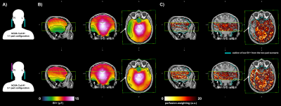 |
The impact of B1+ on the optimisation of high-resolution ASL acquisitions at 7T
Sriranga Kashyap1, Roy A. M. Haast2, Thomas F. Kirk3, An T. Vu4,5, Denizhan Kurban1, Ron Hellenbrand1, Christopher J. Wiggins6, Alard Roebroeck1, Ali R. Khan2, David A. Feinberg1,7,8, Benedikt A. Poser1, and Dimo Ivanov1
1Department of Cognitive Neuroscience, Maastricht University, Maastricht, Netherlands, 2Centre for Functional and Metabolic Mapping, Western University, London, ON, Canada, 3University of Oxford, Oxford, United Kingdom, 4University of California, San Francisco, CA, United States, 5San Francisco Veteran Affairs Health Care System, San Francisco, CA, United States, 6Scannexus B.V., Maastricht, Netherlands, 7Advanced MRI Technologies, Sebastopol, CA, United States, 8Helen Wills Neuroscience Institute, University of California, Berkeley, CA, United States
In this 7T study, we examined potential causes of apparent absence of perfusion in the right inferior temporal lobe, by (i) assessing the B1+ distributions on two 1Tx/32Rx head-coils to rule out a hardware source, and (ii) optimising the spatial positioning of dielectric pads to achieve a more symmetric B1+ field. We observed that using an optimised dielectric pad positioning allows for a near whole-brain symmetric B1+, thereby, ensuring the adiabatic condition for the inversion pulse is fulfilled, resulting in the robust measurement of perfusion in the right temporal lobes.
|
|||
1418.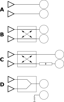 |
Is it feasible to Make More Effective Use of Finite RF Power Resources in pTx Systems Using a Coupling Matrix?
Stephen E. Ogier1, Shaihan Malik1, and Joseph Hajnal1,2
1Biomedical Engineering Department, School of Biomedical Engineering and Imaging Sciences, King's College London, London, United Kingdom, 2Centre for the Developing Brain, School of Biomedical Engineering and Imaging Sciences, King's College London, London, United Kingdom
Parallel transmit using equal numbers of RF power amplifiers and coil elements has emerged as a common way to improve the homogeneity of the transmit field through RF shimming. When performing RF shimming, in many cases the magnitude of the transmit field is limited by the maximum power limit of a single RF amplifier. We propose networks that can be used to share power between transmit channels, increasing the maximum transmit field. These networks are shown to provide substantial flexibility in RF power allocation for certain conditions on the relative phases of the channels.
|
|||
1419.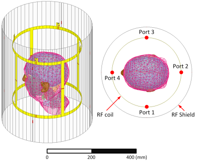 |
Design of transmit array coils by minimizing the modal reflected power values and increasing B1+ efficiency
Ehsan Kazemivalipour1,2, Giorgio Bonmassar3, Laleh Golestanirad4,5, and Ergin Atalar1,2
1Electrical and Electronics Engineering Department, Bilkent University, Ankara, Turkey, 2National Magnetic Resonance Research Center (UMRAM), Bilkent University, Ankara, Turkey, 3AA. Martinos Center, Massachusetts General Hospital (MGH), Harvard Medical School, Boston, MA, United States, 4Department of Radiology, Feinberg School of Medicine, Northwestern University, Chicago, IL, United States, 5Department of Biomedical Engineering, McCormick School of Engineering, Northwestern University, Evanston, IL, United States
The vast majority of RF coil design techniques are based on reducing the coupling level to increase the coils' power efficiency. The assessment of scattering (S) parameters plays a crucial role in these techniques. However, just by checking S-parameters, we cannot be sure that the coil's accepted power generates a field with useful characteristics. Here, we propose to perform the co-simulation on transmit array coils and replace unknown capacitors with power sources. Extraction of the field profiles of the coil's lumped ports/elements could provide the opportunity to involve the field-dependent parameters in the minimization procedure of finding the coil's capacitors.
|
|||
1420.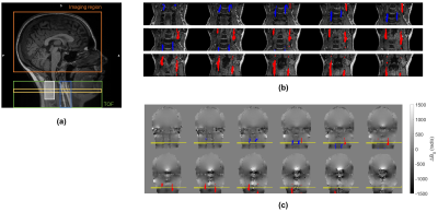 |
Brain perfusion imaging using pseudo-continuous arterial spin labelling MRI: impact of RF coil shimming of the labelling region
Sofia Guterres1, Ana Rodrigues Fouto1, Nuno André Silva2, Pedro Vilela3, and Patrícia Figueiredo1
1Institute for Systems and Robotics - Lisboa and Department of Bioengineering, Instituto Superior Técnico, Universidade de Lisboa, Lisboa, Portugal, 2Hospital da Luz Learning Health, Lisboa, Portugal, 3Hospital da Luz, Lisboa, Portugal
The labelling efficiency of pCASL perfusion imaging sequences can be hampered by B0 field inhomogeneities. We assessed the impact of shimming the labelling region using a commercial RF coil with dedicated shiming channels in the neck, on data acquired from healthy volunteers. Brain perfusion was quantified, as well as the labelling efficiency (based on B0 fieldmaps and numerical simulations). Simulation experiments show that labelling efficiency significantly depends on B0 field inhomogeneity. In vivo experiments, however, showed subtle inhomogeneity reduction within the arteries in the labelling region, yielding no significant improvements in pCASL labelling efficiency or the respective perfusion signal.
|
|||
1421.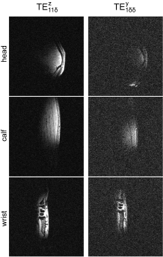 |
The Impact of Quasi-Transverse Electric Modes Excited by Dipole Antennas on Transmit Field in In Vivo Ultrahigh Field MRI
Daniel Wenz1,2 and Rolf Gruetter1,3
1CIBM Center for Biomedical Imaging, Lausanne, Switzerland, 2Animal Imaging and Technology, Ecole Polytechnique Federale de Lausanne (EPFL), Lausanne, Switzerland, 3Laboratory of Functional and Metabolic Imaging (LIFMET), Ecole Polytechnique Federale de Lausanne (EPFL), Lausanne, Switzerland
This work investigates quasi-transverse electric modes which can be excited within rectangular dielectric blocks which are often used to shorten dipole antennas for MRI at 7T. In simulations we studied different parameters which can affect the performance of a dielectrically-shortened dipole antenna. We showed that an optimal geometry for the case when the block and the sample are physically separated differs significantly from the case when perfect direct contact is achieved. Two prototypes were built and used in an in vivo MRI experiments involving one subject showing how different dielectric modes can affect the image quality.
|
|||
1422.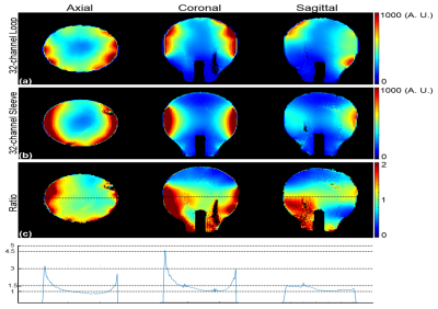 |
A Novel High Density 32-channel Sleeve Antenna Receiver Array for the Human Head Imaging at 10.5 T
Myung Kyun Woo1, Lance DelaBarre1, Matt Waks1, Russell Lagore1, Jeromy Thotland1, Uk-Su Choi2, Andrea Grant1, Steve Jungst1, Nader Tavaf1, Yigitcan Eryaman1, Kamil Ugurbil1, and Gregor Adriany1
1Center for Magnetic Resonance Research, Minneapolis, MN, United States, 2Center for Information and Neural Networks, Osaka, Japan
We designed an elliptically arranged novel 32-channel Sleeve antenna receiver array for human whole brain imaging at 10.5 tesla. To demonstrate the signal-to-noise ratio (SNR) performance of this array, we evaluated and compared it with a standard 32-channel loop receiver array.
|
|||
1423.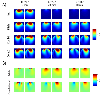 |
Combining Loops and Dipoles to Increase the Signal-to-Noise Ratio in Human Brain MRI at 7T: How to Shorten a Dipole Antenna?
Thomas Dardano1, Rolf Gruetter1,2, and Daniel Wenz2,3
1Laboratory of Functional and Metabolic Imaging (LIFMET), Ecole Polytechnique Federale de Lausanne (EPFL), Lausanne, Switzerland, 2CIBM Center for Biomedical Imaging, Lausanne, Switzerland, 3Animal Imaging and Technology, Ecole Polytechnique Federale de Lausanne (EPFL), Lausanne, Switzerland
Dipole antennas can be used in multi-channel loop/dipole arrays to boost the signal-to-noise ratio in MRI at 7T (f=300MHz). For this purpose, dipole antennas need to be physically shorter. In this work we conducted electromagnetic field simulations and phantom experiments at 7T to compare the performance of an inductively-shortened dipole antenna with a dielectrically-shortened dipole antenna in a loop/dipole combination. We evaluated the performance of both designs in different loading conditions and we found that the dielectrically-shortened dipole antenna performed in a very robust manner providing apparent receive field gains when compared with its inductively-shortened counterpart.
|
|||
1424. |
Optimization of a massive-element self-decoupled transmit array for 7T head imaging
Ming Lu1,2, John C. Gore1,2, and Xinqiang Yan1,2
1Vanderbilt University Institute of Imaging Science, Nashville, TN, United States, 2Department of Radiology and Radiological Sciences, Vanderbilt University Medical Center, Nashville, TN, United States
At high fields, B1 inhomogeneity is one of the major challenges that limit imaging performance. Parallel transmission with an array of coils is a recognized solution to B1 inhomogeneity. The self-decoupled coil is attractive in Tx array design since it exhibits intrinsic inter-element isolation that may ease the coil fabrication, and allows the dipole mode as well as loop mode. In this work, we evaluated a series of self-decoupled coils in terms of these parameters and aimed to provide guidance for the practical building of a high-performance self-decoupled Tx array.
|
|||
1425. |
High-density 72-channel head array at 7Tesla
Mark Gosselink1, Tijl van der Velden1, Hans Hoogduin1, Martijn Froeling1, and Dennis W. J. Klomp1
1University Medical Center Utrecht, Utrecht, Netherlands
A 64 channel head coil receiver is presented that operates with low noise coupling inside a continuously tuned 8-channel transceiver coil. When used as a 72-channel receiver the setup outperforms the default 32 channel receiver array in acceleration, demonstrated by high-resolution MPRAGE images of the human brain.
|
|||
1426. |
A 30-element transmit array for 7 Tesla brain imaging with array compressed parallel transmission
Charlotte Sappo1,2, Gary R Drake1,3, Xinqiang Yan1,3, and William A Grissom1,2,3,4
1Vanderbilt University Institute of Imaging Science, Nashville, TN, United States, 2Biomedical Engineering, Vanderbilt University, Nashville, TN, United States, 3Radiology, Vanderbilt University, Nashville, TN, United States, 4Electrical and Computer Engineering, Vanderbilt University, Nashville, TN, United States
A dense array is desirable for ultra-high field in parallel transmission to better mitigate B1+ field inhomogeneities and control specific absorption rate. Most ultra-high field scanners are limited by the existing number of transmit channels. This limitation can be overcome using array-compressed parallel transmission (acpTx) which enables a small number of channels to drive a large number of coils. This work describes the construction of a 30-loop head transmit array using a mixed overlapped and self-decoupled design with a widely-used commercial 32-channel receive insert for human imaging applications.
|
|||
1427.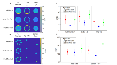 |
SNR of Flexible Versus Rigid Coil Arrays for Knee MRI
Jeremiah Hess1, Marianne Black2, Feliks Kogan2, and Brian Hargreaves1,2,3
1Department of Bioengineering, Stanford University, Stanford, CA, United States, 2Department of Radiology, Stanford University, Stanford, CA, United States, 3Department of Electrical Engineering, Stanford University, Stanford, CA, United States
Flexible extremity coil-arrays for knee imaging in MRI can improve coil positioning and comfort to patients in the scanner and wrap tightly around the knee, placing coil elements close to anatomy of interest. Despite this, flex coils have not been widely adapted due to perceived superiority of the SNR of transmit-receive coils. In this study, we analyzed the SNR of flex coil-arrays versus a rigid coil-array in phantom and in vivo to determine which coil-arrays had improved SNR. Preliminary results suggest that flexible coil-arrays show comparable or increased SNR and generally more uniform SNR over rigid coil-arrays.
|
|||
1428.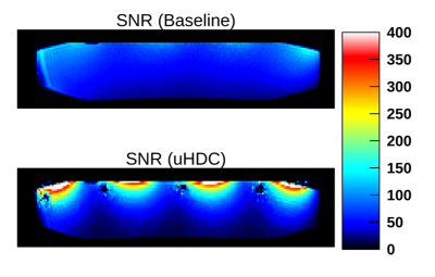 |
Reduction of coupling and noise by ultrahigh dielectric constant (uHDC) materials for phase array coil at 3T
Navid PourramzanGandji1, Christopher T. Sica2, Gary W. Yang3, Hannes Wiesner4, Soo Han Soon4, Xiao-Hong Zhu4, Michael Lanagan5, Wei Chen4, and Qing X. Yang1
1Neurosurgery, Pennsylvania State University, Hershey, PA, United States, 2Radiology, Pennsylvania State University, Hershey, PA, United States, 3Pennsylvania State University, Hershey, PA, United States, 4Center for Magnetic Resonance Research, Department of Radiology, University of Minnesota, Minneapolis, MN, United States, 5Materials Science and Engineering, Pennsylvania State University, State College, PA, United States
Prior work has examined the effects of uHDC material upon phased array coils. Several beneficial effects have been observed: reduction of per-channel noise and inter-channel noise correlations, and enhancement of SNR. Due to the high dielectric constant the electric field will be confined to the dielectric material, which leads to a reduction of the primary noise source, the conservative electric field. As a result the non-conservative field becomes more dominant within the sample, with a resulting increase of B1 field intensity and more substantial SNR enhancement.
|
|||
1429.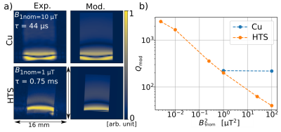 |
A novel characterization method of HTS non-linear electrical properties using MRI
Aimé Labbé1, Isabelle Saniour1, Rose-Marie Dubuisson1, Jean-Christophe Ginefri1, Luc Darrasse1, and Marie Poirier-Quinot1
1Université Paris-Saclay, CEA, CNRS, Inserm, BioMaps, Orsay, France
Decoupling is a major technological challenge of High-Temperature Superconducting (HTS) surface coils. A promising strategy is to exploit nonlinearities in the HTS coil electric response to achieve decoupling. In this work, we present a new characterization method allowing the evaluation of these nonlinearities using MRI. We first present the theoretical model behind this approach; we then proceed to use it on 3D images of a water phantom in the vicinity of a copper coil and then a HTS coil to validate the model through the extraction of the quality factor Q, compared to the one measured by alternative methods.
|
|||
1430.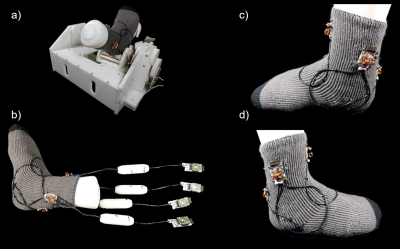 |
Robot Assisted Dynamic Ankle Joint Imaging with a Wearable 4-Channel High Impedance Coil at 1.5T MRI
Matthäus Poniatowski1, Ilan Elias2, Mirsad Mahmutovic1, Gurinder Multani1, Sam-Luca J.D. Hansen1, Markus W. May1, Alexander M. König3, Jens H. Figiel3, Andreas H. Mahnken3, and Boris Keil1
1Institute of Medical Physics and Radiation Protection, TH Mittelhessen University of Applied Sciences, Gießen, Germany, 2Motionrad GmbH, Berlin, Germany, 3Department of Diagnostic and Interventional Radiology, Philipps-University Marburg, Marburg, Germany
Limitations of conventional MRI include its lack of ability to observe controlled joint motion and biomechanics in real-time. Although clinically evident, many injuries, instabilities, and dysfunctions of the musculoskeletal system are frequently not depicted on conventional static MRI. To enable dynamic MRI for joints, an in-bore motion-assisted device and a wearable coil array was designed, constructed, and validated. The combination of robotic assisted joint motion, a tight-fitting coil array that does not restrict the joint's range of motion, and accelerated imaging enabled dynamic MRI of the ankle.
|
The International Society for Magnetic Resonance in Medicine is accredited by the Accreditation Council for Continuing Medical Education to provide continuing medical education for physicians.