Digital Posters
Toolbox: Software & Phantoms
ISMRM & SMRT Annual Meeting • 15-20 May 2021

| Concurrent 2 | 19:00 - 20:00 |
3345.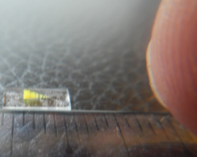 |
Cube set for the quantification of 3-dimensional spatial resolution in MR- micro imaging and microscopy (a = 128 - 4 µm) using 2-photon lithography
Andreas Georg Berg1,2, Stefan Hengsbach3, and Klaus Bade3
1Center for Medical Physics and Biomedical Engineering High field MR-Center, Medical University of Vienna, Vienna, Austria, 2High-Field MR-Center (MRCE), Medical Universits of Vienna, Vienna, Austria, 3Karlsruhe Nano Micro Facility (KNMF), Karlsruhe Institute of Technology (KIT), Leopoldshafen-Eggenstein, Germany
Quality-control for systematic improvements in 3D-isotropic spatial resolution of the MR-imaging system up to the microscopic range becomes increasingly relevant not only for preclinical imaging but also for High-Field-MR human scanners. The design of a prototype phantom, comprising a set of 3D-cubes and an exemplary MR-microscopic evaluation is presented. The extraordinary challenges on accurate bar-width-to-cavity ratios for the cube range from periodicity a=128µm down to a=4µm are dealt with using 2-photon-lithography as manufacturing technology. The spatial resolution for 3D isotropic imaging can be checked qualitatively and quantitatively by interpolation of the Modulation-Transfer-Function demonstrated on a 3D-GRE sequence as an example.
|
|||
3346.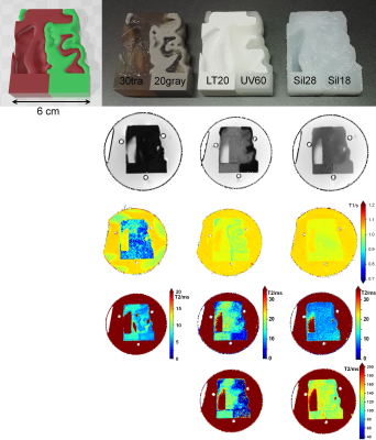 |
En Route to multiphasic anthropomorphic MR phantoms: An additive manufacturing approach applying silicone 3D-printing techniques
Wolfgang Kilian1, Rüdiger Brühl1, Yasser Abdulhadi1, and Bernd Ittermann1
1Physikalisch-Technische Bundesanstalt (PTB), Berlin, Germany
Stable and well characterized anthropomorphic MR-phantoms for quantitative MRI of T1 and T2 measurements are still lacking. We investigate the potential application of additive manufacturing of silicone to produce multi-compartment MR-phantoms with well characterized relaxation properties. Various two-component silicone types with different mixture ratios were investigated. Additionally, the influence of admixing colors to modulate relaxation times was studied. First bi-phasic test samples were designed, produced by different commercial 3D printing sources, and characterized.
|
|||
3347.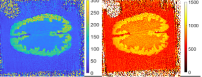 |
En route to multiphasic anthropomorphic MR phantoms: A new mold-based approach applying gel-based preparation to real MR-datasets geometries
Adriano Troia1, Umberto Zanovello1, Luca Zilberti1, Matteo Cencini2, Michela Tosetti2,3, David Kilian4, Martina Capozza5, Wolfgang Kilian6, and Tuğba Dışpınar Gezer7
1INRIM, Turin, Italy, 2IRCCS Stella Maris, Pisa, Italy, 3IMAGO7 Foundation, Pisa, Italy, 4Centre for Translational Bone, Joint and Soft Tissue Research, Faculty of Medicine and University Hospital TUD, Dresden, Germany, 5Department of Molecular Biotechnology and Health Sciences UNITO, Turin, Italy, 6Physikalisch-Technische Bundesanstalt, Braunschweig, Germany, 7National Metrology Institute, TUBITAK, Istanbul, Turkey
Anthropomorphic phantoms for MRI imaging are rapidly evolving also thanks to the increase of 3D printing technologies. Their realization may represent an important support for the implementation of advanced quantitative imaging technique such as Magnetic Resonance Fingerprinting (MRF)or Electrical Properties Tomography (EPT). Even if formulations of gel based tissue-mimicking materials have been widely explored, generally they lacked in achieving the simultaneous tuning of electrical and relaxation properties of the mimicked tissues. In this study, customized moulds of white and grey matter are combined to realize brain-like gel phantoms in which relaxation times, conductivity and permittivity have been measured.
|
|||
3348.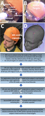 |
A Method for Alignment of an Augmented Reality Display of Brain MRI With The Patient’s Head
Christoph Leuze1, Supriya Sathyanarayana1, Bruce L Daniel1, and Jennifer A McNab1
1Radiology, Stanford University, Stanford, CA, United States
We present a method for alignment of augmented reality display of brain MRI with the patient’s real-world head with potential applications to an AR-neuronavigation system that relies on a see-through display.
|
|||
3349.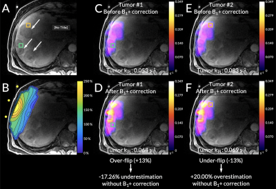 |
Specialized Computational Methods for Denoising, B1 Correction, and Kinetic Modeling in Hyperpolarized 13C MR EPSI Studies of Liver Tumors
Philip Meng-en Lee1, Hsin-Yu Chen1, Jeremy W Gordon1, Zihan Zhu1, Peder EZ Larson1, Nicholas Dwork1, Mark Van Criekinge1, Lucas Carvajal1, Michael A Ohliger1, Zhen J Wang1, Duan Xu1, John Kurhanewicz1, Robert A Bok1, Rahul
Aggarwal2, Pamela N Munster2, and Daniel B Vigneron1
1Department of Radiology & Biomedical Imaging, University of California, San Francisco, San Francisco, CA, United States, 2Department of Medicine, University of California, San Francisco, San Francisco, CA, United States
The inhomogeneous B1 excitation profile of 13C surface transmit/receive coils provides high SNR near the surface, but results in a spatially-varying B1+. For accurately quantifying the pyruvate to lactate conversion rate (kPL), the flip angle needs to be corrected based on the B1 excitation profile. Simultaneously, random noise in hyperpolarized spectral data obscures peaks of downstream metabolites. In this work, we developed and tested a specialized computational pipeline incorporating denoising and a B1 excitation field correction method that improved quantitative kinetic rate analyses of hyperpolarized 13C MRSI scans of liver tumor patients acquired with a T/R 13C surface coil.
|
|||
3350.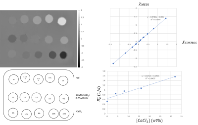 |
A magnetic susceptibility phantom with large range of negative/positive values for quantitative validations
Alexey Dimov1, Kelly Gillen1, and Yi Wang1
1Radiology, Weill Cornell Medicine, New York, NY, United States
A magnetic susceptibility phantom with a large range of negative to positive susceptibility values relative to water is demonstrated for validating quantitative susceptibility mapping (QSM) and related quantitative MRI techniques. The phantom consists of vials with various concentrations of paramagnetic Gadolinium contrast agent (Gd) and diamagnetic calcium chloride (CaCl2) solutions. Compared to previously reported phantoms, this phantom is easy to construct and highly stable and has minimal effects of unwanted air bubbles.
|
|||
3351.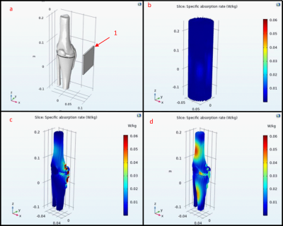 |
3D Printed MRI Knee Phantom for Imaging Safety and Protocol Studies at Ultrahigh Fields
Leo Konst Marecki1, Eric Konst Marecki1, and Xiaoliang Zhang1
1Biomedical Engineering, SUNY University at Buffalo, Buffalo, NY, United States
Most utilized phantoms for determining the SAR compose of a uniform material to determine the properties of each coil and the exposure time to remain below the FDA guidelines and ensure patient safety. Accurate modeling of the tissue permittivity, conductivity, and geometry will provide more accurate methods to measure the SAR of each organ for a variety of RF coil systems. Our results showed that utilizing 3D printed Nylon containers and Polyvinylpyrrolidone (PVP), NaCl, and water solutions can be used to model the geometry and tissue characteristics of the human knee.
|
|||
3352.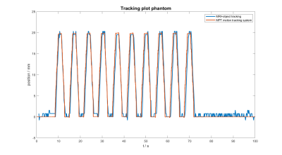 |
Development of an anthropomorphic torso and left ventricle phantom for flow and respiratory motion simulation
Tito Körner1, Stefan Wampl1, Marcos Wolf1, Martin Meyerspeer1, Maxim Zaitsev1,2, Wolfgang Birkfellner1, and Albrecht Schmid1
1High Field MR Center, Center for Medical Physics and Biomedical Engineering, Medical University of Vienna, Vienna, Austria, 2Medical Physics, Department of Radiology, University Medical Center Freiburg, Freiburg, Germany
Development of motion compensation strategies is challenging, especially when involving volunteer measurements due to high demands to subject compliance and low reproducability. In this work a human shaped torso phantom, capeable of simulating respiratory motion of a left ventricle phantom including blood flow simulation is presented. A tracking algorithm is applied in postprocessing to acquired images containing simulated respiratory motion. Comparison to motion tracking data from an external sensor show good agreement proving the setup to be an environment with high reproducability and hence ideal for the purpose to develop and evaluate motion compensation strategies.
|
|||
3353.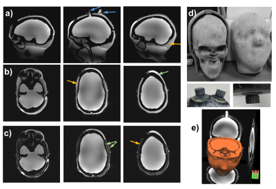 |
3D printed head-shaped phantom with lipid layer and brain-mimicking metabolites for 7 Tesla MRI and MRSI
Rita Schmidt1,2, Ghil Jona3, and Edna Furman-Haran2,3
1Neurobiology, Weizmann Institute of Science, Rehovot, Israel, 2The Azrieli National Institute for Human Brain Imaging and Research, Weizmann Institute of Science, Rehovot, Israel, 3Life Sciences Core Facilities, Weizmann Institute of Science, Rehovot, Israel
Moving to ultra-high fields (≥7T), the inhomogeneity of both RF and static magnetic fields increases, which motivates to design a realistic head-shaped phantom. In this study, a 3D-printed head-shaped phantom with brain mimicking metabolites and lipid layer examined for 7T MRI and MRSI. The phantom was designed to resemble the brain with respect to B0 and B1 distributions, metabolites and lipid layer. We examined it for 1H MRS and MRSI, especially in the lipid layer vicinity. We also demonstrated in EPI that the Fat Suppression pulse flip angle can be optimized to minimize the lipid artifact and reduce the SAR.
|
|||
3354.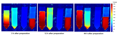 |
Oil-in-water emulsions for tissue simulation in magnetic resonance imaging: Determination of MR-properties of emulsifiers
Victor Fritz1, Petros Martirosian1, Jürgen Machann1,2, Rolf Daniels3, and Fritz Schick1
1Section on Experimental Radiology, University of Tübingen, Tübingen, Germany, 2Institute for Diabetes Research and Metabolic Diseases of the Helmholtz Centre Munich at the University of Tübingen, Tübingen, Germany, 3Institute of Pharmaceutical Technology, University of Tübingen, Tübingen, Germany
The aim of this work was to develop stable and homogeneous oil-in-water emulsions for tissue simulation in MRI. For this purpose, three different emulsifiers (polysorbate 60, sodium dodecyl sulfate (SDS), and soy lecithin) were examined for their stabilizing ability. In addition, their potential impact on the MR-measurements was investigated. Sufficient stability can be achieved using both emulsifier, polysorbate and lecithin. Emulsions stabilized by SDS showed a visually lower stability. Due to its sufficient stabilizing ability, promising relaxometric properties (r1,lecithin=0,11wt%-1s-1, r2,lecithin =0,62wt%-1s-1), and no additional spectral resonances, lecithin is suggested as the preferred emulsifier for use in MRI.
|
|||
3355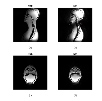 |
1:1 Scale Agar-Agar Paramagnetic Phantom for Brain and Cervical Spine MRI
Elif Aygun1,2, Ahmet Rahmetullah Cagil1,2, and Emine Ulku Saritas1,2,3
1Department of Electrical and Electronics Engineering, Bilkent University, Ankara, Turkey, 2National Magnetic Resonance Research Center (UMRAM), Bilkent University, Ankara, Turkey, 3Neuroscience Graduate Program, Bilkent University, Ankara, Turkey
Phantoms doped with paramagnetic materials are commonly used for quality assessment of MRI protocols and image reconstruction techniques. These phantoms are typically prepared using cylindrical containers and mimic the T1 /T2 characteristics of human tissue. In this work, we propose a 1:1 scale human head and neck agar-agar phantom prepared using a 3D human model. The phantom mimics human tissue characteristics while incorporating the skull and cervical vertebrae. The MRI images show that the phantom closely matches human anatomy, and the susceptibility artifacts in EPI images successfully mimic those seen during in vivo imaging of the brain and spine.
|
|||
3356.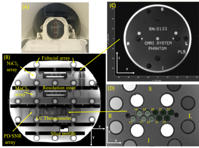 |
MRI System Phantom: assessing the MR visible thermometer, scanner geometric distortion corrections, and effect of fill conductivity
Stephen E Russek1, Kathryn E Keenan1, Karl F Stupic1, Teryn S Wilkes2, Ramesh Karki3, and Todor Karaulanov4
1NIST, Boulder, CO, United States, 2Intermountain Neuroimaging Consortium, University of Colorado, Boulder, CO, United States, 3University of Colorado Anschutz, Radiological Sciences, Aurora, CO, United States, 4CaliberMRI, Inc., Boulder, CO, United States
Using the MRI system phantom we demonstrate the utility of the embedded MR-readable thermometer to measure and correct for temperature variations, demonstrate the use of the fiducial array to characterize scanner geometric distortions and the efficacy of software distortion corrections, and finally look at the effect of the electromagnetic properties of the phantom fill on quantitative measurements.
|
|||
3357.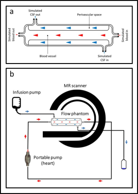 |
A novel MRI phantom to study the fluid dynamics of the glymphatic system: a proof-of-concept study
Endre Grøvik1,2, Elisabeth Lysvik1, Robin Bugge1, Kyrre Emblem1, Trine Hjørnevik1, Svein-Are Vatnehol1, and Tryggve Storås1
1Oslo University Hospital, Oslo, Norway, 2University of South-Eastern Norway, Drammen, Norway
The proposed glymphatic system is hypothesized to be a waste-clearance system of the cerebrospinal fluid (CSF) through the perivascular and interstitial spaces of the brain. The details of this system, and its contribution to the delivery of nutrients and eliminating waste products, are yet to be established. Here we present a tailored MRI phantom to simulate the fluid dynamics of the CSF in the perivascular space. This glymphatic ultra-slow flow phantom facilitates testing of MRI flow measurements in a controlled environment, enabling optimization of MRI scan parameters to allow accurate measurements of glymphatic flow in the human brain.
|
|||
 |
3358.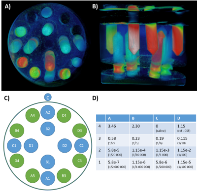 |
Reaching low-gadolinium concentration detection through sequences optimization on a dedicated phantom
Emilie Poirion1, Corentine Marie2, Marine Boudot de La Motte3, Chloé Dupont4, Jean-Claude Sadik1, and Julien Savatovsky1
1Hospital Foundation A. de Rothschild, Imaging department, Paris, France, PARIS, France, 2Sorbonne University, Paris Brain Institute, Paris, France, Paris, France, 3Hospital Foundation Rothschild,Neurology department, Paris, France, Paris, France, 4Hospital Foundation A. de Rothschild, Pharmaceutical department, Paris, France, PARIS, France
Gadolinium-enhanced imaging provides valuable information in clinical practice for the diagnosis of several diseases. Conventional sequences fail to detect low concentrations of gadolinium, such as those encountered in abnormal meninges. We designed a phantom containing 16 diluted gadobutrol tubes to compare and optimize sequences, based on normalized signal and contrast-to-noise ratio, for each gadolinium concentration. We performed a preliminary study comparing T1, FLAIR and FLAIR with optimized parameters on this phantom. The optimized FLAIR sequence shows an efficient detection of low concentrations, with a higher CNR than other sequences. Preliminary in-vivo experiment shows promising results for leptomeningeal abnomalities detection.
|
||
3359.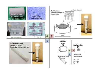 |
Commercially available astriction cotton as a anisotropic DTI phantom: Comparisons with a hand-bundled fibers and a glass capillary plate
Koji Sakai1, Yasuhiko Tachibana2, Toshiaki Nakagawa3, Hiroyasu Ikeno3, Takayuki Obata2, and Kei Yamada1
1Kyoto Prefectural University of Medicine, Kyoto, Japan, 2National Institute of Radiological Sciences, Chiba, Japan, 3Kyoto Prefectural University of Medicine Hospital, Kyoto, Japan
We compared DTI measures among three different anisotropic diffusion phantoms: a commercially available astriction cotton; a glass capillary plate; a hand-bundled polyethylene fibers. The purpose of this study was to examine whether the cotton is useful as an anisotropic diffusion phantom. Differences in ADC and FA were evaluated by comparing coefficients of variation (CVs). No significant difference was seen for the largest eigen value: 2.06 - 2.14 x 10⁻³ mm2/sec. Clear differences in FA were seen among the three DTI phantoms (p = 0.0079). CVs from five acquisitions for ADC and FA from every phantoms were all less than 2%.
|
|||
3360.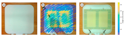 |
OpenCV based RF field mapping for MR coil assessment
Egor Kretov1, Zhao Kaixuan 1,2, Charles Grassin3, and Thoralf Niendorf1
1Max Delbrück Center for Molecular Medicine, Berlin, Germany, 2School of Biomedical Engineering, Southern Medical University, Guangzhou, China, 3Independent Researcher, Paris, France
This work presents a cost-effective near-field RF mapping approach for accurate tracking of a field probe. The method is based on the OpenCV library. It serves as a practical tool for the rapid assessment and characterization of MR coils and arrays. The only equipment required for the setup is a field probe, a regular webcam, and a paper QR code label, which renders this technique highly accessible and easy-to-use.
|
|||
3361.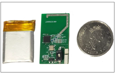 |
A Simple Device for Real-Time Detection of Head and Body Movements in a Mock Scanner, for Screening and Training Subjects
Fadi Ayad1 and Amir Shmuel2
1Biomedical Engineering, McGill University, Montreal, QC, Canada, 2McConnell Brain Imaging Centre, Montreal Neurological Institute, Montreal, QC, Canada
Head and body movements introduce artifacts in structural, diffusion, and functional MRI. With the advent of imaging at ultra-high field, head and body movements limit our capacity to obtain high-resolution, high-quality data. We have developed a small wearable device for acquiring data on head and body movements and transmitting it wirelessly to a computer for real-time analysis while a subject is trained in a mock MRI scanner. This device works in parallel to an in-bore camera used for movement detection, to support subject screening and training to remain still in preparation for MRI.
|
|||
3362.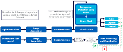 |
3-plane Localizer-aided Background Removal in Magnetic Resonance (MR) Images Using Deep Learning
Apoorva Agarwal1, Megha Goel1, and Jignesh Dholakia1
1GE Healthcare, Bengaluru, India
This study presents a new Deep-Learning (DL) based strategy for background computation in MR images. 3-plane Localizer scans have been used for background-subtraction in all subsequent scans of same MR Examination. This is accomplished by obtaining foreground-background masks for Localizer images using U-Net model and applying Image Resampling techniques on the obtained mask to compute background for subsequent scans. Comparison with existing algorithms demonstrates that proposed method prevails in accuracy, effectiveness and provides improved visual contrast. It can also be used universally across anatomies and MR pulse-sequences as opposed to other methods requiring anatomy/sequence-specific tuning and adaptive parameter adjustments.
|
|||
3363.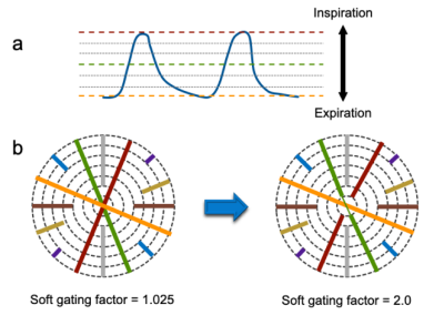 |
Phantom Experiments for Optimal Soft Gating Parameter in Free-Breathing Hepatobiliary Phase MRI with KWIC Reconstruction
Tomohiro Noda1, Keitaro Sofue2, Ryuji Shimada1, Yuichiro Somiya1, Shintaro Horii1, Yoshiko Ueno2, Naoki Yoshida1, Yu Ueda3, Akiko Kusaka1, and Takamichi Murakami2
1Center of Radiology and Radiation Oncology, Kobe University Hospital, Kobe, Japan, 2Department of Radiology, Kobe University Graduate School of Medicine, Kobe, Japan, 3Philips Japan MR Clinical Science, Tokyo, Japan
In free-breathing radial k-space sampling with KWIC reconstruction, soft gating method can be adopted to reduce motion artifacts. We investigated optimal soft gating parameters for free-breathing hepatobiliary phase MRI. A programmable respiratory motion phantom with varying motion distance of 10, 20, 30 mm was imaged using six values of soft gating factor (SGF). Image contrast and sharpness were analyzed by calculating contrast ratio (CR) and full width at half maximum (FWHM). The CR and FWHM were not inferior compared with the reference standard images in any of the motion distances when the SGF was set at more than 1.4.
|
|||
3364.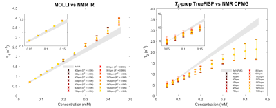 |
A phantom-based method for MRI relaxation time mapping data validation and harmonization
Davide Cicolari1, Domenico Lizio2, Patrizia Pedrotti3, Monica Teresa Moioli2, Alessandro Lascialfari1, Manuel Mariani1, and Alberto Torresin2,4
1Department of Physics, University of Pavia, Pavia, Italy, 2Department of Medical Physics, ASST Grande Ospedale Metropolitano Niguarda, Milan, Italy, 3Department of Cardiology, ASST Grande Ospedale Metropolitano Niguarda, Milan, Italy, 4Department of Physics, University of Milan, Milan, Italy A method for the MRI relaxation time measurement validation and harmonization is proposed: it relies on a phantom composed of vials filled with different concentrations of MnCl2 aqueous solutions, whose relaxation times are characterized as a function of both concentration and temperature, employing NMR techniques. The accuracy and the precision of fast mapping sequences developed for cardiac applications are better quantified through, respectively, the phantom characterization and the SD maps analysis. Scan-dependent recalibrations of the relaxation time maps can be performed relying on the ground-truth NMR values of the phantom, aiming to clinical intra- and inter-center harmonization. |
The International Society for Magnetic Resonance in Medicine is accredited by the Accreditation Council for Continuing Medical Education to provide continuing medical education for physicians.