Digital Posters
Multimodal Imaging & Auxiliary Devices
ISMRM & SMRT Annual Meeting • 15-20 May 2021

Digital Posters
Multimodal Imaging & Auxiliary Devices
| Concurrent 3 | 19:00 - 20:00 |
4262.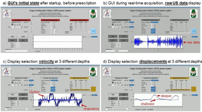 |
A compact and clonable ultrasound-based sensor system to monitor physiological motion
Bruno Madore1, Cheng-Chieh Cheng2, and Frank Preiswerk3
1Radiology, Brigham and Women's Hospital, Harvard Medical School, Boston, MA, United States, 2Computer Science and Engineering, National Sun Yat-sen University, Kaohsiung, Taiwan, 3Amazon Robotics, Westborough, MA, United States Physiological motion continues to be a problem in MRI and PET, as it can cause blurring and other types of artifacts. We developed a compact, clonable ultrasound-based sensor system that monitors internal motion directly. Alternative means of motion monitoring often involve hardware (including the scanner itself) that is attached to floors, walls and/or ceilings and thus cannot follow the patient from one room to another, from one procedure to the next. The present sensors attach to the skin, they can follow the patient, and as such they may help link different procedures in ways that take internal motion into account. |
|||
4263. |
Acoustically transparent and low-profile head coil for high precision magnetic resonance guided focused ultrasound at 3 T
Isabelle Saniour1, Fraser Robb2, Victor Taracila2, Jana Vincent2, Henning U. Voss1, Michael G. Kaplitt3, J. Levi Chazen1, and Simone Angela Winkler1
1Weill Cornell Medicine, New York, NY, United States, 2MR Engineering, GE Healthcare, Aurora, OH, United States, 3Department of Neurological Surgery, Weill Cornell Medicine, New York, NY, United States
Magnetic resonance guided focused ultrasound (MRgFUS) is a non-invasive therapeutic modality for neurodegenerative diseases that allows real-time imaging of the targeted regions. However, MR image quality is poor and severely limits the technology due to the use of the body coil for focal targeting. Acoustic simulations demonstrated an acoustic transparency (signal loss <1%) of the coil when used for thalamic sonication. In vivo results showed increase of the SNR over the body coil by a factor of 7.3 and 7.6 in a brain image with and without the presence of the transducer, respectively.
|
|||
4264. |
A dedicated 3-ch breast coil for Microwave Tomography at 3T
Xiaoyu Yang1, Thomas Eastlake1, Tsinghua Zheng1, Shireen Geimer2, Timothy Raynolds2, Paul Meaney2, and Keith Paulsen2
1Innovation, Quality Electrodynamics, LLC, Mayfield Village, OH, United States, 2Thayer School of Engineering, Dartmouth College, Hanover, NH, United States
MR breast imaging has high sensitivity but not high specificity. Microwave Tomography (MT) provides complimentary permittivity information that may improve specificity. MT has poor spatial resolution. Combining MR and MT may address both spatial and specificity issues. Previous MT and MR work demonstrated the MT-MR combination potential. However, there is no dedicated MR coil available for MT-MR combination. The only available coils are the whole-body coil or a single-loop coil that have poor MR image quality. A dedicated 3-ch MRI coil is proposed here for superior MRI imaging quality which enables the next step MT-MR volunteer evaluation.
|
|||
4265. |
A Dedicated Multichannel Head Coil Array for PET Insert on 3 T MRI
Jo Lee1,2, Ziru Sang1,2, Yongfeng Yang1,2, Xiaoliang Zhang3, and Ye Li1,2
1Paul C. Lauterbur Research Center for Biomedical Imaging, Shenzhen Institutes of Advanced Technology, Chinese Academy of Sciences, Shenzhen, China, 2Shenzhen Key Laboratory for MRI, Shenzhen, China, 3Department of Biomedical Engineering, State University of New York, Buffalo, NY, United States
Simultaneous positron emission tomography/ magnetic resonance imaging(PET/MR) provides advantages in clinical and research applications of human’s brain. The two systems, MRI and PET, can work complementary to provide more information for medical diagnosis. As most research and clinical institutes already have MRI systems, an insert PET and matched MRI coil would be a more economical choice than a PET/MR system. In this study, we designed and built a dedicated quadrature birdcage/47Rx head coil array for PET insert on human’s brain study. The flip-angle maps and SNR performances were measured through a dedicated phantom. Anatomical images were scanned on a volunteer.
|
|||
4266.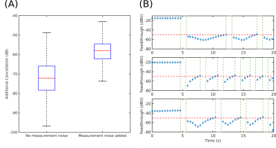 |
Vector Modulator Based Automated Active Compensation of Direct Feedthrough in Magnetic Particle Imaging
Bilal Tasdelen1,2, Mustafa Utkur1,2, Ergin Atalar1,2, and Emine Ulku Saritas1,2
1Department of Electrical and Electronics Engineering, Bilkent University, Ankara, Turkey, 2National Magnetic Resonance Research Center (UMRAM), Ankara, Turkey
In magnetic particle imaging (MPI), simultaneous excitation and signal acquisition leads to direct feedthrough interference. While this interference can be mitigated up to some extent with passive compensation, its time-varying nature necessitates active compensation methods to achieve the sensitivity levels needed for applications such as stem cell tracking. We have recently proposed an active compensation technique for MRI, which uses a vector modulator and a lookup-table-based algorithm for reducing the direct feedthrough in the analog domain. Here, we adapt this technique to MPI, demonstrating a successful recovery of the fundamental frequency and a significant increase in detection sensitivity.
|
|||
4267.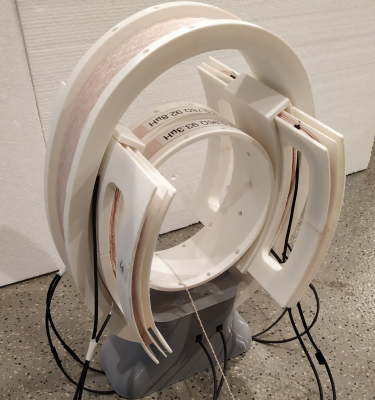 |
Interventional Magnetic Particle Imaging Open-bore Scanner Design
Michael Seeg1, Martin Rueckert1, Stefan Herz2, Thomas Kampf1,3, Thorsten Bley2, Volker Behr1, and Patrick Vogel1
1Experimental Physics V, Julius-Maximilians-University, Wuerzburg, Germany, 2Department of Diagnostic and Interventional Radiology, University Hospital, Wuerzburg, Germany, 3Department of Diagnostic and Interventional Neuroradiology, University Hospital, Wuerzburg, Germany
X-ray-guided endovascular interventions are important treatment approaches for many cardiovascular diseases such as myocardial infarction, stroke or occlusive peripheral arterial disease. However, they pose the risk of radiation exposure to patients and staff. Magnetic particle imaging may provide a radiation-free alternative for diagnostic and image guided treatment. A new open bore scanner concept, based on a traveling field free line and super paramagnetic iron oxide tracers is developed especially for interventional treatments. Due to the open design the scanner provides good accessibility to the patient.
|
|||
4268. |
MR safe electromagnetic direct current motor
Lorne W Hofstetter1, J Rock Hadley1, Robb Merrill1, and Dennis L Parker1
1Department of Radiology and Imaging Sciences, University of Utah, Salt Lake City, UT, United States
Combining the unparalleled soft-tissue imaging capabilities of MR imaging with the precision of robotic-assisted surgeries has the potential to revolutionize image-guided surgical interventions. However, strong magnetic fields generated by the MR system prevent the use of conventional robotic servo motor technologies. Here we present a direct current electromagnetic motor design that uses the electromagnetic interaction between the MR B0 field and currents in the rotor windings to generate rotational mechanical actuation. The motor stall torque and interactions caused by simultaneous motor operation and MR imaging are characterized. This new motor may be useful for robotic assisted interventions performed under MR-guidance.
|
|||
4269.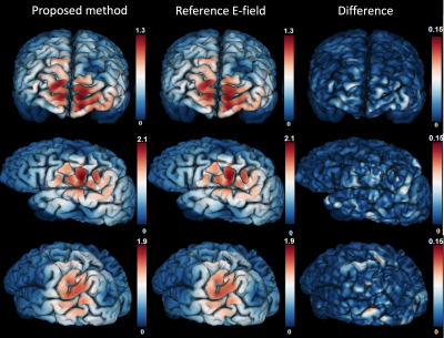 |
Real-time electric field estimation in transcranial magnetic stimulation using deep learning and magnetic resonance imaging
Guoping Xu1,2, Yogesh Rathi2,3, Joan A Camprodon3,4, and Lipeng Ning2,3
1Wuhan Institute of Technology, Wuhan, China, 2Brigham and Women's Hospital, Boston, MA, United States, 3Harvard Medical School, Boston, MA, United States, 4Massachusetts General Hospital, Boston, MA, United States
Transcranial magnetic stimulation (TMS) is an effective treatment approach for mental disorders. Accurate and rapid prediction of TMS-evoked electric field (E-field) in brain tissue is important for accurate targeting and to understand the mechanism of treatment response. The standard method for E-field prediction is based on physical modeling which usually takes long computational time. In this work, we introduce a method based on deep neural networks (DNNs) for real-time E-field prediction. We show that the trained DNN can predict high-precision whole-brain E-field in 0.24 seconds. Moreover, diffusion-MRI based tissue conductivity tensor can improve the prediction accuracy of E-field.
|
|||
4270.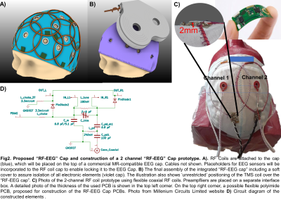 |
A wearable “RF-EEG Cap” for full head coverage concurrent TMS/EEG/fMRI experiments at 3T: a feasibility study
Lucia Navarro de Lara1,2, Padmavathi Sundaram 1,2, Lena Nohava3, Elmar Laistler3, Mohammad Daneshzand1,2, Lawrence Wald1,2, Jason Stockmann1,2, and Aapo Nummenmaa1,2
1Martinos Center - MGH, Charlestown, MA, United States, 2Harvard Medical School, Boston, MA, United States, 3High Field MR Center, Center for Medical Physics and Biomedical Engineering, Medical University of Vienna, Vienna, Austria
To enable concurrent non-invasive stimulation (TMS) and whole-head multimodal imaging (EEG-fMRI), we propose to apply flexible RF coil technology to build a first-of-its-kind TMS compatible integrated multimodal imaging array, the “RF-EEG cap”. The proposed system allows unrestricted positioning of the TMS coil across the entire scalp. We built a 2-channel prototype and conducted a feasibility study analyzing the effects of a TMS coil and EEG-electrodes on the imaging quality (SNR/B0 maps), the in-bore EEG data quality and EPI timeseries stability. Our results indicate that the flexible coaxial RF technology is a feasible choice to build the proposed “RF-EEG Cap”.
|
|||
4271.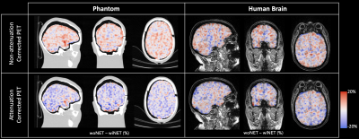 |
Quantification of HD-EEG net attenuation on 18F-FDG static PET: a phantom and human brain study
Erica Silvestri1,2,3, Marco Castellaro1,4, Alessandra Zorz5, Andrea Bettinelli 1,5, Cristina Campi6, Marta Paiusco5, Diego Cecchin2,7, and Alessandra Bertoldo1,2
1Department of Information Engineering, University of Padova, Padova, Italy, 2Padova Neuroscience Center, University of Padova, Padova, Italy, 3Department of Neuroscience, University of Padova, Padova, Italy, 4Department of Neurosciences, Biomedicine and Movement Sciences, University of Verona, Verona, Italy, 5Department of Medical Physics, Veneto Institute of Oncology IOV - IRCCS, Padova, Italy, 6Department of Math, University of Padova, Padova, Italy, 7Department of Medicine, Unit of Nuclear Medicine, University of Padova, Padova, Italy
Simultaneous trimodal EEG-PET-MR imaging can be a tool to delve deeper in the healthy human brain functioning. One of the aspects scarcely investigated is the interplay between HD-EEG net and PET signal detection and reconstruction. In this work, we assess the impact of HD-EEG on both attenuation corrected (AC) and non-attenuation corrected (NAC) PET images. We found that HD-EEG net attenuates PET signal in NAC images in both phantom and human brain, respectively of 3.01%(±3.69%) and of 0.53%(±5.89%). AC images show an opposite behaviour: attenuation correction factor tends to be overestimated in CT- and UTE-derived UMAP obtained with HD-EEG net.
|
|||
4272.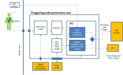 |
Triggering pulse generation by non-contact monitoring of heart rate using an antenna with 600 MHz resonance
Kazuya Okamoto1, Takafumi Ohishi2, Sojuro Kato1, Ryuichi Nanaumi3, Kenji Oyama3, and Kazuhiko Fukutani3
1Advanced MRI Development PJ Team, Canon Medical Systems Corp., Kawasaki, Japan, 2Advanced Technology Research Department, Research and Development Center, Canon Medical Systems Corp., Kawasaki, Japan, 3Medical Products Technology Development Center, R&D Headquarters, Canon Inc., Tokyo, Japan
New non-contact cardiac and respiratory gating method have been developed, which includes an antenna monitoring heart and respiration movement, and independent triggering pulse generation unit with signal processing software. The resonance frequency of the dipole antenna was tuned to around 550MHz on the chest. The triggering pulse generation unit detected and digitized the S11 signals from the antenna. IIR filter removed the low frequency component and a template matching technique was used to seek the triggering points at the inflection point of filtered waveform. As a result, the prospective gating cardiac imaging was successfully performed for volunteers.
|
|||
4273.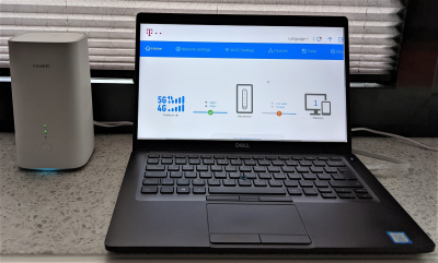 |
MRI data transmission via fifth-generation (5G) cellular networks
Nicolai Spicher1, Ramon Barakat1, and Thomas Martin Deserno1
1Peter L. Reichertz Institute for Medical Informatics, TU Braunschweig and Hannover Medical School, Braunschweig, Germany In the last years, research in MRI has expanded towards portable technology, e.g., trailer units carrying conventional scanners or bedside scanners with low-field strengh. The increased portability and shift away from the centralized hospital environment requires wireless data transmission with appropriate data rates to unfold its full potential. In this work, we made use of current fifth-generation (5G) cellular networks for transmitting DICOM data. We demonstrate the potential of reaching more than 50 Mbit/s upload speed using consumer-grade hard- and software. This holds great potential for cellular transmission of data from portable scanners, allowing to increase access to MRI technology. |
|||
4274.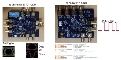 |
Test Bed for High Speed Serial Wireless MRI Data Studies
Audrey Chan1, Fraser Robb2, John Pauly3, Shreyas Vasanawala4, and Greig Cameron Scott5
1Victoria University of Wellington, Wellington, New Zealand, 2GE Healthcare, Aurora, OH, United States, 3Stanford University, Stanford, CA, United States, 4Radiology, Stanford University, Stanford, CA, United States, 5Electrical Engineering, Stanford University, Stanford, CA, United States
We developed a test system to perform pseudo-random binary sequence (PRBS) generation and clock/data recovery to assess high speed serial link performance of wireless and optical signal integrity for MRI data interfacing. We demonstrate applications using up to 1Gbps signal rates and show impairments for wireless amplitude shift keyed data transmission, as well as plastic fiber optical interfacing.
|
|||
4275.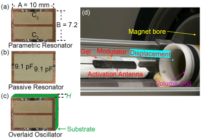 |
Wireless Powered Frequency Encoding of Locally Detected MR Images for Remote Signal Transmission
Wei Qian1 and Chunqi Qian1
1Michigan State University, East Lansing, MI, United States
With a compact and batteryless design, the wireless powered parametric oscillator can simultaneously down-convert and frequency encode locally detected MR signals, and wirelessly transmit them to remotely coupled external receivers, thus maintaining superior sensitivity of localized detectors over larger distance separations.
|
|||
4276.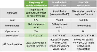 |
Single board computer as a satellite-linked, deep learning capable pocket MR workstation: a feasibility study
Keerthi Sravan Ravi1,2, John Thomas Vaughan Jr.2, and Sairam Geethanath2
1Biomedical Engineering, Columbia University, New York, NY, United States, 2Columbia Magnetic Resonance Research Center, New York, NY, United States
This work shows the feasibility of employing a Raspberry Pi (RPi) single-board computer as a deep learning capable MR workstation. RPi’s ability to run a Tensorflow-Lite optimized brain tumour segmentation model is demonstrated. A comparison of data upload across fixed broadband, cellular broadband (LTE, 3G) and satellite terminal methods of internet access is presented. Finally, the setup described in this work is compared with a conventional fixed MRI workstation and a portable MRI workstation.
|
|||
4277.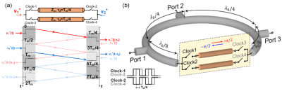 |
STAR MRI With Non-Magnetic, Integrated Circulator based on Switched Transmission Lines
Aravind Nagulu1, Ahmed Kord1, Gehua Tong1, Michael Garwood2, Lance DelaBarre2, Djaudat Idiyatullin2, SungMin Sohn3, J. Thomas Vaughan1, and Harish Krishnaswamy1
1Columbia University, New York, NY, United States, 2University of Minnesota, Minneapolis, MN, United States, 3Arizona State Univeristy, Tempe, AZ, United States
Traditional MRI employs time division duplexing between the receiver (RX) and transmitter (TX) to avoid RX saturation from TX self-interference. Large switching time between the TX and RX modes demands large TX power and constricts the imaging to tissues with large relaxation times. In this work, we propose a magnetic-free, switch-transmission line circulator which is fully compatible with MRI systems and achieve simultaneous transmit and receive (STAR) MRI.
|
|||
4278. |
A 5.7mW 1.5T Silicon Germanium Pre-Amp, and its Effects on Image Distortion
Christopher Vassos1, Fraser Robb2, Shreyas Vasanawala3, John Pauly1, and Greig Scott1
1Electrical Engineering, Stanford University, Stanford, CA, United States, 2GE Healthcare, Aurora, OH, United States, 3Radiology, Stanford University, Stanford, CA, United States
A 5.7mW Silicon Germanium Pre-Amplifier was constructed, characterized, and its potential impact on image quality demonstrated. The amplifier demonstrates significant power savings relative to current implementations at the cost of increased device non-linearity and decreased input impedance. A benchtop measurement setup was constructed to simulate coils signals in conjunction with a physical receive chain. Measured image distortion began at peak input powers of -25dBm. This corresponds to high bandwidth, high dynamic range imaging sequences. For small coil sizes this distortion may not play a role, reducing the power barrier to wireless receive arrays.
|
|||
4279. |
Hybrid Pair Ratio Adjustable Power Splitter using off-shelf components and easy-to-replace microstrip phase shifter
Yue Zhu1,2, William A Grissom1,2,3,4, John C Gore1,2,3,4, and Xinqiang Yan1,2
1Vanderbilt University Institute of Imaging Science, Nashville, TN, United States, 2Department of Radiology and Radiological Sciences, Vanderbilt University, Nashville, TN, United States, 3Biomedical Engineering, Vanderbilt University, Nashville, TN, United States, 4Electrical and Computer Engineering, Vanderbilt University, Nashville, TN, United States
A low loss ratio adjustable power splitter (RAPS) was designed with a Wilkinson Splitter, a pair of transmission lines, and a hybrid coupler. There are some potential issues with the previous RAPS: 1. Build a high-performance RAPS circuit with lumped elements is challenging and laborious at 298 MHz. 2. The 100-Ohm RF decoupling resistor poses a safety hazard to the patient. In this abstract, the Wilkinson splitter was replaced with a hybrid coupler, and the adjustable transmission lines are replaced with printed microstrip lines to mitigate the problems mentioned above.
|
|||
4280.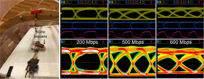 |
High Speed Serial ASK Signal Integrity for Wireless MRI
Greig Cameron Scott1, Audrey Chan2, Wonje Lee3, Fraser Robb4, John Pauly5, and Shreyas Vasanawala6
1Electrical Engineering, Stanford University, Stanford, CA, United States, 2Victoria University of Wellington, Wellington, New Zealand, 3Pediatric Radiology, Stanford University, Stanford, CA, United States, 4GE Healthcare, Aurora, OH, United States, 5Stanford University, Stanford, CA, United States, 6Radiology, Stanford University, Stanford, CA, United States
A major challenge of wireless MRI is how to implement the data link. Here, progress is made using amplitude shift keyed diode ring modulators, serializer/deserializer components and clock/data recovery to demonstrate bit rate transmissions up to 600 Mbps. High gain biquad reflector antennas and UWB antennas are compared for carriers between 3 and 5 GHz. These prototypes also demonstrate bandwidth limitations and electrodynamic interactions in a birdcage test bore on wireless signal integrity. Even if dedicated wireless ICs are not possible, we believe components exist to make an easily integrated modulator for MRI wireless data transmission.
|
|||
4281.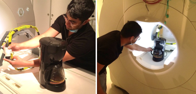 |
A Sip in the Bore: How to Make Coffee with an MRI System
Michael Bock1, Thomas Lottner1, and Ali Caglar Özen1,2
1Radiology - Medical Physics, University Medical Center, Medical Faculty, University Freiburg, Freiburg, Germany, 2German Consortium for Translational Cancer Research (DKTK), Freiburg, Germany
Energy harvesting of the RF fields has been proposed to power small milliwatt RF amplifiers. In this work we demonstrate that the time-varying gradient fields can deliver much higher power levels up to a kW which we used to energize a conventional drip coffee machine. A large pick-up coil was designed in which voltages of 100-200V were induced by rapidly oscillating gradients. The coil was connected to a commercial coffee machine in the magnet, and coffee was prepared in and with the MRI in just 5min.
|
The International Society for Magnetic Resonance in Medicine is accredited by the Accreditation Council for Continuing Medical Education to provide continuing medical education for physicians.