Digital Posters
New Shim Systems & Other Advanced MR Engineering
ISMRM & SMRT Annual Meeting • 15-20 May 2021

Digital Posters
New Shim Systems & Other Advanced MR Engineering
| Concurrent 2 | 17:00 - 18:00 |
3105.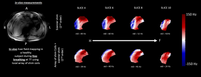 |
In-vivo B0 shimming of the liver using a local array of shim coils in combination with 2nd order spherical harmonics at 7T
Lieke van den Wildenberg1, Quincy van Houtum1, Wybe van der Kemp1, Catalina Arteaga de Castro1, Alex Bhogal1, Paul Chang2, Sahar Nassirpour2, and Dennis Klomp1
1Radiology Department, UMC Utrecht, Utrecht, Netherlands, 2MR Shim GmbH, Reutlingen, Germany
Maximizing shim performance, particularly for larger organs such as the liver, is crucial at ultra-high field MRI due to the increased sensitivity to B0 field inhomogeneity. The conventional shimming in most MRI systems at ultra-high field, provides insufficient correction. However, by combining the available 2nd order spherical harmonic fields with an external array of 16 local shim coils, the magnetic field homogeneity in the liver is improved by as much as 44%.
|
|||
3106.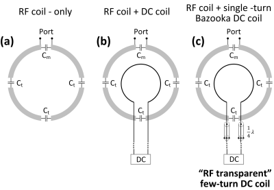 |
“RF transparent” local B0 shim coil
Xinqiang Yan1,2
1Vanderbilt University Institute of Imaging Science, Nashville, TN, United States, 2Department of Radiology and Radilogical Science, Vanderbilt University Medical Center, Nashville, TN, United States
Multiple coils shimming technology has been demonstrated as a promising approach to reduce the B0 inhomogeneity in MRI. However, strong coupling arises when local DC coils are placed close to the imaging area and RF coils. To solve this problem, we proposed a novel local DC shim coil that has little effect on the RF coils, which we called "RF transparent" DC coil. This concept was validated by EM simulation, bench test, and MR experiments. The RF transparent concept will bring much more freedoms to DC coil design (such as many turn and irregular shaped).
|
|||
3107.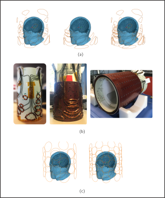 |
Shim Coils Tailored for Correction of B0 Inhomogeneity in the Human Brain (SCOTCH) at Ultra High Field
Bruno Pinho-Meneses1, Jason Stockmann2,3, Edouard Chazel1, Paul-François Gapais1, Eric Giacomini1, Franck Mauconduit1, Alexandre Vignaud1, Michel Luong4, and Alexis Amadon1
1Université Paris-Saclay, CEA, CNRS, BAOBAB, NeuroSpin, Gif-sur-Yvette, France, 2Athinoula A. Martinos Center for Biomedical Imaging, Charlestown, MA, United States, 3Harvard Medical School, Boston, MA, United States, 4Université Paris-Saclay, CEA, Institut de Recherche sur les Lois Fondamentales de l'Univers, Gif-sur-Yvette, France
A Singular Value Decomposition based Multi-Coil Array for B0 shimming of the human brain was designed from a database of 100 δB0 fieldmaps. An optimized 2-layer 36-channel MCA design was obtained and constructed. The system was characterized and measured fieldmaps were used for comparison to expected performance from ideal simulated fields. The static whole-brain and dynamic slice-wise shimming was validated in-vivo and assessed with GRE and EPI acquisitions at 7 Tesla.
|
|||
3108.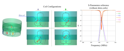 |
Designing a high-density combined RF/B0 shim coil for imaging the brain at 7T
Paul Chang1, Sahar Nassirpour1, Kaizad Rustomji2, Elodie Georget2, Ingmar Voogt3, Aidin Haghnejad3, Evita Wiegers4, Jannie Wijnen4, and Dennis Klomp4
1MR Shim GmbH, Reutlingen, Germany, 2Multiwave Imaging, Marseille, France, 3WaveTronica, Utrecht, Netherlands, 4UMC Utrecht, Utrecht, Netherlands
Increased B0 inhomogeneity at ultra-high fields continues to pose a challenge for many MR applications. Local arrays of shim coils offer a complimentary way of reducing the local inhomogeneities. In this study, practical design considerations for a combined RF/B0 shim 7T head coil were assessed. Numerical simulations were used to find the optimal size and distance of the shim coils relative to Rx loops. These results were then verified in a bench test. Finally, having these considerations in mind, it was shown that by optimizing the arrangement of the shim coils improved B0 homogeneity can be achieved.
|
|||
3109.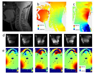 |
Shielded coaxial cable coils for transmit, receive and B0 shimming in a 7T neck array
Vincent O. Boer1, Jan Ole Pedersen2, Hørður Andreasen3, Sadri Güler1,4, Vitaliy Zhurbenko3, Jason Stockmann5, Irena Zivkovic6, and Esben Thade Petersen1,4
1Danish Research Centre for Magnetic Resonance, Centre for Functional and Diagnostic Imaging and Research, Copenhagen University Hospital, Hvidovre, Denmark, 2Philips Healthcare, Copenhagen, Denmark, 3Department of Electrical Engineering, Technical University of Denmark, Kongens Lyngby, Denmark, 4Section for Magnetic Resonance, DTU Health Tech, Technical University of Denmark, Kongens Lyngby, Denmark, 5Athinoula A Martinos Center for Biomedical Imaging, Department of Radiology, Massachusetts General Hospital, Charlestown, MA, United States, 6Electrical Engineering department, Technical University of Eindhoven, Eindhoven, Netherlands
Shielded coaxial RF coils show very low coupling between elements, and are ideal for building transmit-receive arrays. In this work we extended a 6 element transceive array with B0 shim capabilities.
|
|||
3110.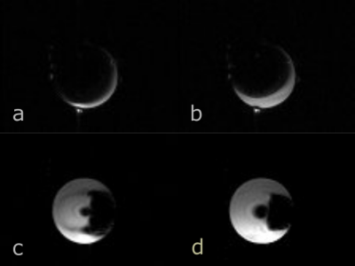 |
Improving MRI Near Metal with Local B0 Shimming using a Unified Shim-RF Coil (UNIC): First Case Study, Hip Prosthesis in Phantom.
Fardad Michael Serry1, Junzhou Chen1,2, Anthony G Christodoulou1,2, Yuheng Huang1,2, Fei Han3, Won Bae4,5, Christine Chung4,5, Richard Handlin1, John Stager1, Matthew Dausch1, Yubin Cai1, Yujie Shan1, Yucen Liu1, Yibin Xie1,
Xiaoming Bi3, Rohan Dharmakumar1,2, Zhaoyang Fan1,6, Debiao Li1,2, Hsin-Jung Yang1, and Hui Han1
1Biomedical Imaging Research Institute, Cedars-Sinai Medical Center, Los Angeles, CA, United States, 2Department of Bioengineering, UCLA, Los Angeles, CA, United States, 3Siemens Medical Solutions USA, Inc., Los Angeles, CA, United States, 4University of California, San Diego, San Diego, CA, United States, 5VA Medical Center, San Diego, CA, United States, 6University of Southern California Department of Radiology, Los Angeles, CA, United States
Metal artifact image degradation can interfere with tissue status assessment. Metal artifact reduction pulse sequences, mainly spin-echo based, and scanner B0-shimming can mitigate the artifacts. We have tested in a metal hip prosthesis phantom improving shimming locally with a novel technology that integrates separate but co-planar RF receive and shim arrays, expanding the freedom to position shim coils and shape B0, independently of the RF coil geometry and pulse sequence. Metal-induced signal-void artifact was reduced in GRE scans, revealing up to 50% additional area around the prosthesis. The sequence-agnostic technology may help improve MRI near metal prostheses and interventional devices.
|
|||
3111.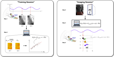 |
Shimming-Toolbox: An open-source software package for performing realtime B0 shimming experiments
Alexandre D'Astous1, Ryan Topfer1, Gaspard Cereza1, Eva Alonso-Ortiz1, Lincoln Craven-Brightman2, Jason Stockmann2,3, and Julien Cohen-Adad1,4
1NeuroPoly Lab, Institute of Biomedical Engineering, Ecole Polytechnique, Montreal, QC, Canada, 2Athinoula A. Martinos Center for Biomedical Imaging, Massachusetts General Hospital, Charlestown, MA, United States, 3Harvard Medical School, Boston, MA, United States, 4Functional Neuroimaging Unit, Centre de recherche de l'Institut universitaire de gériatrie de Montréal, Montreal, QC, Canada
As MRI static fields get increasingly stronger, reducing $$$B_0$$$ inhomogeneities becomes more important but also challenging. When standard active shimming on commercial systems is insufficient for researchers’ needs, alternative approaches such as external shim or hybrid AC/DC coils can be used. However, in order to perform advanced shimming experiments, a proper software ecosystem is required and there is currently no open-source solution for transparent and reproducible shimming experiments. Here we introduce the Shimming Toolbox (https://shimming-toolbox.org), an open-source software package for performing and prototyping static, dynamic and realtime $$$B_0$$$ shimming experiments.
|
|||
3112.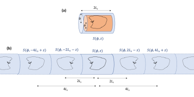 |
Magnetic fields produced by simple coils inside finite-length cylindrical passive shields with end-caps
Richard W. Bowtell1, Michael Packer 2, Peter Hobson 2, James Leggett1, Niall Holmes1, Paul Glover 1, Matthew Brookes1, and Mark Fromhold2
1Sir Peter Mansfield Imaging Centre, University of Nottingham, Nottingham, United Kingdom, 2School of Physics and Astronomy, University of Nottingham, Nottingham, United Kingdom
Cylindrical shields formed from material of high permeability are commonly used in providing low-magnetic field environments for experimental research. Cylindrical coils are often sited inside such shields to produce controlled patterns of field variation, but interaction of the coils with the mu-metal distorts the fields. Here we show how to analytically calculate the fields that are produced by simple coils inside a finite-length, cylindrical mu-metal shield with end-caps, and report experimental measurements on two different coils (loop and saddle), demonstrating excellent agreement with the theory. Optimal spacings of Helmholtz-type coils inside finite-length shields are also derived using the analytic expressions.
|
|||
3113.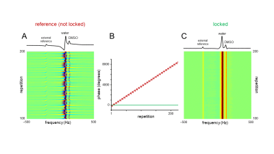 |
Hardware developed for phase and frequency locking of interleaved MRI and DMI studies
Terence W. Nixon1, Yanning Liu1, Henk M. DeFeyter1, Scott McIntyre1, and Robin A. de Graaf1
1Yale University, New Haven, CT, United States Deuterium metabolic imaging (DMI) is a powerful new method to supplement the anatomical and functional information of MRI with a metabolic component. A reduction in overall scan time by interleaving MRI and DMI is clinically relevant, whereas research applications can benefit from complimentary information obtained through interleaved acquisition. Because most MR scanners only allow acquisition of one frequency, additional hardware is required to enable interleaved acquisition. Here we present hardware for the upconversion of 2H data into the 1H receive path and present a solution to achieve effective phase and frequency locking. |
|||
3114. |
Simultaneous transmit/receive for Bloch-Siegert encoding: a feasibility study
Baosong Wu1, Sajad Hosseinnezhadian2, Yonghyun Ha2, Kartiga Selvaganesan2, Charles Rogers III2, Kasey Hancock2, Gigi Galiana2, and R. Todd Constable2
1Department of Radiology and Biomedical Imaging, Yale School of Medicine, NEW HAVEN, CT, United States, 2Department of Radiology and Biomedical Imaging, Yale School of Medicine, New Haven, CT, United States
This work presents the feasibility of Bloch-Siegert (BS) frequency encoding for magnetic resonance (MR) applications requiring a simultaneous transmit and receive system. RF frequency encoding technique is referred to receiving MR signal while existing one offset-field radiation, which can be considered as a radio frequency interference(RFI). A prototype was built demonstrating first MR experiment and preliminary data using this method. Our system is currently a research technology with the goal of generating a low-field and point-of-care medical imaging system using novel RF encoding.
|
|||
3115.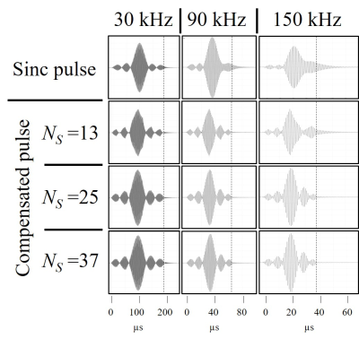 |
RF sinc pulse distortion compensation using multiple square pulses at 1 MHz.
Yonghyun Ha1, Kartiga Selvaganesan1, Baosong Wu1, Kasey Hancock1, Charles Rogers III1, Sajad Hosseinnezhadian1, Gigi Galiana1, and R. Todd Constable1
1Department of Radiology and Biomedical Imaging, Yale School of Medicine, New Haven, CT, United States
The response times of RF coils in MRI are long when operating at low B0 field, making it difficult to achieve the desired pulse shape in the coil. Compensation pulses are shown that were optimized using a series of square pulses. Both the duration and amplitude of the compensation elements were calculated with Q-factor which was calculated from the ring-down time measured using a sniffer coil. The SNRs of the echo signal acquired using compensated pulse was compared with those of signal obtained with uncompensated pulses and showed significant improvements of 51.5%.
|
|||
3116.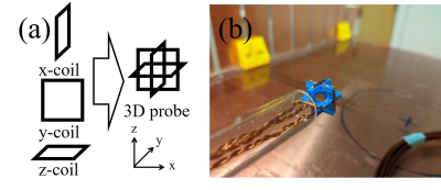 |
Automatic 3D B1 field mapping using 3D printer and digital oscilloscope for gradient-free MRI system
Yonghyun Ha1, Kartiga Selvaganesan1, Sajad Hosseinnezhadian1, Baosong Wu1, Kasey Hancock1, Gigi Galiana1, and R. Todd Constable1
1Department of Radiology and Biomedical Imaging, Yale School of Medicine, New Haven, CT, United States
For gradient-free RF spatial encoding, MR image based B1 mapping methods cannot be used due to the absence of gradient coils. However, it is important to get a B1 map particularly if Bloch-Siegert shift encoding is used. In this work, a method to automatically measure the amplitude of B1 field is introduced. Three perpendicular pickup loops were used to measure the B1 field in three orthogonal axes simultaneously. The position of the pickup loops was controlled by 3D printer, and the peak-to-peak voltage of the coupled signal was automatically saved using PC oscilloscope.
|
|||
3117.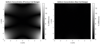 |
Inhomogeneity and ramping effects in field-cycled quantitative molecular MRI
Matthew A. McCready1, William B. Handler1, Francisco Martinez2,3, Timothy J. Scholl2,3, and Blaine A. Chronik1,2
1Physics and Astronomy, Western University, London, ON, Canada, 2Medical Biophysics, Western University, London, ON, Canada, 3Robarts Research Institute, Western University, London, ON, Canada
Delta relaxation enhanced magnetic resonance (dreMR) is a field-shifting quantitative molecular imaging method. The dreMR method may be used to produce images with signal proportional to concentration of contrast agents with longitudinal relaxivity dispersion. Here we find that field inhomogeneities and ramping periods, which were previously ignored, cause significant errors in dreMR images. These errors include apparent non-zero signal from dispersion-free tissue, and apparent change in signal due to contrast agents. We show that these effects can be mitigated by use of an improved homogeneity design method, and increased slew rates.
|
|||
3118. |
Very low field MRI for brain imaging
Samson Lecurieux Lafayette1 and Claude Fermon1
1SPEC - CEA Saclay - Université Paris Saclay, Saclay, France
Classic high field MRI is a powerful tool in healthcare. However, it is an expensive and complex installation and it might be useful to supply a less convenient device adapted to a lot of usage not requiring high field potential. We have developed a very low field MRI working at between 1mT and 10mT. Main static field is providing by a water-cooled resistive magnet. In order to achieve an iso spatial resolution of 2mm in a reasonably short time of acquisition, we have worked on adapted sequences including compressed sensing and optimized detection with parallel acquisition.
|
|||
3119.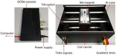 |
Comparison of SNR between a low-field (0.26T) Tabletop-MRI and a clinical high-field (3T) scanner
Robert Kowal1, Enrico Pannicke2, Marcus Prier1, Ralf Vick2, Georg Rose3, and Oliver Speck1
1Department of Biomedical Magnetic Resonance, Otto von Guericke University, Magdeburg, Germany, 2Chair of Electromagnetic Compatibility, Otto von Guericke University, Magdeburg, Germany, 3Chair in Healthcare Telematics and Medical Engineering, Otto von Guericke University, Magdeburg, Germany The SNR-performance of a low-field (0.26T) Tabletop-MRI-system was experimentally compared to a high-field (3T) clinical scanner. The SNR of simple FID sequences were evaluated for RF-units with identical coil and sample geometries as well as T/R-switch designs. The SNR was calculated from time signals and put into perspective to compute the relative SNR-performance of the Tabletop-system which was measured at 11.3%. Additional compensations for several differing experimental conditions were carried out. The full comparison suggests limiting factors in the equivalent measurement in the clinical scanner which can result in more SNR loss than expected. |
|||
3120.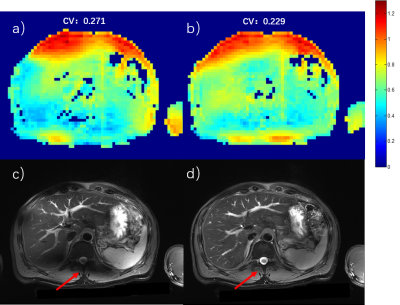 |
Initial Experience of Body Imaging at 5T
Zhenhua Shen1, Xuchen Zhu1, Shihong Han1, Fuyi Fang1, Wei Luo1, Shao Che1, Zidong Wei1, Jinguang Zong1, Yongquan Ye2, Bo Li1, Shuheng Zhang1, Anthony Vu2, Weiguo Zhang2, and Guobin Li1
1United Imaging Healthcare, Shanghai, China, 2UIH America, Inc., Houston, TX, United States
For the first time, routine clinical body imaging with large FOV on a whole-body 5T MRI system is demonstrated. With multi-channel RF parallel transmission hardware architecture and static RF shimming techniques, the uniformity of the RF transmission field is shown to be well controlled for imaging quality guarantee. Preliminary results show great promise for body imaging at 5T.
|
|||
3121.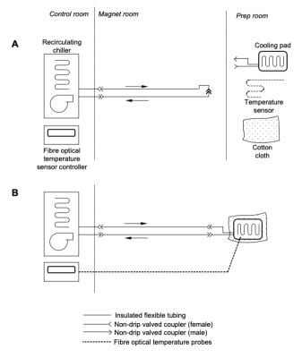 |
A sample temperature control system for post mortem MRI
Sebastian Walter Rieger1, Karla Miller1, Peter Jezzard1, and Wenchuan Wu1
1Wellcome Centre for Integrative Neuroimaging, University of Oxford, Oxford, United Kingdom
In post mortem MRI, the absence of body temperature autoregulation can lead to significant local heating in the sample from RF absorption and heat from the environment. This can interfere with scanning (such as diffusion measurements) and in unfixed samples, accelerate tissue decomposition. In this work, a temperature control system is presented which enables prolonged scanning of post mortem samples at a stable temperature while preserving tissue.
|
|||
3122.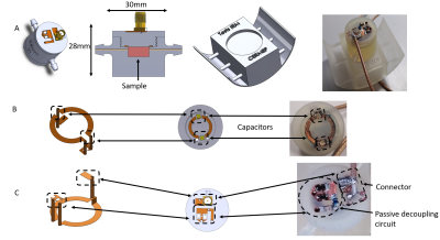 |
Compact MRI bioreactor for real-time monitoring 3D printed tissue-engineered constructs.
Jean-Lynce GNANAGO1,2,3,4,5,6, Tony GERGES1,2,3,4,5,6, Laura Chastagnier1,2,4,6,7,8,9,10, Emma Petiot2,4,6,7,8,9,10, Vincent SEMET1,2,4,5,6, Philippe Lombard1,2,3,4,5,6, Christophe Marquette1,2,4,6,7,8,9,10, Michel Cabrera1,2,3,4,5,6, and Simon Auguste Lambert1,2,3,4,5,6
1Université Claude Bernard Lyon 1, VILLEURBANNE, France, 2INSA LYON, VILLEURBANNE, France, 3Ecole Centrale Lyon, Ecully, France, 4CNRS, VILLEURBANNE, France, 5AMPERE UMR 5005, VILLEURBANNE, France, 6Université de Lyon, VILLEURBANNE, France, 73d.FAB, VILLEURBANNE, France, 8CPE Lyon, VILLEURBANNE, France, 9ICBMS, VILLEURBANNE, France, 10UMR 5246, VILLEURBANNE, France
Tissue engineering for regenerative medecine is a growing field which faces structural and functional challenges at different scales. Real-time quantitative 3D characterization of tissues both in vitro and in vivo would help biologists assessing their methods. Magnetic Resonance Imaging (MRI) offers the possibility to perform such characterization non-invasively. We propose here a 7T MRI coil integrated within a perfusion tissue engineering bioreactor to perform tissue assessments throughout its growth. The MRI bioreactor is built using 3D printing and plastronics. This works resulted in a successful observation of a bioprinted tissue with a 75 µm in plane resolution.
|
|||
3123.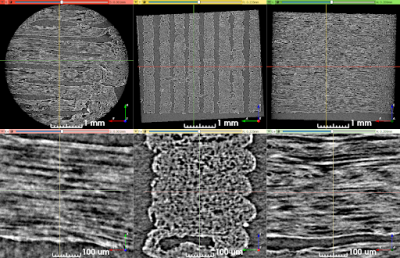 |
Characterization of 3AM diffusion MRI phantoms via microscopy, and phase-contrast micro-CT
Farah Mushtaha1,2, Tristan K. Kuehn1,3, Omar El-Deeb4, Seyed A. Rohani3, Luke W. Helpard3, Hanif Ladak2,3,5, Amanda Moehring6, Corey A. Baron1,2,3,7, and Ali R. Khan1,2,3,7
1Robarts Research Institute, London, ON, Canada, 2Medical Biophysics, Schulich School of Medicine and Dentistry, Western University, London, ON, Canada, 3School of Biomedical Engineering, Western University, London, ON, Canada, 4Neuroscience, Western University, London, ON, Canada, 5Department of Electrical and Computer Engineering, Western University, London, ON, Canada, 6Biology, Western University, London, ON, Canada, 7The Brain and Mind Institute, Western University, London, ON, Canada
Validating diffusion MRI (dMRI) representations and models of brain tissue is challenging because there is no reference ground-truth for in vivo scans. We investigate the microstructural characteristics of 3D printed axon-mimetic (3AM) phantoms as a dMRI validation tool using fluorescence microscopy and phase-contrast micro computed tomography (micro-CT). Both microscopy and micro-CT yielded pore diameters of ~ 8 μm. We constructed a 2-compartment microstructural model using microscopy and micro-CT data to simulate 3AM dMRI scans, and compared the observed metrics from in-vivo scans and simulation data.
|
|||
3124.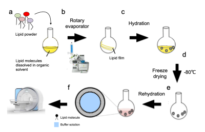 |
A phantom system for evaluating the effect of lipid and iron composition on qMRI parameters
Rona Shaharabani1, Shir Filo1, Oshrat Shtangel1, and Aviv Mezer1
1The Edmond and Lily Safra Center for Brain Science, The Hebrew University of Jerusalem, Jerusalem, Israel, Jerusalem, Israel
Neurodegenerative diseases such as Alzheimer, Parkinson, and Multiple sclerosis are often linked to abnormal changes in lipids and iron. Lipids are the main components of any membrane, including myelin, and iron is an essential brain’s trophic. The interaction of lipids and iron with water protons is considered to be a major contributor to the brain’s MRI signals. Here we examine the combined effects of the iron and lipid compositions on quantitative MRI parameters using a novel phantom system. We quantitatively characterize the R2* dependence on lipid and iron concentrations.
|
The International Society for Magnetic Resonance in Medicine is accredited by the Accreditation Council for Continuing Medical Education to provide continuing medical education for physicians.