Digital Posters
SAR & RF Heating
ISMRM & SMRT Annual Meeting • 15-20 May 2021

| Concurrent 4 | 17:00 - 18:00 |
2298.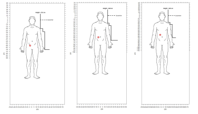 |
Compact 3T MRI for patients with implanted devices: Software tool to display MR fields at a specified location
Lydia Jean Bardwell Speltz1,2, Yunhong Shu2, Myung-Ho In2, Nolan Meyer1,2, Erin Gray2, Diana Lanners2, Yihe Hua3, Robert E Watson2, John Huston III2, Thomas KF Foo3, and Matt A Bernstein2
1Mayo Clinic Graduate School of Biomedical Sciences, Mayo Clinic, Rochester, MN, United States, 2Department of Radiology, Mayo Clinic, Rochester, MN, United States, 3GE Global Research, Niskayuna, NY, United States
Many implanted devices are labeled MR Conditional, meaning specific, labeled conditions must be met to ensure safe scanning. We developed a software tool for use with a high-performance, compact 3T (C3T) scanner that verifies relevant MR conditions can be met at the location of the device (e.g., abdomen), even if those conditions are exceeded at the level of the anatomy being scanned (e.g., brain). MR parameters assessed include main magnetic field strength, gradient slew rate, RF amplitude (B1+2), and dB/dz. The limited extent of the fields with C3T suggests high-performance exams can sometimes be obtained without compromising patient safety.
|
|||
2299.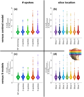 |
Patient specific parallel transmit pulses are patient position dependent while safety models are fixed: safety implications
Emre Kopanoglu1
1CUBRIC, School of Psychology, Cardiff University, Cardiff, United Kingdom
Pulses designed using patient-specific B1+-maps are inherently patient position dependent, while safety models (available on scanners) used for local SAR supervision are not. The effect of this positional mismatch on SAR estimation was investigated for 1-/2-/3-/4-/5-spoke pulses. The results showed substantial underestimation of local SAR: the actual local SAR at off-centre positions was observed to be up to 4.6-fold higher compared to the peak estimated using a centred model. This behaviour was worse for more spokes and consistent across slices between cerebellum and crown. Using multiple distributed models reduced the likelihood of SAR underestimation, but at the cost of over-restrictiveness.
|
|||
2300.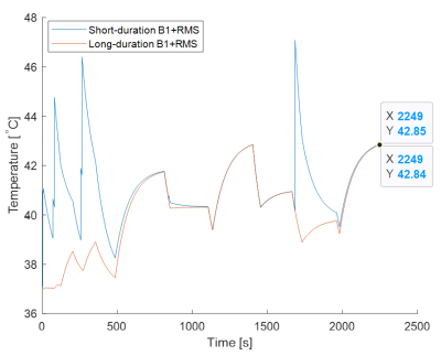 |
Assessing the In-Vivo RF Heating Effects of Short-Duration B1+RMS in MRI Sequences
Negin Behzadian1 and Shiloh Sison2
1Research and Development, Abbott, Sylmar, CA, United States, 2Research and Development, Abbott, Sunnyvale, CA, United States
Exposure to the RF energy of an MRI shall be limited to prevent potential harm to biological tissues. Whole-body SAR, averaged over any 6-minute interval, shall be limited to 2W/kg and 4W/kg under the limits of Normal and First Level Controlled Operating Mode, respectively, and is further restricted to twice the operating mode limit over any 10-second period. Our study investigates whether similar short-duration assessments are necessary for the alternative RF exposure metric of B1+RMS, in the context of RF heating safety of cardiac protocols with leaded cardiac implants.
|
|||
2301.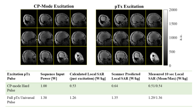 |
Validation of SAR Management Procedure for Dynamic pTx RF Waveforms Using a Self-Built Coil at 7 Tesla
Sydney Nicole Williams1, Sarah Allwood-Spiers2, Paul McElhinney1, Yuehui Tao3, John E. Foster2, Shajan Gunamony1,4, and David A. Porter1
1Imaging Centre of Excellence, University of Glasgow, Glasgow, United Kingdom, 2MRI Physics, NHS Greater Glasgow & Clyde, Glasgow, United Kingdom, 3Siemens Healthcare Ltd., Glasgow, United Kingdom, 4MR CoilTech Limited, Glasgow, United Kingdom
Parallel transmit (pTx) can reduce B1+ field inhomogeneity present at 7T, but needs additional consideration for specific absorption rate (SAR) monitoring due to the superimposed electromagnetic fields. For self-built pTx coils, validation includes comparing electromagnetic field simulations with experimental B1+ maps and thermometry, previously presented for static pTx in a self-built 8Tx/32Rx coil. We present full-waveform pTx validation with two further tests: comparison of measured and prescribed RF waveforms, and calculation of local SAR with virtual observation points (VOPs) for all pTx pulses with comparison to scanner predictions and measurements. After validation, dynamic pTx is performed in vivo.
|
|||
2302.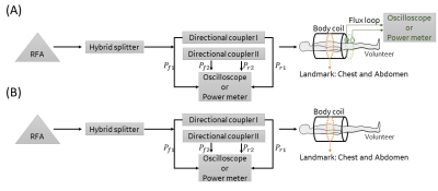 |
Individualized and accurate SAR characterization method based on an equivalent circuit model in MRI system
Weiman Jiang1, Fan Yang1, and Kun Wang1
1GE Healthcare, Beijing, China
This work proposed a novel SAR characterization method based on an equivalent circuit model and the circuit’s frequency response analysis. Comparing to well recognized pulse energy method defined in NEMA MS 8, this method doesn’t need the flux loop fixed on transmit coil. Meanwhile, it can monitor SAR accurately. The Root Mean Square (RMS) error and maximum error of the novel method relative to the method in NEMA MS 8 are 4.96% and 9.47% respectively. This method could avoid design complexity of integrating the flux loop and has potential to be easily realized in most MRI scanners.
|
|||
2303.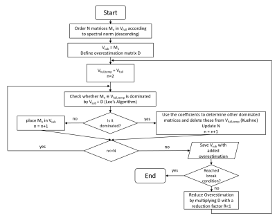 |
A local SAR compression algorithm with improved compression, speed and flexibility
Stephan Orzada1, Thomas M. Fiedler1, Harald H. Quick2,3, and Mark E. Ladd1,2,4,5
1Medical Physics in Radiology (E020), German Cancer Research Center (DKFZ), Heidelberg, Germany, 2Erwin L. Hahn Institute for MRI, University Duisburg-Essen, Essen, Germany, 3High-Field and Hybrid MR Imaging, University Hospital Essen, Essen, Germany, 4Faculty of Physics and Astronomy, University of Heidelberg, Heidelberg, Germany, 5Faculty of Medicine, University of Heidelberg, Heidelberg, Germany
For parallel transmit systems, control of local SAR is very important to ensure safety. For pulse calculation and online supervision, compression of the SAR matrices is used to reduce calculation effort. The original clustering method by Eichfelder et al. was later outperformed by a method proposed by Lee et al. We propose an enhancement to Lee’s algorithm that further increases compression efficiency, speed and flexibility by iteratively reducing the overestimation.
|
|||
2304.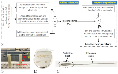 |
Validation of a Temperature Prediction Workflow for Imaging Complete Deep Brain Stimulation Systems
Alireza Sadeghi-Tarakameh1, Nur Izzati Huda Zulkarnain1, Noam Harel1, and Yigitcan Eryaman1
1Center for Magnetic Resonance Research (CMRR), University of Minnesota, Minneapolis, MN, United States
We validate a previously proposed temperature prediction workflow for a complete DBS Systems undergoing MRI. Accuracy of the workflow is investigated for different termination conditions using an extension cable and an implantable pulse generator (IPG) device. The workflow accurately predicted the temperature time-course during the MRI scan for different trajectories and terminations.
|
|||
2305.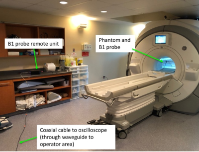 |
Radiofrequency peak B1+ Survey of commercial 3T MR Systems
Xin Huang1, Vick Chen1, and Shiloh Sison1
1Abbott Laboratories, Sunnyvale, CA, United States
To determine the appropriate peak B1+ field levels, an RF peak B1+ survey is performed on commercial 3T MR systems loaded with ASTM phantom filled with saline. The peak B1+ averaged over center slab inside the 3T commercial MR systems can be as high as 27 µT. The 3T MRI safety assessment should include values up to this level.
|
|||
2306.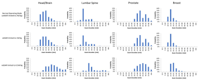 |
Impact of 1.5T SAR Limits on the MRI Scan Time for Implantable Devices
Yuqing Wan1, Nathan Ooms2, Paul Nguyen1, and Guangqiang (Jay) Jiang1
1Axonics Modulation Technologies, Irvine, CA, United States, 2Purdue University, West Lafayette, IN, United States
Implant device manufacturers often specify a RF exposure limit based on specific absorption rate (SAR) or B1+rms and a continuous active scan time in the device magnetic resonance imaging (MRI) labelling. Due to the restriction of the labeled SAR, MR sequence parameters may need to be adjusted to reduce RF power, resulting in prolonged scan time. The patient examination duration may be extended by up to 67% when compared to standard protocols. The overall session may be further prolonged due to additional wait time requirements from certain implanted devices, which can result in great inconvenience and extra cost to patients.
|
|||
2307.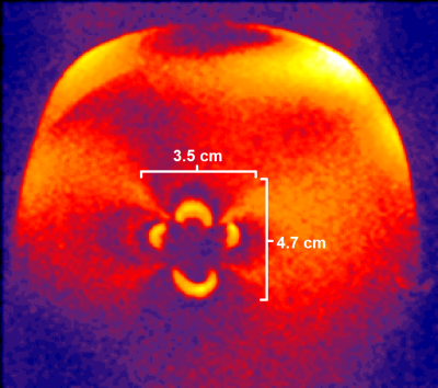 |
RF safety and image quality testing of deep brain stimulation electrodes with 3T MRI
Gaurav Verma1, Paul Min2, MyungHo In2, Jungho Cha3, Akbar Alipour1, Charlotte Elorette4, Lazar Fleysher5, Priti Balchandani1, Helen Mayberg3, and Ki Sueng Choi3
1Radiology, Icahn School of Medicine at Mount Sinai, New York, NY, United States, 2Radiology, Mayo Clinic, Rochester, MN, United States, 3Center for Advanced Circuit Therapeutics, Icahn School of Medicine at Mount Sinai, New York, NY, United States, 4Neuroscience, Icahn School of Medicine at Mount Sinai, New York, NY, United States, 5Diagnostic, Molecular and Interventional Radiology, Icahn School of Medicine at Mount Sinai, New York, NY, United States
Radio frequency (RF) safety and image quality testing was performed in the presence of deep brain stimulation (DBS) electrodes using gel acrylamide phantoms and two 3T MRI scanners. Two electrodes, (Abbott and Medtronic) were tested with sequences including GRE, T1-weighted magnetization-prepared rapid gradient echo (MPRAGE), and T2-weighted turbo spin-echo (TSE). Safety testing showed low heating when electrodes were aligned along the B0 field (Z-axis) of the magnet, but higher heating when the electrodes were angled to the main field. Image quality testing showed smaller ferromagnetic susceptibility artifacts when electrode was parallel with B0 field and normal to transmit field.
|
|||
2308.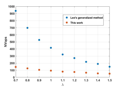 |
Improved compression of SAR matrices by a reformulation of the generalized virtual observation point compression scheme
Vincent Gras1 and Nicolas Boulant1
1DRF/Joliot/Neurospin, CEA - Université Paris Saclay, Gif sur Yvette, France
Parallel transmission to date is the most promising technology to tackle the RF field inhomogeneity problem in MRI at ultra-high field. The Virtual Observation Point compression technique was a cornerstone in the field to reduce drastically the number of SAR matrices to handle in exam supervision and RF pulse design. After its original discovery, it was further improved to boost the compression. This work describes a reformulation of the problem which simplifies substantially the numerical search and allows reducing further the number of matrices by a factor ~5 or more.
|
|||
2309.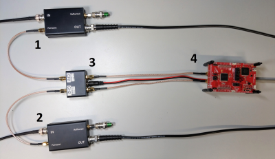 |
Stand-Alone Hardware SAR Monitor based on low cost Electronic Standard Components
Marcus Prier1,2, Max Joris Hubmann1,2, Enrico Pannicke1,2, and Oliver Speck1,2
1Otto-von-Guericke University, Magdeburg, Germany, 2Research Campus STIMULATE, Magdeburg, Germany
A low cost, stand-alone RF power monitor was developed that fulfills the requirements given in the standard 60601-2-33 and is based on electrical standard components. It consists of two dual directional couplers, two RMS envelope filters and a microcontroller. A fixed power limit can be programmed or patient dependent power limits can be communicated from a host computer. Evaluation measurements show power measurement and RFPA blanking switching times accuracies with an error less than 1%. A sampling rate of 130kSamples/s indicates usability for relatively short or moderate complex shaped RF waveforms.
|
|||
2310.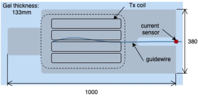 |
Is a Local Tx Coil sufficient for Guidewire Safety in MRI?
Felipe Godinez1,2, Greig Scott3, Joseph V Hajnal1,2, and Shaihan J Malik1,2
1Biomedical Engineering Department, School of Biomedical Engineering and Imaging Sciences, King's College London, London, United Kingdom, 2Centre for the Developing Brain, School of Biomedical Engineering and Imaging Sciences, King's College London, London, United Kingdom, 3Department of Electrical Engineering, Stanford, Stanford, CA, United States
Imaging guidewires in MRI is known to have heating hazards associated. In this work we show initial data that supports the use of local Tx coils over whole body coils for the safe imaging of standard guidewires with MRI. This was tested in vitro using parallel transmit (PTx) techniques to generate a scenario for a proper comparison between the two coil types. It was found that a local Tx coil produces less heating at the guidewire tip, than a body coil under comparable conditions.
|
|||
2311.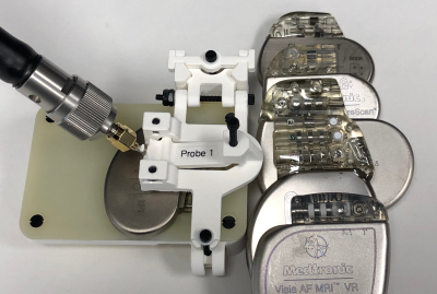 |
Input Impedance Comparison of MR-Conditional Cardiac Implantable Pulse Generators at the 1.5T MR Frequency of 63.87 MHz
Jason Meyers1, David Prutchi1, and Ramez Shehada2
1Impulse Dynamics (USA) Inc., Marlton, NJ, United States, 2Medical Technology Laboratories, La Mirada, CA, United States
The MR environment poses a tissue heating hazard to patients with cardiac IPGs (implantable pulse generators), such as pacemakers and defibrillators, due to RF currents circulating in the loop formed by the IPG, a transvenous lead, and the tissue. Heating will be limited by the impedance of this loop at the MR frequency (63.87 MHz for 1.5T). The IPG input impedance (a portion of this loop) was measured in six MR-Conditional IPG’s. All have a comparable, low impedance, suggesting that device manufacturers are not intentionally adding impedance and that some interchangeability may be possible without changing RF-induced heating.
|
|||
2312.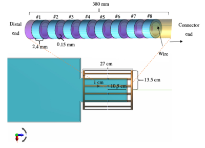 |
Computational simulations of heating in the vicinity of an 8-contact depth EEG electrode: the impact of model simplification
Hassan B Hawsawi1,2, Ozlem Ipek3,4, David W Carmichael5,6, and Louis Lemieux1
1Clinical and Experimental Epilepsy, University College London, London, United Kingdom, 2Administration of Medical Physics, King Abdullah Medical City, Makkah, Saudi Arabia, 3King’s College London, London, United Kingdom, 4CIBM-AIT, Ecole Polytechnique Federale de Lausanne, Lausanne, Switzerland, 5Department of Biomedical Engineering, King’s College London, London, United Kingdom, 6Developmental Imaging and Biophysics Section, UCL Great Ormond Street Institute of Child Health, London, United Kingdom
Electromagnetic (EM) simulations offer the possibility of assessing RF-induced heating around intracranial EEG (icEEG) electrodes across a variety of placement scenarios in a shorter time than experimental phantom-based temperature measurements. However, given the sub-millimetric dimensions of the wires and contacts the range of spatial scales spans many orders of magnitude, leading to high computational demands. In this study, we assessed the effect of model simplification for a typical 8-contact depth EEG electrode on the estimated heating patterns.
|
|||
2313.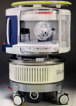 |
Safety Considerations in Neuroimaging of Neonatal and Pediatric Patients Using Portable Low Field MRI
Mark Smith1, Harry Hu2, Ram Krishnamurthy1, John Pitts3, and Mai-Lan Ho1
1Nationwide Children's Hospital, Columbus, OH, United States, 2Hyperfine, Dublin, OH, United States, 3Hyperfine, Cleveland, OH, United States
In 2020, a portable 64mT ultra-low field MRI system designed for point of care bedside use received 510K clearance (Hyperfine, Guilford, CT). Our pediatric institution (Nationwide Children’s Hospital) has acquired one of these systems to meet neuroimaging needs for critically ill NICU or PICU patients who cannot tolerate transport to the MRI department. Prior to scanning patients, safety testing for displacement and heating was conducted on monitoring hardware that will be connected to the patient during the bedside MRI. The monitoring hardware was found safe to stay connected to the patient during the bedside MRI.
|
|||
2314.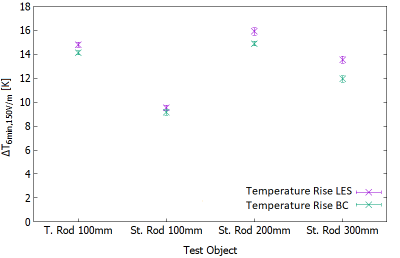 |
Influence of E-Field Homogeneity and Drift for Testing Medical Implants for RF-induced heating at 64MHz according to ASTM-F2182 and ISO/TS 10974
Finya Ketelsen1,2, Vincent Hammersen1, Kevin Kröninger2, and Gregor Schaefers1,3
1MRI-STaR - Magnetic Resonance Institute for Safety, Technology and Research GmbH, Gelsenkirchen, Germany, 2TU Dortmund University, Dortmund, Germany, 3MR:comp GmbH, Testing Services for MR Safety & Compatibility, Gelsenkirchen, Germany
Medical implants of all kind must be tested for RF-induced heating to determine their MR safety and compatibility. Test conditions concerning E-field homogeneity and E-field drift during assessment time are defined in ISO/TS 10974 and ASTM-F2182. This study evaluates the influence of E-field drift and E-field homogeneity on RF-heating. It shows that both effects can lead to a temperature underestimation. Even for small implants whose volume is in a homogenous area within ±1dB, this underestimation can occur. System instability and implant size and position can further increase the underestimation of RF-induced heating and should be carefully considered.
|
|||
2315.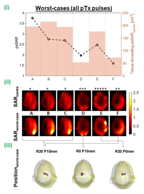 |
Parallel-transmit coil dimensions affect SAR sensitivity to motion at 7T.
Alix Plumley1, Philip Schmid1, and Emre Kopanoglu1
1Cardiff University Brain Research Imaging Centre, Cardiff, United Kingdom
Subject motion in parallel-transmit (pTx) causes channels’ electric field interference patterns to change, influencing SAR distributions. This can cause safety limits to be exceeded when SAR-constrained pulses are designed for one position. Here, we consider effects of pTx coil dimensions on SAR sensitivity to motion by simulating 6 differently-sized coil models, and evaluating SAR at 19 displaced positions. Our results agree with those previously reported for the similar-sized coil, but SAR sensitivity was generally lower for larger coils, and higher for smaller coils, with maximum motion-induced local-SAR increase of 3.8-fold and 1.6-fold for the smallest and largest coil models, respectively.
|
|||
2316.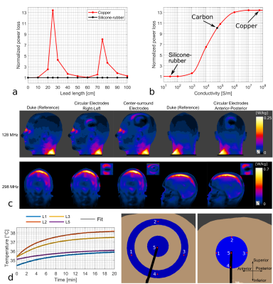 |
Safety evaluation with respect to RF-induced heating of a new setup for Transcranial Electric Stimulation during MRI
Fróði Gregersen1,2,3,4, Cihan Göksu2,5, Gregor Schaefers6,7, Rong Xue4,8,9, Axel Thielscher1,2, and Lars Hanson1,2
1Section for Magnetic Resonance, DTU Health Tech, Technical University of Denmark, Kgs Lyngby, Denmark, 2Danish Research Centre for Magnetic Resonance, Centre for Functional and Diagnostic Imaging and Research, Copenhagen University Hospital, Amager and Hvidovre, Denmark, 3Sino-Danish Center for Education and Research, Aarhus, Denmark, 4University of Chinese Academic of Sciences, Beijing, China, 5High-Field Magnetic Resonance Center, Max-Planck-Institute for Biological Cybernetic, Tübingen, Germany, 6MRI-STaR-Magnetic Resonance Institute for Safety, Technology and Research GmbH, Gelsenkirchen, Germany, 7MR:comp GmbH, MR Safety Testing Laboratory, Gelsenkirchen, Germany, 8State Key Laboratory of Brain and Cognitive Science, Beijing MRI Center for Brain Research, Institute of Biophysics, Chinese Academy of Sciences, Beijing, China, 9Beijing Institute for Brain Disorders, Beijing, China
Combining transcranial electrical stimulation (TES) with MRI offers various interesting research opportunities, but also introduces safety concerns. Coupling between the RF field and highly conductive TES leads can lead to skin burns. These safety issues are usually mitigated with the use of safety resistors and controlled lead paths that reduce the power absorbed by the leads. However, these methods introduce practical limitations for combined TES/MRI experiments, such as limited stimulation currents and cable stray fields corrupting MR current density imaging. We overcome these limitations by using low-conductivity silicone-rubber as TES leads. Simulations and temperature measurements are used for safety assessment.
|
|||
2317.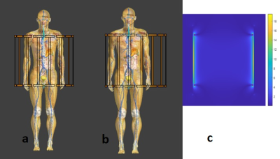 |
Unloaded RF transmit coil B1+ maps do not correlate with SAR hotspots
Xin Chen1 and Michael Steckner1
1Canon Medical Research USA Inc., Mayfield Village, OH, United States
MRI vendors have been asked to provide B1+ contour plots of unloaded whole body transmit coils in order to show technologists/radiographers where not position a patient for SAR hotspot/RF burn avoidance purposes. Two flaws with this strategy are demonstrated by modeling Duke at the abdominal landmark in the centered and off-set position: 1) B1+ maps correlate poorly to SAR hotspots, 2) loaded B1+ maps are significantly different relative to unloaded B1+ maps. Knowledge of the birdcage coil end-ring position in conjunction with spacing pads will improve RF burn safety outcomes.
|
The International Society for Magnetic Resonance in Medicine is accredited by the Accreditation Council for Continuing Medical Education to provide continuing medical education for physicians.