Digital Posters
fMRI Using Animal Models: Methods
ISMRM & SMRT Annual Meeting • 15-20 May 2021

Digital Posters
fMRI Using Animal Models: Methods
| Concurrent 3 | 15:00 - 16:00 |
2910.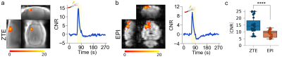 |
fMRI with a Zero Echo Time (ZTE) Pulse Sequence
Martin John MacKinnon1,2,3,4, Yuncong Ma1,2,4, Sheng Song1,2,4, Tzu-Hao Harry Chao1,2,4, Tzu-Wen Winnie Wang1,2,4, SungHo Lee2,4, SungHo Lee1,2,4, Wei-Tang Chang2,5, and Yen-Yu Ian Shih1,2,4
1Center for Animal MRI, University of North Carolina at Chapel Hill, Chapel Hill, NC, United States, 2Biomedical Research Imaging Center, University of North Carolina at Chapel Hill, Chapel Hill, NC, United States, 3The Joint Department of Biomedical Engineering, University of North Carolina at Chapel Hill, Chapel Hill, NC, United States, 4Department of Neurology, University of North Carolina at Chapel Hill, Chapel Hill, NC, United States, 5Department of Radiology, University of North Carolina at Chapel Hill, Chapel Hill, NC, United States
Conventional fMRI studies, carried out with the gold-standard echo-planar imaging (EPI), are confounded by the deleterious effects of the sequence’s limitation – its sensitivity to magnetic field inhomogeneities and high acoustic noise. The properties of short acquisition delay sequences, that also have minimal incrementing of gradients during spatial encoding, such as MB-SWIFT and ZTE, render them resistant to the aforementioned confounding factors. We study the feasibility of using ZTE to detect functional activations with endogenous contrast using a simple rat forepaw electrical stimulation paradigm. We show that ZTE-fMRI has a 67% greater sensitivity than the gold-standard BOLD-weighted EPI.
|
|||
2911.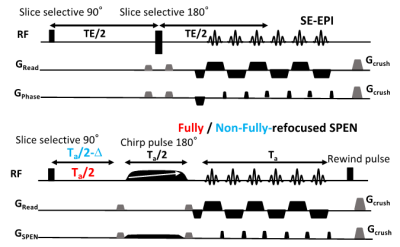 |
Functional MRI of mice olfactory bulbs using novel fMRI methodologies at 15.2T
Odélia Jacqueline Chitrit1, Qingjia Bao1, Silvia Chuartzman2, Noga Silkha2, Tali Kimchi2, and Lucio Frydman1
1Department of Chemical and Biological Physics, Weizmann institute of Science, Rehovot, Israel, 2Department of Neurobiology, Weizmann institute of Science, Rehovot, Israel
Spatiotemporal Encoding (SPEN) MRI was used in fully- and non-fully-refocused modes, to capture the activation of olfactory bulbs (OBs) in mice, in response to odors. At the 15.2T field employed, responses on the order of 10% could easily be observed in male OBs; clear but weaker responses were observed in females. In all cases image quality largely exceeded that arising in GE or even SE EPI, with compromises between image quality and fMRI activation “tunable” by SPEN’s refocusing conditions. The high spatial and temporal resolutions achievable in these scans can shed light into this important behavioral cue of social animals.
|
|||
2912.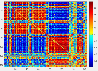 |
High Resolution EPI-based rs-fMRI Performed at 21.1 T
David C. Hike1,2, Lauren C. Daley1,2, Frederick A Bagdasarian1,2, Shannon Helsper1,2, and Samuel Colles Grant1,2
1National High Magnetic Field Laboratory, Florida State University, Tallahassee, FL, United States, 2Chemical & Biomedical Engineering, FAMU-FSU College of Engineering, Tallahassee, FL, United States
This study utilizes resting-state (rs)-fMRI and graph theory as methods for detecting functional recovery following ischemia. rs-fMRI was performed at 21.1 T on an ischemic rat model and naïve controls. Current data shows correlations in multiple areas of the brain indicating differences between hemispheres for the parameters observed.
|
|||
2913.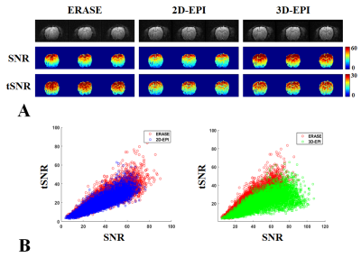 |
Comparison of 2D-EPI, 3D-EPI and ERASE in terms of physiological noise, SNR and tSNR
Jae-Kyun Ryu1,2 and Jang-Yeon Park2,3,4
1Biomedical Institute for Convergence at SKKU, Sungkyunkwan University, Suwon, Korea, Republic of, 2Center for Neuroscience Imaging Research, Institute for Basic Science, Suwon, Korea, Republic of, 3Department of Biomedical Engineering, Sungkyunkwan University, Suwon, Korea, Republic of, 4Department of Intelligent Precision Healthcare Convergence, Sungkyunkwan University, Suwon, Korea, Republic of
Recently we proposed an ultrafast 3D gradient-echo imaging sequence using spatiotemporal-encoding (SPEN), called ERASE (Equal-TE Rapid Acquisition with Sequential Excitation), which has high tolerance to B0 inhomogeneities. Here, we investigated the noise characteristics, SNR, and tSNR of ERASE with rat brain imaging at 9.4T at high spatial resolution, in comparison with 2D and 3D gradient-echo EPI. While ERASE provided better tSNR than both of them, it provided intermediate SNR between them. The ratio of physiological and thermal noise (σP/σ0) and lambda (λ) were smaller in ERASE than 2D and 3D-EPI mainly due to its robustness against B0 inhomogeneity.
|
|||
2914.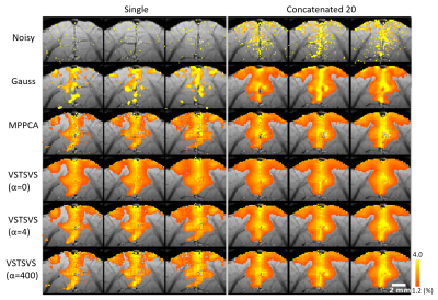 |
Denoise functional magnetic resonance imaging with variance-stabilizing transformation and optimal singular value shrinkage (VST-SVS)
Wei Zhu1, Xiaodong Ma1, Xiao-Hong Zhu1, Kamil Uğurbil1, Wei Chen1, and Xiaoping Wu1
1University of Minnesota, Minneapolis, MN, United States
High-resolution fMRI is largely hindered by random thermal noise. In this study, we propose a denoising method to reduce such noise in magnitude fMRI data. The proposed method synergistically combines: 1) variance-stabilizing transformation to convert Rician data to Gaussian-like data, 2) principle-component-analysis-based denoising algorithm with optimal singular value shrinkage to remove noise, and 3) patch-based implementation with tunable Gaussian weighting to tradeoff between functional sensitivity and specificity. Our results using synthetic and in-vivo cat task fMRI data show that the denoising method can effectively remove Rician noise, promoting functional fidelity and sensitivity in comparison to existing denoising methods.
|
|||
 |
2915.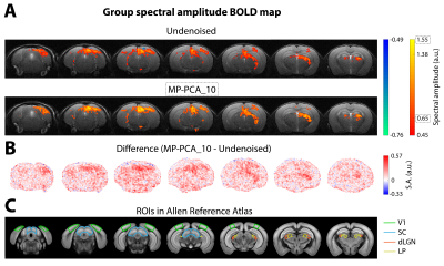 |
Enhanced conventional and ultrafast responses in preclinical functional MRI using MP-PCA denoising
Francisca F. Fernandes1, Rita Gil1, Jonas L. Olesen2,3, Sune N. Jespersen2,3, and Noam Shemesh1
1Champalimaud Research, Champalimaud Centre for the Unknown, Lisbon, Portugal, 2Center of Functionally Integrative Neuroscience (CFIN) and MINDLab, Department of Clinical Medicine, Aarhus University, Aarhus, Denmark, 3Department of Physics and Astronomy, Aarhus University, Aarhus, Denmark
The MP-PCA denoising approach is an objective way to reduce fluctuations caused by thermal noise. In rodents, thermal noise in the coils is an important fMRI confounder that can reduce the accuracy of activation mapping. Here, we develop the MP-PCA denoising scheme for rodent fMRI. Data was obtained from 3 mice using conventional multislice and ultrafast acquisitions (1s and 50ms temporal resolution, respectively), during visual stimulation. MP-PCA denoising generated strong Fourier spectral amplitude increases in BOLD maps of 20% and 85% and tSNR increases of 100% and 275% for multislice and ultrafast data, respectively.
|
||
2916.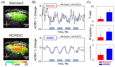 |
High Resolution Functional Mapping of Orientation Domains in the Cat Visual Cortex using Denoising with NORDIC
Shinho Cho1, Steen Moeller1, Mehmet Akçakaya2, Logan Dowdle1, Luca Vizioli1, Djaudat Idiyatullin1, Wei Chen1, and Kâmil Uğurbil1
1Center for Magnetic Resonance Research and Department of Radiology, University of Minnesota, Minneapolis, MN, United States, 2Center for Magnetic Resonance Research and Department of Electrical and Computer Engineering, University of Minnesota, Minneapolis, MN, United States
Animal model studies in functional brain imaging (fMRI) require extremely high spatial resolutions, thus being challenged by the low signal-to-noise ratio (SNR). Recent advances in fMRI denoising provide gains by suppressing thermal noise. Here we demonstrate significant improvements of fMRI results by NORDIC (NOise reduction with DIstribution Corrected PCA) de-noising; increased functional CNR and T-statistics, yielded the high signal stability of single fMRI acquisition with reproducible quantification of brain responses.
|
|||
2917.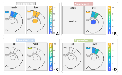 |
Automatic detection of BOLD oscillations in the anesthetized brain
Henriette Lambers1, Lydia Wachsmuth1, Ping Zheng1, and Cornelius Faber1
1Translational Research Imaging Center, Clinic for Radiology, University Hospital Muenster, Muenster, Germany
Periodic BOLD oscillations caused by vasomotion can superimpose temporal signal fluctuations in fMRI measurements and may affect brain network analysis. In order to establish a reliable detection of BOLD oscillations, we developed an automatic oscillation detection tool with an accuracy of 96 %. With this program, we examined high temporal resolution MR data consisting in total of 1.259.400 images. We found that BOLD oscillations occur brain wide starting between two and four hours after initiation of medetomidine sedation.
|
|||
2918.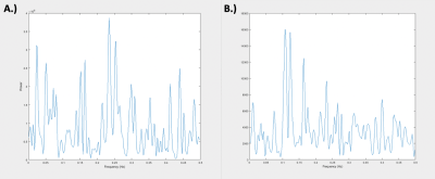 |
Global Signal vs. Global Noise in Rat rs-fMRI
Nmachi Anumba1,2, Wenju Pan1,2, Eric Maltbie1,2, and Shella Keilholz1,2
1Biomedical Engineering, Emory University, Atlanta, GA, United States, 2Biomedical Engineering, Georgia Institute of Technology, Atlanta, GA, United States
Regression of the BOLD “global signal” in human rs-fMRI studies to reduce widespread noise is a controversial practice since the signal may also contain neural activity. Rodent rs-fMRI studies offer an opportunity to better disentangle the neural and nonneural contributors to the global signal. However, global signal in rodents has been relatively little studied. We performed a voxel-wise analysis to examine the global signal’s spatial distribution. We found that, as in humans, global signal contributions vary spatially within the rat brain and that certain attributes of this signal are unique to the brain when compared to nonneural sources of noise.
|
|||
2919.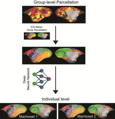 |
Accurate Brain Parcellation of Individual Marmosets Based on Awake Resting-State fMRI Data and Deep Neural Networks
Xiaoguang Tian1, Zhifeng Liang2, Afonso C Silva1, and Cirong Liu2
1Dept. of Neurobiology, University of Pittsburgh, Pittsburgh, PA, United States, 2Institute of Neuroscience, Chinese Academy of Sciences, Shanghai, China
As a prerequisite for understanding how the brain works, it has been a long-sought goal to subdivide (parcellate) the brain into a mosaic of anatomically- and functionally-defined parcels (areas). However, reaching a consensus parcellation has been hindered by inaccuracies in aligning brain areas across subjects. Here, we developed a novel cortical parcellation approach using resting-state fMRI data collected in a population of awake marmosets to accurately map the functional brain organization of individuals.
|
|||
2920.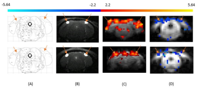 |
Functional cerebral blood volume imaging of the mouse visual cortex using vascular space occupancy
Naman Jain1, Atena Akbari1, Markus Barth1,2, and Kai-Hsiang Chuang1,3
1Centre for Advanced Imaging, The University of Queensland, Brisbane, Australia, 2School of Information Technology and Electrical Engineering, The University of Queensland, Brisbane, Australia, 3Queensland Brain Institute, The University of Queensland, Brisbane, Australia
Cerebral Blood Volume (CBV) – weighted vascular-space-occupancy (VASO) functional MRI (fMRI) has shown superior spatial specificity compared to the conventional gradient-echo blood-oxygen-level-dependent (BOLD) contrast in humans [1]. Although VASO fMRI has been used for CBV imaging in rats and monkeys, the applicability of this method in the mouse brain has not been investigated due to the technical challenges. In this study we examine the feasibility of VASO fMRI of the visual cortex in the anesthetized mouse.
|
|||
2921.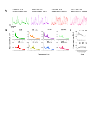 |
Combined RS-fMRI and calcium recordings show stabile brain states in mice after switching anesthetic regimen
Bruno Pradier1,2, Lydia Wachsmuth1, Daniel Segelcke2, Nina Nagelmann1, Esther Pogatzki-Zahn2, and Cornelius Faber1
1Department of Clinical Radiology, University Hospital Münster, Münster, Germany, 2Department of Anesthesiology, University Hospital Münster, Münster, Germany
Resting-state fMRI in mice became a popular method to investigate brain circuits. However, uncertainty resides over the dynamic effects of anesthetics on brain states and functional connectivity. Using a multimodal approach with optical calcium recordings and RS-fMRI, we assessed brain states and networks while switching anesthetic regimen from 1% isoflurane to a combined 0.2% isoflurane/medetomidine anesthesia. We find that brain states reach a steady state after a short transition time and detected that functional connectivity changes were strongest relative to the 1% isoflurane condition. We conclude that brain states and networks are stable from 30 minutes after switching anesthetic regimen.
|
|||
2922.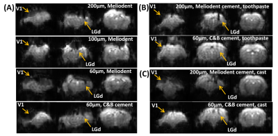 |
Optimize Simultaneous Multi-channel Calcium Recording and fMRI in Mouse Brain
Shabnam Khorasani Gerdekoohi1, Pankaj Sah2, and Kai-Hsiang Chuang1,3
1Queensland Brain Institute, Brisbane, Australia, 2Quuensland Brain Institute, Brisbane, Australia, 3Center for Advanced Imaging, Brisbane, Australia · Susceptibility artifacts of EPI induced by fiber photometry were successfully reduced at 9.4T MRI. · High quality event-related BOLD and calcium responses were measured using short visual stimuli. · Neural to hemodynamic transfer functions of a cortical region (V1) and a subcortical region (LGd) were calculated. |
|||
2923.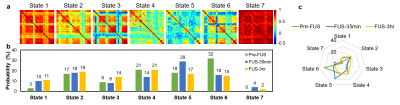 |
Dynamic Functional Connectivity of Focused Ultrasound-induced Neuromodulation in Normal Rat Model
Yu-Chieh Hung1, Yi-Cheng Wang1, Hao-Li Liu2, and Hsu-Hsia Peng1
1Biomedical Engineering and Environmental Sciences, National Tsing Hua University, Hsinchu, Taiwan, 2Electrical Engineering, National Taiwan University, Taipei, Taiwan
Non-invasive focused ultrasound (FUS) offered attractive advantages to modulate neuronal activity. The functional connectivity (FC) between different brain regions is a dynamic process during a period of examinations. We aim to explore FUS-induced neuromodulation by dynamic FC. We observed the evolution of dynamic FC at Pre-FUS, FUS-35min, and FUS-3hr for normal rats with FUS sonication at ventral posteromedial and ventral posterolateral thalamic nuclei (VPM/VPL) region in left thalamus. With K-means clustering, we quantitatively evaluated the evolution of altered probability% of dynamic FC in seven states at Pre-FUS, FUS-35min, and FUS-3hr, suggesting the potential of FUS-neuromodulation.
|
|||
2924.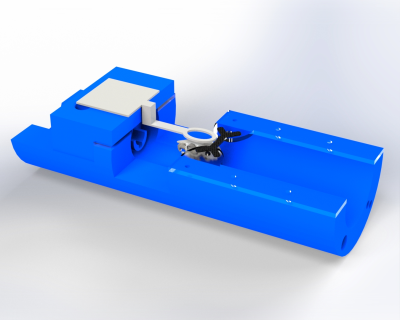 |
Restraint System for Motion Reduction in MRI studies of Awake Mice
Derek Prusener1, Maysam Nezafati1, Gloria Perrin Clavijo1, and Shella Keilholz1
1Biomedical Engineering, Emory University/Georgia Institute of Technology, Atlanta, GA, United States
We designed a head restraint system for image acquisition in awake mouse fMRI studies that minimized head motion. The system consists of a head implant, combined with a customized cradle and head holder. Two distinct head implant designs are tested to evaluate if the motion goal for the system is achieved.
|
|||
2925.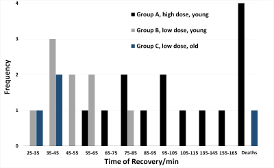 |
Extra low dose pancuronium bromide for fMRI improves survival and recovery times while suppressing translational motion
Muhammad Danial Afiq Bin Abdullah1, Isaac Huen1, Redha Boubertakh1, Xing Qi Teo1, and Kuan Jin Lee1
1SBIC, Agency for Science, Technology and Research (A*STAR), Singapore, Singapore
Minimising motion artefacts for pre-clinical studies requires the use of pancuronium bromide (PB), a muscle relaxant paired with the use of mechanical ventilators. Current literature is limited in its description of the mice recovery from the effects of PB. We detail a recovery process using low dose PB which improves survival while keeping translational motion suppressed.
|
|||
2926.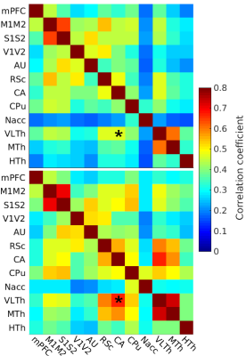 |
Light sedation with short habituation time for large-scale fMRI studies in rat
Lenka Dvořáková1, Petteri Stenroos1, Ekaterina Zhurakovskaya1, Raimo Salo1, Jaakko Paasonen1, and Olli Gröhn1
1A.I.V. Institute for Molecular Sciences, University of Eastern Finland, Kuopio, Finland
As anesthesia is a serious confounding factor in pre-clinical fMRI, there is increasing interest in awake fMRI. However, awake protocols tend to be time-consuming and laborious, making large studies unfeasible. Therefore, we investigated whether a light sedation protocol with a short habituation period can provide comparable data to fully awake protocol. We measured 102 rats under light sedation and compared the FC to both on-site measured awake data and open awake rat fMRI database. Our results show, that apart from slightly modified thalamic connectivity, light sedation provides results that are comparable to the data obtained in the awake state.
|
The International Society for Magnetic Resonance in Medicine is accredited by the Accreditation Council for Continuing Medical Education to provide continuing medical education for physicians.