Digital Posters
fMRI Using Animal Models: Applications
ISMRM & SMRT Annual Meeting • 15-20 May 2021

Digital Posters
fMRI Using Animal Models: Applications
| Concurrent 3 | 15:00 - 16:00 |
 |
2927.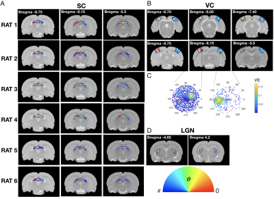 |
MRI based retinotopy of the rat visual pathway
Joana Carvalho1, Francisca F. Fernandes1, and Noam Shemesh1
1Champalimaud Centre for the Unknown, Lisbon, Portugal
Unravelling the fine-grained organization of the visual system is essential for understanding the foundations of vision in health and disease. Receptive field (RF) properties and retinotopic maps across the visual pathway were estimated non-invasively at unprecedented resolution, for the first-time in rodents. We tailored a set-up to deliver complex stimuli in preclinical scanners, and combined BOLD-fMRI with biologically-inspired models to investigate how RFs vary across cortical depth and hierarchy. We show that Lateral Geniculate Nucleus (LGN), Visual Cortex (VC) and Superior Colliculus (SC) are retinotopically organized and that RF sizes vary across cortical layers, spiking at layers IV and VI.
|
||
2928.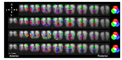 |
HIGH RESOLUTION ODOR MAPPING IN AWAKE MOUSE OLFACTORY BULB USING CONTRAST ENHANCED FMRI
Christopher Cover1, Alexander Poplawsky1, Sujatha Nallama1, and Mitsuhiro Fukuda1
1Department of Radiology, University of Pittsburgh, Pittsburgh, PA, United States
The rodent olfactory bulb provides an ideal system to investigate cell- and layer-specific contributions to neurovascular coupling using noninvasive fMRI. However, anesthesia is regularly used to minimize motion, which is known to interfere with neurovascular coupling and confounds the neural interpretation of the results. Here, we introduce a technique for reliable awake rodent fMRI of the olfactory bulb at high spatial resolutions (100 x 100 x 300 μm3). Activation maps of four unique odors are consistent with previously published 2-deoxyglucose autoradiography studies. Awake rodent olfactory fMRI is a reliable technique to reproduce neural-specific odor maps at laminar resolutions.
|
|||
2929.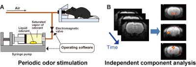 |
Functional MRI study of olfactory responses evoked by musk odor in the mouse whole brain under medetomidine anesthesia
Yumiko Tsubakihara1, Mitsuhiro Takeda1, Sosuke Yoshinaga1, and Hiroaki Terasawa1
1Faculty of Life Sciences, Kumamoto University, Kumamoto, Japan
Muscone, a musk odorant component that attracts male mice, binds to olfactory receptors on the olfactory epithelium. Subsequently, activation signals are transferred from the receptors to the olfactory bulb, and then to the olfactory cortex and other brain regions. To detect such odor-evoked activation pathways associated with the perception of odorants and induced behaviors, we employed a method that combines periodic odor stimulation and independent component analysis. In this study, we used medetomidine as the anesthesic. Since medetomidine reportedly influences neural activity in a dose-dependent manner, we investigated the muscone-evoked activations at different levels of medetomidine anesthesia.
|
|||
2930.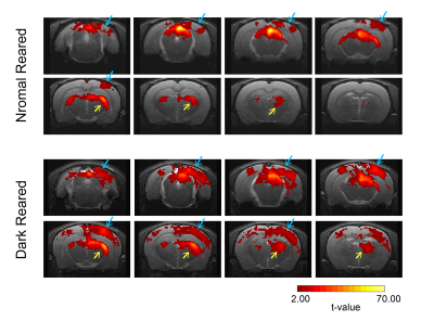 |
Lack of visual critical period of plasticity induces BOLD modulations in the rat binocular subregion of the primary visual cortex
Rita Gil1, Frederico Severo1, and Noam Shemesh1
1Champalimaud Centre for the Unknown, Lisbon, Portugal
Experience induced visual cortical (VC) plasticity is key for adequate pathway maturation and circuitry refinement. Competing inputs from each eye arriving to the thalamic lateral geniculate nucleus and, later, to VC, are necessary to shape and develop visual binocularity. Without such competition, an increased responsiveness and number of binocular cortical neurons has been reported. Here, we investigated monocular stimulation in rats that had been dark reared and compared their cortical BOLD responses with normal reared animals. Increased BOLD responses appeared along the VC of the dark reared group, suggesting a lack of pathway maturation and binocular integration.
|
|||
2931.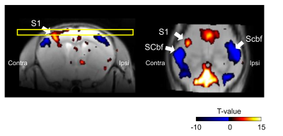 |
Correlation of negative and positive BOLD signal change in the cerebral cortex by somatosensory stimulation in mice
Tomokazu Tsurugizawa1, Boucif Djemai2, and Kazumi Kasahara1
1Human Informatics and Interaction Research Institute, National Institute of Advanced Industrial Science and Technology (AIST), Tsukuba, Japan, 2NeuroSpin/CEA-Saclay, Gif-sur-Yvette, France
In the current study, we aim to investigate the spatiotemporal profile of negative BOLD response evoked by somatosensory stimulation in mouse. Positive BOLD response was observed in contralateral primary somatosensory cortex (S1) by forepaw somatosensory stimulation, while negative BOLD response was observed in the bilateral barrel field of somatosensory cortex (Scbf). The amplitude of BOLD response in Scbf was negatively correlated with positive BOLD response in S1, while negative BOLD response lasted longer than positive BOLD response. These results indicate that origin of negative BOLD response could be neuronal activity, but neurovascular coupling is not similar as positive BOLD response.
|
|||
2932.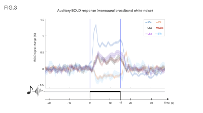 |
Monaural auditory stimulation induces negative BOLD in the rat auditory pathway
Frederico Severo1, Rita Gil1, and Noam Shemesh1
1Champalimaud Centre for the Unknown, Lisboa, Portugal
fMRI has been used to characterize the auditory pathway in rodents in the past, but unlike other modalities, these studies haven’t shown any prominent negative responses in any structure along the auditory pathway. Our findings reveal, for the first time, negative BOLD signals induced by monaural stimulation with broadband white noise in the ipsilateral inferior colliculus and contralateral striatum. These findings may shed light into the intercollicular dynamics in sound processing in the rat brain upon, e.g., sound localization or spatial navigation.
|
|||
2933.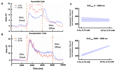 |
The Effect of Somatostatin+ Interneurons on the Negative BOLD Response.
Rik Lodevicus Elizabeth Maria Ubaghs1, Roman Böhringer1, Markus Marks1, Mehmet Fatih Yanik1, and Benjamin Friedrich Grewe1
1Institute of Neuroinformatics University and ETH Zurich, Zurich, Switzerland
How the exact interaction between excitatory and inhibitory neurons shapes the fMRI BOLD response is currently not well understood. As such, this often hinders any precise conclusions about the causal involvement of a brain area during a specific task. Here we show a demonstration of stimulus-dependent excitatory and inhibitory processing that could explain previously reported observations in BOLD polarity (Niranjan et al., 2016). We show evidence that the activity of Somatostatin+ interneurons is related to a negative inflection in the BOLD signal even when there is a simultaneous increase in local excitatory activation.
|
|||
2934.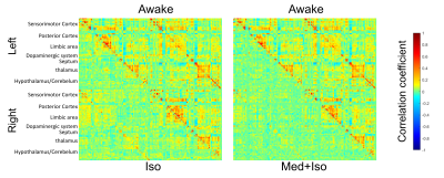 |
Awake fMRI shows an impact of anesthesia on resting state functional connectivity in mice
Tomokazu Tsurugizawa1
1Human Informatics and Interaction Research Institute, National Institute of Advanced Industrial Science and Technology (AIST), Tsukuba, Japan
This study aims to develop awake fMRI protocol to compare the resting state functional connectivity between awake state and anesthetized state (isoflurane or medetomidine + isoflurane) in the same mouse. The typical structure of resting state functional connectivity did not alter under ISO or MED+ISO anesthesia, but the connectivity strength and fractional amplitude of low frequency fluctuation in the cerebral cortex and the thalamic nuclei altered under anesthesia. These results indicate the availability of anesthesia on the resting state fMRI although the impact of anesthesia on neuronal activity should be considered.
|
|||
2935.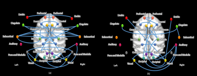 |
The brain activity of Rhesus monkeys in anesthesia
Yifan Miao1
1Institute of Biophysics, CAS., Beijing, China
In this study, we have observed a dose-dependent difference the brain activity and functional connectivity of Rhesus monkeys during propofol-induced anesthesia. The overall change of ALFF values was insignificant, but under lighter level there was an increase in the prefrontal and occipital lobes and in the frontal lobe inversely. The FC values across hemispheres were highly synchronized under deep anesthesia, and gradually decreased under lighter. The pattern of change in functional connectivity values showed a significant decrease followed by a small increase, which may have some reference significance to reveal the brain network formation mechanisms during resting state.
|
|||
2936.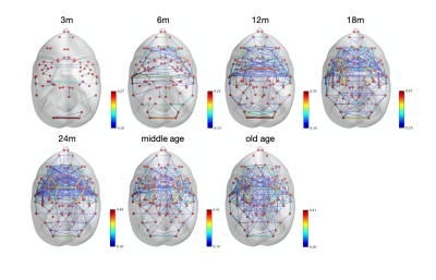 |
Life-span development of brain functional connectivity in common marmosets
Rina Ito1,2, Yuji Komaki2, Fumiko Seki2, Mayu Iida1,2, Mitsuki Rikitake1,2, Marin Nishio1,2, Junichi Hata1,3, and Takako Shirakawa1
1Department of Radiological Sciences, Human Health Sciences, Tokyo Metropolitan University, Tokyo, Japan, 2Live imaging Center, Central Institute for Experimental Animals, Kanagawa, Japan, 3Jikei University Graduate School of Medicine, Tokyo, Japan
Resting-state functional MRI enables to assess the psycho-neurological disease and developmental disability. The purpose of this study was finding the trend of functional connectivity life-span development in healthy common marmosets. We verified that the number of resting-state networks and its strength increased dramatically with age until common marmosets became adults (the age of 24 months) and declined steadily at older age after 24 months. The results demonstrated mostly the same data as human; therefore, it could apply as control models comparing with psycho-neurological disorder models in further research.
|
|||
2937.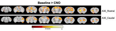 |
Suppression of the default mode network in mouse affects memory consolidation
Zengmin Li1, Dilsher Athwal1, and Kai-Hsiang Chuang1,2
1Queensland Brain Institute, The University of Queensland, St Lucia, Australia, 2Centre for Advance Imaging, The University of Queensland, Brisbane, Australia
|
|||
2938.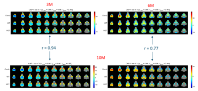 |
Resting-state co-activation patterns (CAPs) accurately predict pre- and manifest- stage Huntington’s disease in mice.
Mohit H Adhikari1, Tamara Vasilkovska1, Dorian Pustina2, Longbin Liu2, Roger Cachope2, Haiying Tang2, Celia Dominguez2, Ignacio Munoz-Sanjuan2, Annemie Van der Linden1, and Marleen Verhoye1
1Biomedical Sciences, University of Antwerp, Antwerp, Belgium, 2CHDI Foundation, Princeton, NJ, United States
Magnetic Resonance Imaging (MRI) biomarkers hold great potential to become key in understanding Huntington’s Disease (HD) pathophysiology as well as disease progression. We investigated longitudinal changes in spatiotemporal properties of transient brain-wide co-activation patterns (CAPs) during the brain’s resting-state (RS) in a mouse model of HD. We found early differences in the temporal components of two biologically prominent CAPs in the diseased mice and showed, using supervised learning, that spatial features of CAPs accurately distinguish the diseased animals from healthy. Our findings show the promise of RS-CAPs in the development of MRI-based biomarkers of HD.
|
|||
2939.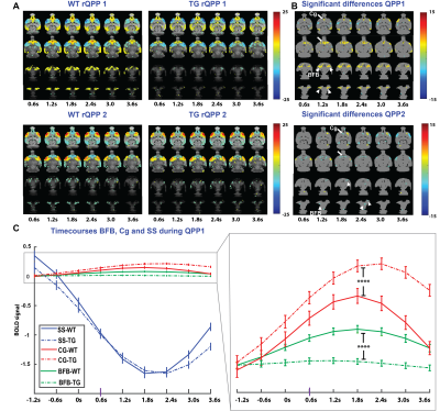 |
Spatiotemporal aberrations in resting-state quasi-periodic patterns in 4-month-old TgF344-AD rats
Monica van den Berg1, Mohit Adhikari1, Georgios A. Keliris1, and Marleen Verhoye1
1Bio-Imaging Lab, University of Antwerp, Wilrijk, Belgium
To identify spatiotemporal network alterations at an early stage of AD, we’ve performed rsfMRI and analysed quasi-periodic patterns in 4-month old TgF344-AD rats. We observed spatiotemporal alterations in whole brain network activity, mainly in the basal forebrain and cingulate cortex. Moreover, occurrences of QPPs were significantly lower in TgF344-AD rats. Co-activation of basal forebrain and cingulate cortex, present in wild-type rats, was greatly reduced in TgF344-AD rats. These results highlight the important role of the BFB in orchestrating brain networks and indicate a potential signature to identify early onset changes at the network level.
|
|||
2940.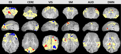 |
Detecting functional connectivity changes in a pig traumatic brain injury model using resting-state fMRI
Wenwu Sun1, Kelly M. Scheulin2, Sydney E. Sneed2, Madison M. Fagan2, Savannah R. Cheek2, Christina B. Welch2, Morgane E. Golan2, Frankin D. West2, and Qun Zhao1
1Department of Physics and Astronomy, University of Georgia, Athens, GA, United States, 2Regenerative Bioscience Center, University of Geogia, Athens, GA, United States
Functional magnetic resonance imaging (fMRI) has great potential to evaluate how networks respond and compensate for network dysfunction caused by traumatic brain injury (TBI). In this study, sparse dictionary learning (sDL) and independent component analysis (ICA) were applied to resting-state fMRI (rs-fMRI) data, collected from a group of piglets 1-day (D1) and 7-days (D7) after TBI. Activation maps were generated using group ICA and group sDL, both with dual regression. Voxel-wise permutation tests were then applied to identify changes to six resting-state networks (RSNs). Consistency was observed through the two methods, indicating functional network activity changes after injury.
|
|||
 |
2941.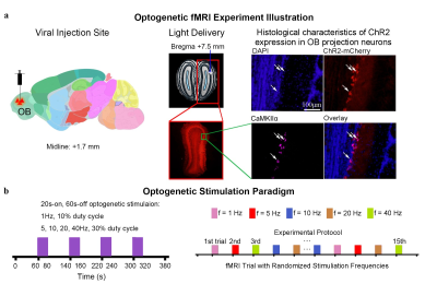 |
Optogenetic fMRI interrogation of the olfactory system
Teng Ma1,2,3, Xunda Wang1,2, Eddie C. Wong1,2, Pit Shan Chong4, Sanchal Sanchayyan4, Lee Wei Lim4, Pek-Lan Khong3, Ed X. Wu1,2, and Alex T. L. Leong1,2
1Laboratory of Biomedical Imaging and Signal Processing, The University of Hong Kong, Hong Kong SAR, China, 2Department of Electrical and Electronic Engineering, The University of Hong Kong, Hong Kong SAR, China, 3Department of Diagnostic Radiology, Li Ka Shing Faculty of Medicine, The University of Hong Kong, Hong Kong SAR, China, 4School of Biomedical Sciences, Li Ka Shing Faculty of Medicine, The University of Hong Kong, Hong Kong SAR, China
The olfactory system is one of the major sensory systems and has been often linked to numerous non-olfactory regions pivotal for sensory perception and higher-order cognition. However, the extent of where olfactory signals are distributed brain-wide remains poorly described. By optogenetically stimulating the excitatory projection neurons in olfactory bulb (OB), we interrogated the brain-wide functional organization of the olfactory system using fMRI. We revealed that OB neural activity propagated to regions that are associated with higher-order cognition, reward-related behaviors and multisensory processing.
|
||
2942.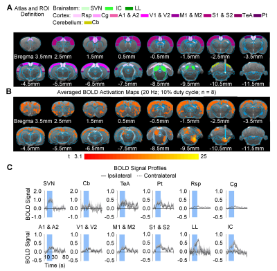 |
Brain-wide functional organization of the central vestibular pathways: an optogenetic fMRI study
Eddie C. Wong1,2, Teng Ma1,2,3, Xunda Wang1,2, Pek-Lan Khong3, Ed X. Wu1,2, and Alex T.L. Leong1,2
1Laboratory of Biomedical Imaging and Signal Processing, The University of Hong Kong, Hong Kong SAR, China, 2Department of Electrical and Electronic Engineering, The University of Hong Kong, Hong Kong SAR, China, 3Department of Diagnostic Radiology, Li Ka Shing Faculty of Medicine, The University of Hong Kong, Hong Kong SAR, China
The vestibular system is essential to our sense of balance and spatial orientation. fMRI mapping of the vestibular system has been challenging due to the physical constraints limiting a subject’s ability to perform motion, balance, and orientation related tasks within an MRI scanner. As such, our present knowledge of the brain-wide cortical and subcortical regions that participate in processing the vestibular sense is scarce. Here, we combined fMRI and optogenetic stimulation of vestibular excitatory neurons to visualize two distinct brain-wide functional organization of central vestibular pathways originating from two major vestibular subnuclei, the superior vestibular nucleus and medial vestibular nucleus.
|
|||
2943.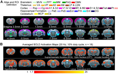 |
The role of central vestibular system in circadian rhythms?
Alex T. L. Leong1,2, Christopher Man1,2, Yong Gu3, and Ed X. Wu1,2
1Laboratory of Biomedical Imaging and Signal Processing, The University of Hong Kong, Hong Kong SAR, China, 2Department of Electrical and Electronic Engineering, The University of Hong Kong, Hong Kong SAR, China, 3Institute of Neuroscience, CAS Center for Excellence in Brain Science and Intelligence Technology, Chinese Academy of Sciences, Shanghai, China
The vestibular system is critical for bodily functions, yet it remains an underappreciated sense. Recently, seminal studies of the prolonged exposure to microgravity in space showed significant alteration in vestibular activity and circadian rhythms. However, little is known regarding the role of the central vestibular system in regulating the circadian clock. Here, we deploy fMRI and optogenetic stimulation of vestibular excitatory neurons to visualize the neural targets of the vestibular system in circadian circuits. We reveal that the central vestibular system provides inputs to the suprachiasmatic nucleus, optic nerve and locus coeruleus, notable regions that drive and regulate circadian rhythms.
|
|||
2944.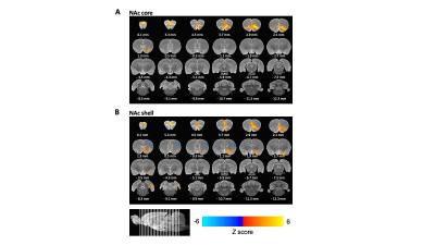 |
Direct Mapping of the Nucleus Accumbens Core and Shell using Deep Brain Stimulation with functional Magnetic Resonance Imaging in rats
Hyeon-Joong Kim1, Ryan S Clement2, Roger B Bagwell2, Tirko N Natasha2, Yen-Yu Ian Shih1, and Sung-Ho Lee1
1Center for Animal MRI, Biomedical Research Imaging Center, University of North Carolina, Chapel Hill, NC, United States, 2Actuated medical, Bellefonte, PA, United States
Nucleus accumbens (NAc) is known as the backbone of the reward circuit, which the core and shell appear to be dominant. To directly map their distinct functional connectivity structure, we employed simultaneous Deep Brain Stimulation (DBS) at NAc during functional Magnetic Resonance Imaging (fMRI) and demonstrated the difference of the functional organization between core and shell. MR compatible 16ch microprobe was utilized to stimulate multiple regions during single scan session. We expect this technique could be useful to dissect the therapeutic circuit of DBS in NAc in the preclinical approach.
|
|||
2945.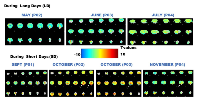 |
Hypercapnic challenge, BOLD fMRI and immunohistochemistry to examine the in-vivo presence of adult neurogenesis in the sheep hypothalamus
Pierre-Marie Chevillard1, Martine Migaud1, and Nathalie Just1
1NhyRVana, INRAE, Nouzilly, France
In the present work, we used hypercapnia measured with BOLD fMRI in ewes responding to photoperiodism to obtain a potential in-vivo marker of the presence of adult neurogenesis in their hypothalamus. Immunohistochemical methods were also used to analyse the relationships between blood vessels and Sox2 stained neural stem cells.
|
The International Society for Magnetic Resonance in Medicine is accredited by the Accreditation Council for Continuing Medical Education to provide continuing medical education for physicians.