Digital Posters
High-Resolution fMRI
ISMRM & SMRT Annual Meeting • 15-20 May 2021

| Concurrent 3 | 19:00 - 20:00 |
3365.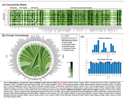 |
Functional connectome of arousal and motor brainstem nuclei using 7 Tesla resting-state fMRI in living humans
Kavita Singh1, Simone Cauzzo1,2, Maria Guadalupe Garcia Gomar1, Matthew Stauder1, Nicola Vanello3, Claudio Passino2,4, and Marta Bianciardi1
1Brainstem Imaging Laboratory, Department of Radiology, Athinoula A. Martinos Center for Biomedical Imaging, Boston, MA, United States, 2Sant'Anna School of Advanced Studies, Institute of Life Sciences, Pisa, Italy, 3Dipartimento di Ingegneria dell’Informazione, University of Pisa, Pisa, Italy, 4Fondazione Toscana Gabriele Monasterio, Pisa, Italy
For a holistic understanding of sleep, arousal and associated motor processes, we investigated the resting-state functional connectivity of 18 arousal and motor brainstem nuclei in living humans by the use of high spatial-resolution 7 Tesla resting-state fMRI, as well as a recently developed in-vivo probabilistic atlas of these nuclei in standard space. Further, we verified the translatability of our brainstem connectome approach to conventional (e.g. 3 Tesla) fMRI. Results provided comprehensive and augmented brainstem-brain connectome to understand mechanisms of arousal-motor function in health and disease conditions and its translatability in clinical settings.
|
|||
3366.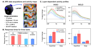 |
Motor preparatory inhibition is reflected as a layer dependent suppression in the human primary motor cortex
Yinghua Yu1,2, Ikuhiro Kida1,2, and Nobuhiro Hagura1,2
1Center for Information and Neural Networks, National Institute of Information and Communications Technology, Osaka, Japan, 2Graduate School of Frontier Biosciences, Osaka University, Osaka, Japan
In humans, corticospinal excitability is reduced when preparing for an action; i.e. motor preparatory inhibition. Functional relevance of this inhibition has been proposed, however, the site where the actual inhibition occurs, either at the primary motor cortex (M1) or at the downstream spinal-circuit, is still unclear. By measuring vascular space occupancy (VASO) with 7T fMRI, we investigated the direct signature of preparatory inhibition of neural activity across M1 cortical layers. Our preliminary analysis showed that preparation was associated with a reduced activity of the superficial layers of M1. Motor preparatory inhibition likely takes place in M1 in a layer-dependent manner.
|
|||
3367. |
Investigating in-vivo function of Zebrin-II stripes of the cerebellum using tactile stimulation and prediction.
Lenno R. P. T. Ruijters1, Nikos Priovoulos1, and Wietske van der Zwaag1
1Spinoza Centre for Neuroimaging, Amsterdam, Netherlands
The functional organization of the cerebellar cortex is largely unexplored in-vivo. However, connectivity studies suggest it is organized in successive Zebrin-II positive and negative stripes that receive descending signals from the cerebrum and ascending input from the peripheral nervous system respectively. To examine this stripe-based functional organization we used a tactile-stimulation experiment with actual (bottom-up) and predicted (top-down) stimulation at 7T. This paradigm gives reliable responses to predicted and experienced stimuli. Our fMRI results suggest that there is a spatial separation in the cerebellum between these two processes in line with the hypothesised relation between function and Zebrin-II stripes.
|
|||
3368. |
Temporal deviant detection in human auditory cortex using high-field fMRI
Miriam Heynckes1, Elia Formisano1,2, Peter De Weerd1, and Federico De Martino1
1Faculty of Psychology and Neuroscience, Maastricht University, Maastricht, Netherlands, 2Maastricht Centre for Systems Biology, Maastricht University, Maastricht, Netherlands
Layer-fMRI probes the mesoscopic neural architecture in humans non-invasively. We studied the behavioral relevance of layer-specific processing by investigating temporal deviance detection in the auditory system. Preliminary results indicate increased response in superficial layers to a temporal deviant, in line with a feedback signal entering superficial layers during deviant detection.
|
|||
3369.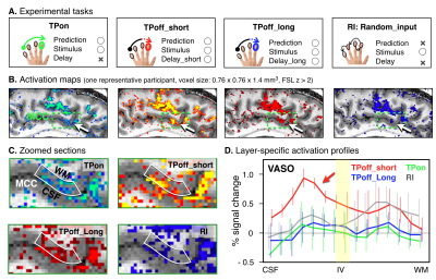 |
Layer-specific activation of prediction in the human midcingulate cortex
Jiajia Yang1,2, Masaki Fukunaga3, Yinghua Yu2,4, Laurentius Huber5, Peter A Bandettini2, and Norihiro Sadato3
1Graduate School of Interdisciplinary Science and Engineering in Health Systems, Okayama University, Okayama, Japan, 2Section on Functional Imaging Methods, National Institute of Mental Health, Bethesda, MD, United States, 3Division of Cerebral Integration, National Institute for Physiological Sciences, Okazaki, Japan, 4Center for Information and Neural Networks, National Institute of Information and Communications Technology, Suita, Japan, 5Department of Cognitive Neuroscience, Maastricht University, Maastricht, Netherlands
The human brain is constantly generating and updating predictions. The midcingulate cortex (MCC) is one of the areas that contribute to prediction processing, while MCC layer-specific function remains unknown. In the present study, we used a tactile index finger prediction task that consists of sequential finger poking during high-resolution (0.76mm) BOLD and VASO fMRI at 7T to investigate how the prediction activity changes across layers in the human MCC. We found the double-peak activity feature across MCC layers for all prediction tasks, and the enhanced activity within MCC superficial layers may reflect the prediction error processing.
|
|||
3370.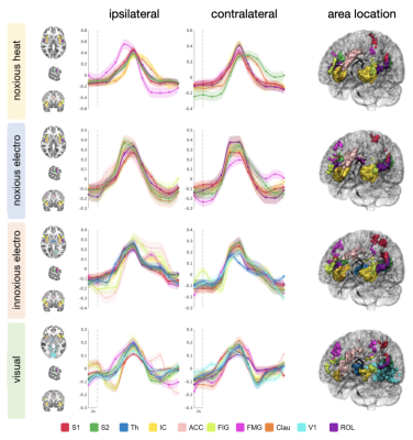 |
Where is the Pain in the Brain? Functional MRI of Saliency versus Nociception at 7.0 Tesla en route to Diagnostic Biomarkers of Pain
Gijs J. Heij1,2, Joao Periquito1, Haopeng Han1, Antje Els1, Thoralf Niendorf1,3, and Henning M. Reimann1
1Berlin Ultrahigh Field Facility (B.U.F.F.), Max Delbrück Center for Molecular Medicine in the Helmholtz Association, Berlin, Germany, 2Spinoza Centre for Neuroimaging, Amsterdam, Netherlands, 3Experimental and Clinical Research Center (ECRC), a joint cooperation between the Charité Medical Faculty and the Max Delbrück Center for Molecular Medicine in the Helmholtz Association, Berlin, Germany
Identifying diagnostic biomarkers of pain is of utmost clinical importance. This is challenging since similar fMRI patterns are elicited by painful and equisalient non-painful stimuli at 3T. To decipher pain-specificity this work uses 7T high-resolution fMRI of (1) salient non-painful versus painful stimuli, (2) different types of painful stimuli in "healthy" controls versus (3) a subject with a genetically-induced pain-free phenotype. We found that canonical HRF modeling is insufficient to reliably capture the shape of elicited BOLD responses that further varied in correlation to perceived saliency.
|
|||
3371.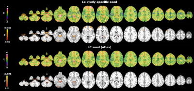 |
In vivo mapping of human locus coeruleus functional connectivity at 7T: a feasibility study
Michela Pievani1, Ileana O. Jelescu2, Joao Jorge3, Olivier Reynaud4, Federica Ribaldi1,5,6, Valentina Garibotto7, Giovanni B. Frisoni1,5, and Jorge Jovicich8
1Laboratory of Alzheimer’s Neuroimaging and Epidemiology, IRCCS Istituto Centro San Giovanni di Dio Fatebenefratelli, Brescia, Italy, 2CIBM - Center for Biomedical Imaging, Ecole Polytechnique Fédérale de Lausanne, Lausanne, Switzerland, 3Systems Division, Swiss Center for Electronics and Microtechnology (CSEM), Nêuchatel, Switzerland, 4Human Neuroscience Platform, Fondation Campus Biotech Geneva, Geneva, Switzerland, 5Memory Clinic and LANVIE - Laboratory of Neuroimaging of Aging, University Hospitals and University of Geneva, Geneva, Switzerland, 6Department of Molecular and Translational Medicine, University of Brescia, Brescia, Italy, 7Division of Nuclear Medicine and NIMTlab, University Hospitals and University of Geneva, Geneva, Switzerland, 8Center for Mind/Brain Sciences, University of Trento, Trento, Italy
The locus coeruleus (LC) is a brainstem nucleus whose functional disruption may be an early signature of Alzheimer’s disease. Potentially due to its small size, mixed results exist about its functional connectivity to core memory, attention and salience networks. This limits a baseline definition for patient studies. Here we use high-resolution high-field resting-state fMRI to investigate the pattern of LC connectivity in healthy young subjects. Preliminary findings show positive correlations with the cerebellum and the frontal cortex. The default mode and frontoparietal networks, but not the salience network, show FC with the brainstem. Data acquisition is ongoing.
|
|||
3372.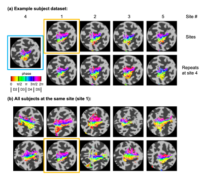 |
Multi-centre, multi-vendor 7 Tesla fMRI reproducibility of hand digit representation in the human somatosensory cortex
Ian D Driver1, Rosa M Sanchez Panchuelo2, Olivier Mougin2, Michael Asghar2, James Kolasinski1, William T Clarke3, Catarina Rua4, Andrew T Morgan5, Adrian Carpenter4, Keith Muir5, David Porter5, Christopher T Rodgers4, Stuart Clare3,
Richard G Wise1,6,7, Richard Bowtell2, and Susan T Francis2
1Cardiff University Brain Research Imaging Centre, School of Psychology, Cardiff University, Cardiff, United Kingdom, 2Sir Peter Mansfield Imaging Centre, University of Nottingham, Nottingham, United Kingdom, 3Wellcome Centre for Integrative Neuroimaging, University of Oxford, Oxford, United Kingdom, 4Wolfson Brain Imaging Centre, Department of Clinical Neurosciences, University of Cambridge, Cambridge, United Kingdom, 5Imaging Centre of Excellence, University of Glasgow, Glasgow, United Kingdom, 6Department of Neuroscience, Imaging and Clinical Sciences, "G. D’Annunzio University" of Chieti-Pescara, Chieti, Italy, 7Institute for Advanced Biomedical Technologies, "G. D’Annunzio University" of Chieti-Pescara, Chieti, Italy
This study compares within- and across-site reproducibility of 7 Tesla somatomotor fMRI using a travelling wave task to map the hand and individual digit localization at high spatial resolution. We performed a “travelling-heads” study acquiring data on Siemens Magnetom, Siemens Terra and Philips Achieva whole-body 7 Tesla MRI systems at five sites. Simple harmonization of the EPI sequence protocol was performed. We show metrics of intra- and inter-site variability of digit representations, with good reproducibility observed across sites. These results demonstrate the potential benefits of multi-site 7T fMRI studies for digit mapping where large cohorts are required.
|
|||
3373.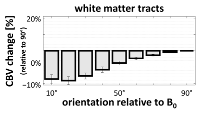 |
Dependence of CBV estimated from 7T Dynamic Susceptibility Contrast data on white matter tract and cortical gray matter orientation relative to B0
Jonathan R. Polimeni1,2, Olivia M. Viessmann1, Qiyuan Tian1, Michaël Bernier1, Meher R. Juttukonda1, Yi-Fen Yen1, and David H. Salat1,3
1Athinoula A. Martinos Center for Biomedical Imaging, Massachusetts General Hospital/Harvard Medical School, Charlestown, MA, United States, 2Division of Health Sciences and Technology, Massachusetts Institute of Technology, Cambridge, MA, United States, 3Neuroimaging Research for Veterans Center, VA Boston Healthcare System, Boston, MA, United States
Previous studies have reported an orientation effect in DSC data within the white matter in which CBV estimates vary systematically with the orientation of local fiber orientation. Other studies have reported similar orientation effects in BOLD fMRI data both with respect to white-matter fiber orientation and gray-matter cortical orientation. Here we extend these findings by investigating orientation effects in gray and white matter in DSC-based CBV estimates. We find strong effects in both brain regions consistent with both groups of prior studies, corroborating previous interpretations that the observed BOLD effects in the white matter are vascular in origin.
|
|||
3374.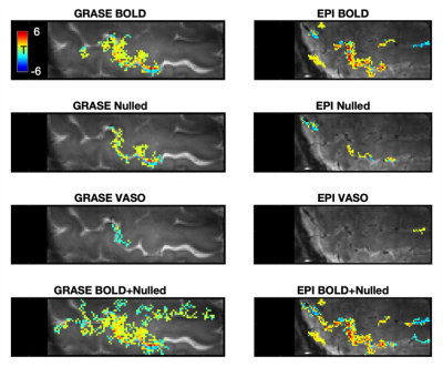 |
Combining BOLD and CBV for enhanced fMRI CNR
An Thanh Vu1,2, Alexander JS Beckett3,4, Jennifer Townsend3,4, Salvatore Torrisi3,4, and David Feinberg3,4
1Radiology, University of California, San Francisco, San Francisco, CA, United States, 2San Francisco Veteran Affairs Health Care System, San Francisco, CA, United States, 3Advanced MRI Technologies, Sebastopol, CA, United States, 4Helen Wills Neuroscience Institute, University of California, Berkeley, Berkeley, CA, United States
In this 7T study, we investigate the constructive combination of the BOLD and CBV weighted timeseries of EPI and GRASE based Vascular Space Occupancy (VASO) sequences for enhanced fMRI CNR. Activations from this BOLD+CBV combination resulted in 9% greater t-values as well as, on average, 39% more above-threshold voxels, relative to traditional BOLD. Although this combination method resulted in reduced microvascular specificity for EPI based VASO, for GRASE based VASO, the middle layers were predominantly enhanced, suggesting minimal loss in specificity. Advantageously, this technique does not require re-acquisition of data or modifications to pulse sequence parameters of the VASO sequence.
|
|||
3375.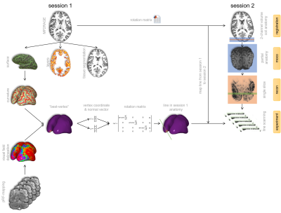 |
Combining functional and structural information for optimal planning during ultrahigh temporal resolution line-scanning
Jurjen Heij1, Luisa Raimondo1, Jeroen C.W. Siero1,2, Serge O Dumoulin1,3, Wietske van der Zwaag1, and Tomas Knapen1,3
1Spinoza Centre for Neuroimaging, Amsterdam, Netherlands, 2Radiology, University Medical Centre Utrecht, Utrecht, Netherlands, 3Experimental and Applied Psychology, VU University, Amsterdam, Netherlands
Depth-resolved fMRI is an emerging field growing in popularity given the potential of separating feedforward from feedback signals. When employing line-scanning methods, we sacrifice coverage in order to sample BOLD-responses with ultra-high temporal and spatial resolution. With a limited field-of-view being targeted, one needs to know precisely where to place the line. Here we describe a multi-session approach that combines functional and structural information for an optimal acquisition of line-scanning data based on user-defined properties. We show its feasibility of obtaining hemodynamic response functions of a specific cortical patch.
|
|||
3376.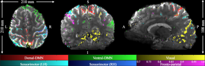 |
Mapping of Whole-brain Resting-State Networks with Half-millimetre Resolution using TR-external EPIK at 7T
Seong Dae Dae Yun1, Patricia Pais-Roldán1, Nicola Palomero-Gallagher2,3,4, and N. Jon Shah1,5,6,7
1Institute of Neuroscience and Medicine 4, INM-4, Forschungszentrum Juelich, Juelich, Germany, 2Institute of Neuroscience and Medicine 1, INM-1, Forschungszentrum Juelich, Juelich, Germany, 3C. & O. Vogt Institute for Brain Research, Heinrich-Heine-University, Duesseldorf, Germany, 4Department of Psychiatry, Psychotherapy and Psychosomatics, Medical Faculty, RWTH Aachen, Aachen, Germany, 5Institute of Neuroscience and Medicine 11, INM-11, JARA, Forschungszentrum Juelich, Juelich, Germany, 6JARA - BRAIN - Translational Medicine, Aachen, Germany, 7Department of Neurology, RWTH Aachen University, Aachen, Germany
Mapping of resting-state networks has been performed in numerous studies to investigate overall brain function and its underlying connectivity. However, attempts to acquire resting-state fMRI data with high spatial resolution have been hampered by the current technical limitations. The spatial resolution from recent submillimetre-resolution fMRI studies still remains around 0.7 × 0.7 × 0.7 mm3, with only partial brain coverage. This work employs a novel high-resolution fMRI method, TR-external EPIK, to perform resting-state fMRI with half-millimetre in-plane resolution and whole-brain coverage at 7T. Various resting-state networks over the whole-brain have been identified with high mapping fidelity onto the cortical ribbon.
|
|||
3377.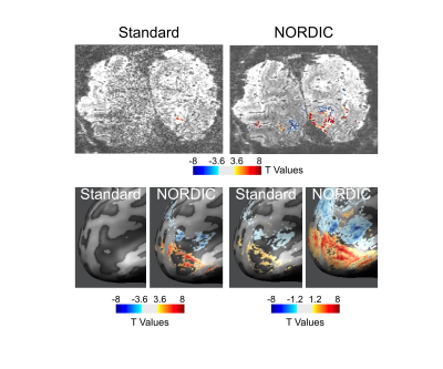 |
New Frontiers of Human Neuroscience: 0.5 mm isotropic 7T in Vivo Human fMRI
Luca Vizioli1,2, Logan T Dowdle1, Steen Moeller1, Essa Yacoub1, and Kamil Ugurbil1
1CMRR, University of Minnesota, minneapolis, MN, United States, 2Department of Neurosurgery, University Of Minnesota, minneapolis, MN, United States
The goal of 0.1μL voxel resolution for human fMRI set by the BRAIN Initiative has thus far been nearly impossible to reach, due to the overwhelming thermal noise contribution at such high spatial resolutions. Here we show that, with the recently developed denoising algorithm NORDIC, we are able to achieve robust functional activation to visual stimuli at 0.5 mm isotropic (0.125μL) voxel resolution at 7T field strengths with only 20 minutes of data, a results that is unobtainable with conventional data reconstruction. Preliminary layer findings show a central peak, suggesting that this may dramatically improve quantification of depth-dependent activation profiles.
|
|||
3378.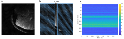 |
Multi-echo line-scanning for ultra-high spatiotemporal resolution: optimal settings for BOLD sensitivity enhancement
Luisa Raimondo1, Jurjen Heij1, Tomas Knapen1,2, Serge O. Dumoulin1,3, Jeroen C.W. Siero1,4, and Wietske van der Zwaag1
1Spinoza Centre for Neuroimaging, Amsterdam, Netherlands, 2VU University, Amsterdam, Netherlands, 3Experimental and Applied Psychology, VU University, Amsterdam, Netherlands, 4Radiology, University Medical Centre Utrecht, Utrecht, Netherlands
We present multi-echo line-scanning fMRI results in humans. The potential of this technique lies in the combination of both high spatial and temporal resolution while sacrificing spatial coverage outside the region of interest. We reached a 250 μm resolution along the line direction with a temporal resolution of ~100 ms. We compared BOLD sensitivity and tSNR of 5 different multi-echo acquisitions to select the optimal protocol, and three methods for echo combination. Although differences were small, the 5 echo protocol and tSNR-weighted combination were found to yield the highest BOLD sensitivity in a visual cortex ROI.
|
|||
3379.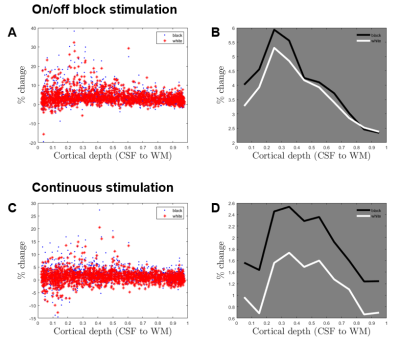 |
Laminar analysis of luminance-dependent visual activation in human V1 with voxel centroid mapping method
Hankyeol Lee1, Seong-Gi Kim1,2, and Kâmil Uludağ1,2,3
1Center for Neuroscience Imaging Research, Institute for Basic Science, Suwon, Korea, Republic of, 2Department of Biomedical Engineering, Sungkyunkwan University, Suwon, Korea, Republic of, 3Techna Institute & Koerner Scientist in MR Imaging, University Health Network, Toronto, ON, Canada
Luminance-dependent laminar activation analysis in human V1 is presented. White and black dot-shaped visual stimuli were used to evoke BOLD responses in the early visual cortex. Traditional on/off block stimulation paradigm and continuous stimulation paradigm without interleaved rest periods were used. With voxel centroid mapping, where each voxel’s relative cortical depth is estimated, activation ROIs across multiple image slices and subjects can be analyzed collectively with minimum smoothing and no resampling of functional data. Results show black-dominant responses in V1, with laminar profiles from continuous stimulation emphasizing the role of middle layers by minimizing the effect of ascending vein drainage.
|
|||
3380.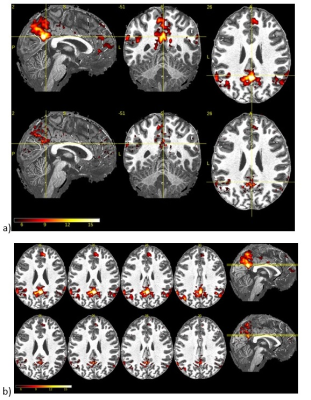 |
Optimization of simultaneous multislice (SMS) technique for submillimeter whole brain functional MRI at 7T
Baolian Yang1, Graeme McKinnon2, and Brice Fernandez3
1MR, GE Healthcare, Waukesha, WI, United States, 2GE Healthcare, Waukesha, WI, United States, 3GE Healthcare, Buc, France
Through the optimization of simultaneous multislice (SMS) technique, a 0.8mm3 whole brain resting state fMRI data with minimum distortion was acquired to show the benefit of doing fMRI study at ultra-high field.
|
|||
3381.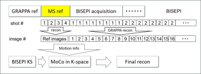 |
Shot-wise separate motion correction and ICA denoising for BISEPI high-resolution fMRI study at 7T
Guoxiang Liu1,2, Adnan Shah1,2, Takashi Ueguchi1,2, and Seiji Ogawa1,3
1CiNet, NICT, Osaka, Japan, 2Graduate School of Frontier Biosciences, Osaka University, Osaka, Japan, 3Tohoku Fukushi University, Sendai, Japan
Shot-wise separate motion correction is presented for BISEPI (Block-Interleaved Segmented EPI)-based high-resolution fMRI at 7T. Artifacts caused by subject motion during longer acquisition time of one volume than normal multi-shot EPI reduces the performance of BISEPI. The proposed k-space motion correction (KMoCo) method calculates and corrects the motion during the shot-wise acquisition of each volume separately. We also performed ICA denoising on the KMoCo data. The proposed method improves temporal SNR, the number of detected active voxels, and localization of brain activity in high-resolution fMRI at 7T
|
The International Society for Magnetic Resonance in Medicine is accredited by the Accreditation Council for Continuing Medical Education to provide continuing medical education for physicians.