Digital Posters
fMRI Engineering, Acquisition & Analysis
ISMRM & SMRT Annual Meeting • 15-20 May 2021

Digital Posters
fMRI Engineering, Acquisition & Analysis
| Concurrent 3 | 13:00 - 14:00 |
 |
2673.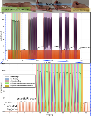 |
A custom MR-compatible data glove for fMRI of the human motor cortex at 7T
Shota Hodono1,2, Donald Maillet1, Jin Jin2,3, David Reutens1,2, and Martijn A. Cloos1,2
1Centre for Advanced Imaging, The University of Queensland, Brisbane, Australia, 2ARC Training Centre for Innovation in Biomedical Imaging Technology, The University of Queensland, Brisbane, Australia, 3Siemens Healthcare Pty Ltd, Brisbane, Australia
We present a custom-built MR-compatible data glove to capture hand motion during concurrent fMRI experiments at 7
|
||
2674.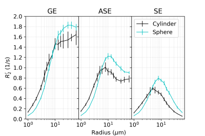 |
Creating a robust BOLD fMRI phantom using microspheres: How well do microspheres approximate microvasculature?
Jacob Chausse1, Avery J. L. Berman2, Jonathan R. Polimeni2, and J. Jean Chen1,3
1Rotman Research Institute, Baycrest Health Sciences, North York, ON, Canada, 2A. A. Martinos Center for Biomedical Imaging, Harvard Medical School, Massachusetts General Hospital, Boston, MA, United States, 3Department of Medical Biophysics, University of Toronto, Toronto, ON, Canada
BOLD phantoms are important for the evaluation of fMRI acquisition techniques, but validating numerical simulations based on conventional random-cylinder models of the microvasculature is difficult because available phantoms lack the microstructure required to emulate these geometries. In comparison, a phantom using randomly positioned microspheres is a more feasible alternative, but it is still unclear how well microspheres approximate cylinders in their transverse relaxation behaviour. In this work, we use 3D Monte Carlo simulations to compare the behaviour of the two perturber types at 3 T.
|
|||
2675.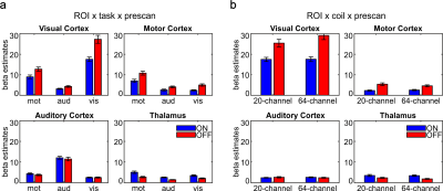 |
Recommendations of choice of head coil and prescan normalize filter depend on region of interest and task
Tina Schmitt1 and Jochem Rieger1,2,3,4
1Neuroimaging Unit, School of Medicine and Health Sciences, Carl-von-Ossietzky University of Oldenburg, Oldenburg, Germany, 2Neuroimaging Unit, Carl-von-Ossietzky University of Oldenburg, School of Medicine and Health Sciences, Oldenburg, Germany, 3Applied Neurocognitive Psychology, Department of Psychology, School of Medicine and Health Sciences, Carl-von-Ossietzky University of Oldenburg, Oldenburg, Germany, 4Cluster of Excellence “Hearing4all”, Carl-von-Ossietzky University of Oldenburg, Oldenburg, Germany
The performance of the 20- and the 64-channel head coil and the influence of the prescan normalize filter was evaluated in a standard fMRI setting using a 3T Siemens Prisma MRI. Larger beta estimates and tSNR occurred in auditory cortex and thalamus with prescan normalize, in contrast to the visual and motor cortex with larger beta estimates and tSNR without prescan normalize. In line with the results of the MRIQC tool, the 20-channel head coil with prescan normalize is better suitable for functional measurements, especially in deeper brain areas. The 64-channel head coil is the best choice for anatomical scans.
|
|||
2676.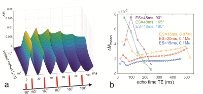 |
The vessel size specificity and sensitivity of rapid CPMG sequences in functional BOLD imaging
Klaus Scheffler1,2, Joern Engelmann1, and Rahel Heule1,2
1Max Planck Institute for Biological Cybernetics, Tuebingen, Germany, 2University of Tuebingen, Tuebingen, Germany
For reduced refocusing flip angles, the peak of the vessel size sensitivity curve is shifting towards larger radii with increasing echo time. The BOLD sensitivity is largely independent of the refocusing flip angle down to about 40°. CPMG or GRASE can be used with low refocusing flip angles without significant loss of sensitivity to BOLD and without the need for centric reordering. Signals acquired before or after the spin echo time point show contributions from larger vessels similar to gradient echo sequences. This effect is reduced for longer echo times.
|
|||
2677. |
Coherence-resolved Looping Star – improvements for silent multi-gradient echo structural and functional neuroimaging
Nikou Louise Damestani1, David John Lythgoe1, Ana Beatriz Solana2, Brice Fernandez3, Steven Charles Rees Williams1, Fernando Zelaya1, and Florian Wiesinger2
1Department of Neuroimaging, King's College London, London, United Kingdom, 2ASL Europe, GE Healthcare, München, Germany, 3ASL Europe, GE Healthcare, Paris, France
We have previously reported acoustically silent functional MRI using a novel pulse sequence known as Looping Star, which has been successfully implemented across distinct paradigms in spite of limitations to image quality and signal-to-noise. Here, we present a recent important improvement of Looping Star and demonstrate its robust performance for both functional imaging (in terms of visual BOLD fMRI) and structural imaging (in terms of T2*-weighted and quantitative susceptibility mapping).
|
|||
2678.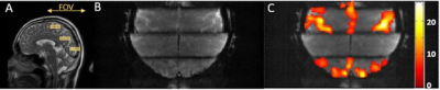 |
Simultaneous Multi-Segment (SMSeg) EPI for Functional MRI
Kaibao Sun1, Zheng Zhong1,2, Muge Karaman1,2, Qingfei Luo1, and Xiaohong Joe Zhou1,2,3
1Center for MR Research, University of Illinois at Chicago, Chicago, IL, United States, 2Department of Bioengineering, University of Illinois at Chicago, Chicago, IL, United States, 3Departments of Radiology and Neurosurgery, University of Illinois at Chicago, Chicago, IL, United States
Reduced field-of-view (rFOV) imaging offers several advantages, including decreased image distortion and high spatial resolution. We describe a simultaneous multi-segment (SMSeg) imaging method to extend the benefits of rFOV to multi-segment imaging in multiple focal regions. SMSeg was implemented using a 2D RF pulse whose multiple spatial response replicates excited different focal regions of interest, followed by GRAPPA reconstruction utilizing the coil spatial sensitivity variations in two orthogonal spatial directions. We have applied the SMSeg imaging technique to brain fMRI to visualize activations in multiple areas with minimal image distortion.
|
|||
2679.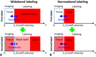 |
Ultra-fast arterial spin labeling with narrow-band velocity-selective (nb-VS) labeling
Jia Guo1
1Bioengineering, University of California Riverside, Riverside, CA, United States
We proposed a novel strategy to improve the temporal resolution and/or SNR efficiency of perfusion imaging using velocity-selective (VS) labeling, by purposely labeling spins within a narrow velocity band. This strategy allows faster recovery/refreshment of the magnetization of arterial spins for improved SNR efficiency and temporal resolution. A few implementation methods of such labeling strategy were explored, using modified Fourier-transform based VS inversion pulses. The SNR efficiency and achievable temporal resolution were examined by ASL signal modeling, demonstrating a good promise for ultra-fast perfusion imaging with high SNR efficiency.
|
|||
2680.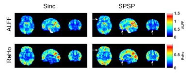 |
Improved resting-state fMRI with through-plane phase precompensated spectral-spatial pulses
Christopher Sica1, Hairong Chen2, Guangwei Du2, Sangam Kanekar1, Jianli Wang1, Qing X Yang3, and Prasanna Karunanayaka1
1Radiology, Penn State College of Medicine, Hershey, PA, United States, 2Neurology, Penn State College of Medicine, Hershey, PA, United States, 3Neurosurgery, Penn State College of Medicine, Hershey, PA, United States
fMRI scans with long echo times are vulnerable to signal loss due to through-plane dephasing caused by susceptibility field gradients (SFG), particularly near the air-tissue interface of the sinus regions. Resting state fMRI techniques that sample networks with connectivity through these artifact-plagued regions could be compromised. Several approaches have been proposed for susceptibility compensation, with one promising approach based upon the use of phase-precompensated spectral-spatial RF pulses. These RF pulses pre-phase the magnetization to cancel out the dephasing caused by SFG’s. Here, we evaluated the ability of spectral-spatial pulses to improve detection of resting state fMRI activity.
|
|||
2681.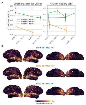 |
Functional imaging of the anterior temporal lobe: effects of acceleration, multi-echo acquisition, and reconstruction methods
Matteo Visconti di Oleggio Castello1, Valentina Borghesani2, Katherine P. Rankin3, Jack L. Gallant1, and An T. Vu3,4
1University of California, Berkeley, Berkeley, CA, United States, 2Université de Montréal, Montréal, QC, Canada, 3University of California, San Francisco, San Francisco, CA, United States, 4San Francisco Veteran Affairs Health Care System, San Francisco, CA, United States
The anterior temporal lobe (ATL) is an important brain structure that is hard to functionally image with EPI sequences due to signal dropout and low SNR. To address this problem, we developed a whole-brain EPI sequence with increased SNR in the ATL. We evaluated single- and multi-echo sequences while varying in-plane and multiband acceleration with different reconstruction methods. Sequence evaluation was based on tSNR and functional metrics from computational neuroscience (explainable variance and encoding model performance). The highest functional SNR in ATL and across the cortex was provided by multi-echo sequences with moderate iPAT and MB acceleration, and LeakBlock reconstruction.
|
|||
2682. |
Development of an optimized approach to spinal cord fMRI based on the combination of an ad hoc acquisition method and data analysis pipeline
Michela Fratini1, Marta Moraschi2, Laura Maugeri1, Silvia Tommasin3, Mauro DiNuzzo2, Julien Cohen-Adad4, Fabio Mangini5, Daniele Mascali6, and Federico Giove2
1CNR-Nanotec, rome, Italy, 2Centro Ricerche Enrico Fermi, Rome, Italy, 3Sapienza University of Rome, Rome, Italy, 4Institute of Biomedical Engineering, Polytechnique Montreal, Montreal, QC, Canada, 5Santa Lucia Foundation, Rome, Italy, 6Centro Ricerche Enrico Fermi, rome, Italy
The spinal cord (SC) is the caudal extension of the Central Nervous System (CNS) and is responsible for several complex functions. Among imaging methods, functional Magnetic Resonance Imaging (fMRI) represents the most promising tool for non-invasive investigation of SC functions/dysfunctions. However, the utilization of SC-fMRI is widely under-exploited, either due to challenges in acquiring good quality data, or to the lack of dedicated analysis tools. In this study, we implemented an optimized experimental approach and defined a pipeline for SC-fMRI data analysis. We validate such pipeline and investigate the impact of acquisition direction on noise removal.
|
|||
2683.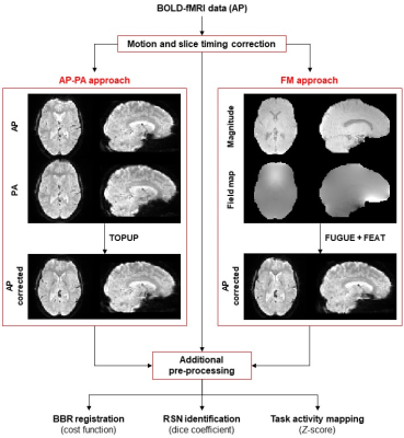 |
The impact of geometric distortions and their correction on fMRI data analyses
Rodolfo Abreu1, Miguel Castelo-Branco1,2, and João Valente Duarte1,2
1Coimbra Institute for Biomedical Imaging and Translational Research (CIBIT), Institute for Nuclear Sciences Applied to Health (ICNAS), University of Coimbra, Coimbra, Portugal, 2Faculty of Medicine, University of Coimbra, Coimbra, Portugal
The impact of geometric distortions on fMRI data analyses has been scarcely investigated, and a direct comparison between fMRI distortion correction approaches has not been performed so far. Here, we found that correcting the distortions from phase-reversed or field map images improved the registration into structural data, the identification of resting-state networks, and the mapping sensitivity of task-related activations, relatively to not correcting the distortions. Accounting for fMRI distortions is recommended, with the use of field map images yielding the best results at the cost of longer scanning times when compared to acquiring a few phase-reversed fMRI volumes.
|
|||
| 2684. | A new method for mapping baseline cerebral oxygen metabolism using breath-hold calibrated fMRI
Michael Germuska1, Rachael Stickland2, Hannah Chandler3, and Richard Wise3,4
1School of Physics and Astronomy, Cardiff University, Cardiff, United Kingdom, 2Department of Physical Therapy and Human Movement Sciences, Northwestern University, Chicago, IL, United States, 3School of Psychology, Cardiff University, Cardiff, United Kingdom, 4Department of Neurosciences, University of Chieti-Pescara, Chieti, Italy
We derive a new expression for the maximum BOLD signal (M) that includes a simple model of oxygen exchange. Using this new expression for M and Monte-Carlo simulations we show that the resting cerebral metabolic rate of oxygen consumption (CMRO2) can be estimated from the resting perfusion (CBF) and maximum BOLD signal (M). The proposed method is demonstrated in-vivo using a repeated breath-holding protocol to measure M. This new gas-free method of estimating CMRO2 offers a pragmatic approach for estimate cerebral oxygen consumption that is simple to perform and well tolerated.
|
|||
2685.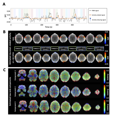 |
Low-rank and sparse simultaneous blind estimation of global fluctuations and neuronal-related activity from fMRI data.
Eneko Uruñuela1, Stefano Moia1, and César Caballero-Gaudes1
1Basque Center on Cognition, Brain and Language, Donostia - San Sebastián, Spain
We propose a novel deconvolution approach to overcome the detrimental effect of global signal fluctuations on the estimates of neuronal-related activity from fMRI data to blindly map single-trial BOLD events without prior information of their timings. We demonstrate that the low-rank and multivariate SPFM algorithm can estimate global signals related to motion in our task, while estimating neuronal-related activity with high fidelity, and benchmark it against sparsity-promoting deconvolution approaches and conventional GLM analysis. This method allows exploring the brain’s functional dynamics during task, naturalistic and resting state paradigms, being less affected by motion and physiologically related fluctuations.
|
|||
2686.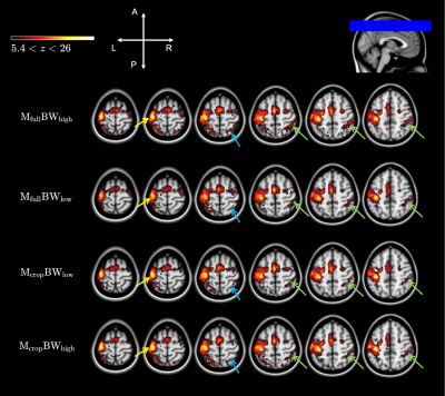 |
Smoothing-matched k-space coverage for enhanced BOLD fMRI
Didi Chi1,2, Rebecca Glarin2, Yasmin Blunck1,2, Catherine E. Davey1,2, and Leigh A. Johnston1,2
1Department of Biomedical Engineering, The University of Melbourne, Parkville, Australia, 2Melbourne Brain Centre Imaging Unit, The University of Melbourne, Parkville, Australia
Spatial smoothing is commonly applied as a pre-processing step in fMRI activation analysis pipelines. We investigate smoothing-matched k-space coverage for multiband EPI, with a particular focus on how varying the sampling speed in a reduced k-space will affect image SNR and resultant activation maps. A block task finger-tapping experiment demonstrates improved SNR from smoothing-matched k-space coverage, and improvements in activation level and specificity in the activation map. Our results demonstrate that dense sampling around the central echo time in multiband EPI produces superior results.
|
|||
2687. |
Open Source Random Matrix Theory Software for the Analysis of Functional Magnetic-Resonance Imaging Examinations
Derek Berger1, Gurpreet S Matharoo1,2,3, and Jacob Levman1
1Computer Science, St. Francis Xavier University, Antigonish, NS, Canada, 2Physics, St. Francis Xavier University, Clydesdale, NS, Canada, 3ACENET, St. Francis Xavier University, Antigonish, NS, Canada
We assess the potential application of RMT-based features for the analysis of functional MRI (fMRI) across diverse datasets. As novel contributions, we (1) assess the potential for RMT-inspired, whole-brain features extracted from voxel-wise functional connectivity, (2) assess these features’ predictive—rather than explanatory—value, (3) investigate the effect of varying RMT analysis methods on the robustness of study findings, and (4) make general-purpose code publicly available for users to extract these features from a wide variety of data. We find preliminary evidence suggesting that RMT-inspired features may have unique potential in analyses of fMRI functional connectivity.
|
|||
2688.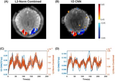 |
1D Convolutional Neural Network for Estimating BOLD Signal from Oscillating Steady State Signal
Mariama Salifu1, Melissa Haskell2, and Douglas C Noll1
1Biomedical Engineering, University of Michigan, Ann Arbor, MI, United States, 2Electrical Engineering and Computer Science, University of Michigan, Ann Arbor, MI, United States
Oscillating steady state imaging (OSSI) for fMRI commonly combines images across a fast oscillation to produce a steady time series suitable for fMRI analysis. The signal magnitude derived from L2-norm combination can be sensitive to frequency variations, which can exacerbate physiological noise, particularly respiration. Here, we use a 1D convolutional neural network (1DCNN) to directly estimate the underlying T2* BOLD response from both simulated and real OSSI fMRI signals. We demonstrated that our technique reduces B0 fluctuations and thermal noise as compared to the L2-norm combined OSSI fMRI signal.
|
|||
| 2689. | CNN-based autoencoder and machine learning model for identifying betel-quid chewers using functional MRI features
Hsin-An Shen1, Ming-Chou Ho2,3, and Jun-Cheng Weng1,4,5
1Department of Medical Imaging and Radiological Sciences, and Bachelor Program in Artificial Intelligence, Chang Gung University, Taoyuan, Taiwan, 2Department of Psychology, Chung Shan Medical University, Taichung, Taiwan, 3Clinical Psychological Room, Chung Shan Medical University Hospital, Taichung, Taiwan, 4Medical Imaging Research Center, Institute for Radiological Research, Chang Gung University and Chang Gung Memorial Hospital at Linkou, Taoyuan, Taiwan, 5Department of Psychiatry, Chang Gung Memorial Hospital, Chiayi, Taiwan
Previous studies indicated that betel-quid chewing may cause brain functional alternations, but it cannot be distinguished with human eyes. We used resting-state functional magnetic resonance imaging as input features for machine learning to classify betel-quid chewers, alcohol- and tobacco-user controls, and healthy control.The results showed that logistic regression has a significant performance on identifying betel-quid chewers. The major advantage to this study is providing a 3D-autoencoder model and machine learning algorithm that can be used to discover the brain alternations in betel-quid chewers for clinical use in the future.
|
|||
2690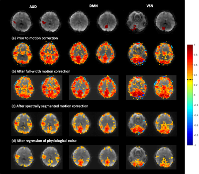 |
Spectrally Segmented Regression of Physiological Noise and Motion in High-Bandwidth Resting-State fMRI Video Permission Withheld
Khaled Talaat1, Bruno Sa de La Rocque Guimaraes1, and Stefan Posse2,3
1Nuclear Engineering, U New Mexico, Albuquerque, NM, United States, 2Neurology, U New Mexico, Albuquerque, NM, United States, 3Physics and Astronomy, U New Mexico, Albuquerque, NM, United States
It has been demonstrated in prior works that whole-band linear nuisance regression can result in the introduction of artifactual connectivity in the high frequency regime in resting-state fMRI. In the present work, an alternative approach is proposed to whole-band linear nuisance regression relying on spectral and temporal segmentation of the motion parameters and the physiological noise signals. The new approach is shown to not only avoid the injection of artifactual connectivity, but it also substantially improves the removal of physiological noise and motion effects throughout the whole frequency spectrum when uncertainties are present in the regression vectors.
|
|||
2691. |
Dephasing the speaking brain: Cleaning covert sentence production activation maps with a phase-based fMRI data analysis
Iñigo De Vicente1, Eneko Uruñuela1, Maite Termenon1, and César Caballero-Gaudes1
1BCBL - Basque Center on Cognition, Brain and Language, Donostia-San Sebastian, Spain
Phase-based denoising methods in BOLD fMRI can remove signal contributions from large draining veins. Here, we investigate the performance of this methodology in a covert sentence production task, where part of the activations observed in the inferior frontal gyrus (e.g. Broca’s region) may reflect macrovascular effects from large perisylvian veins. We found significant signal denoising at the left pars orbitalis, a region in Broca’s area which is located next to the Sylvian fissure that thus contains macrovascular veins. Our results demonstrate that this method is efficient to remove undesired signal contributions and improve the spatial specificity of brain activation maps.
|
|||
2692.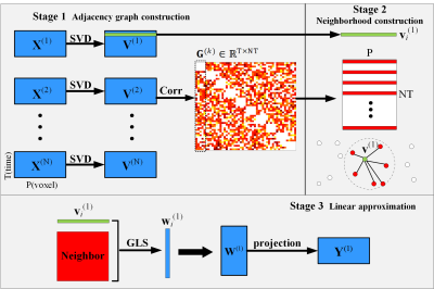 |
An Efficient FMRI Data Reduction Strategy Using Neighborhood Preserving Embedding Algorithm
Wei Zhao1, Huanjie Li1, Yunge Zhang1, Blaise B. Frederick 2, and Fengyu Cong1
1Biomedical Engineering, Dalian University of Technology, Dalian, China, 2Department of Psychiatry, Harvard Medical School, Boston, MA, United States
In neuroscience research, the group analysis using fMRI data for studying functional brain networks/connectivity in brain faces the challenge about information loss during fMRI data dimensionality reduction for increasing dimensionality. Proposed adapted the NPE (Neighborhood Preserving Embedding) stratagem on fMRI datasets, is an effective data reduction method that shows superior performance on efficient data reduction and sufficient information preservation. Our proposed method can strengthen useful group-sharing information and can avoid the limitation of selecting components based on variance of eigenvectors. Therefore, it has better performance on individual and group level outcomes, as well as improvements on the reliability and reproducibility.
|
The International Society for Magnetic Resonance in Medicine is accredited by the Accreditation Council for Continuing Medical Education to provide continuing medical education for physicians.