Digital Posters
Functional Connectivity Methods
ISMRM & SMRT Annual Meeting • 15-20 May 2021

Digital Posters
Functional Connectivity Methods
| Concurrent 3 | 17:00 - 18:00 |
3125.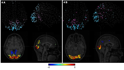 |
Investigating the relationship between temporal SNR and resting-state networks to evaluate the feasibility of fMRI at a low field MR scanner
Arjama Halder1,2, Demetrius Riberio de Paula3, William B Handler1, Andrea Soddu1, and Blaine A Chronik1,2
1xMR Labs, Physics and Astronomy, Western University, London, ON, Canada, 2Medical Biophysics, Western University, London, ON, Canada, 3Donders Institute, Radbound University, Nijmegen, Netherlands
This preliminary investigation evaluates the feasibility of resting-state BOLD fMRI at a 0.5T MR scanner equipped with a high-performance gradient coil. Here, we evaluated the decrement in the number of extracted resting-state networks as tSNR decreased with reduced scanner field strength. The results from this study suggest a positive linear relationship between the calculated correlation coefficients for different resting-state networks and tSNR within a 95% confidence interval. A lower limit on the tSNR at which the networks are detectable can be calculated from this analysis and will be used in experimental studies with the 0.5T scanner.
|
|||
3126.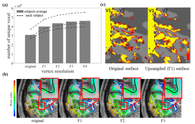 |
Evaluation of spatial blur induced by preprocessing and distortion in UHF fMRI data
Jianbao Wang1,2, Shahin Nasr2,3, Anna Wang Roe1,4, and Jonathan R. Polimeni2,3,5
1Department of Neurology of the Second Affiliated Hospital, Interdisciplinary Institute of Neuroscience and Technology, School of Medicine, Zhejiang University, Hangzhou, China, 2Athinoula A. Martinos Center for Biomedical Imaging, Massachusetts General Hospital, Charlestown, MA, United States, 3Department of Radiology, Harvard Medical School, Charlestown, MA, United States, 4Key Laboratory for Biomedical Engineering, of Ministry of Education, Zhejiang University, Hangzhou, China, 5Division of Health Sciences and Technology, Massachusetts Institute of Technology, Cambridge, MA, United States
Functional MRI is vulnerable to several sources of spatially-varying blurring imposed both at the data acquisition and analysis stages. For studies investigating fine-scale functional organization, any losses in imaging resolution, caused by blurring, dramatically affect the interpretation of the data. Therefore, these often-unavoidable blurring effects should be taken into account. Here we suggest approaches to minimize losses in spatial accuracy and recommend methods for quantifying the imaging resolution. These quantification methods can provide spatial “error bars” to use when evaluating results.
|
|||
3127.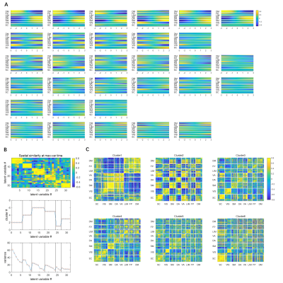 |
Spatiotemporal Trajectories in Resting-state FMRI Revealed by Convolutional Variational Autoencoder
Xiaodi Zhang1, Eric Maltbie1, and Shella Dawn Keilholz1
1BME, Emory University/Georgia Tech, Atlanta, GA, United States
We trained a novel convolutional variational autoencoder to extract intrinsic spatial temporal patterns from short segments of resting-state fMRI data. The network was trained in an unsupervised manner using data from the Human Connectome Project. The extracted latent dimensions not only show clear clusters in the spatial domain that were in agreement with DMN/TPN anticorrelations and principal gradients, but also provide temporal information as well. The method provides a way to extract orthogonal spatial temporal patterns within fMRI data in a short time window, among which many patterns were not previously discovered and are worth investigating in the future.
|
|||
3128.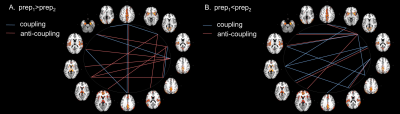 |
Temporal filtering affects time-varying functional connectivity metrics of the human brain
Francesca Saviola1, Stefano Tambalo1, and Jorge Jovicich1
1CIMeC, Center for Mind/Brain Sciences, University of Trento, Rovereto (TN), Italy
The use of temporal filtering is still controversial in resting-state brain functional MRI. In this study, we show that time-varying connectivity patterns largely depend on the type of temporal filtering applied to the data. Our results highlight the importance of properly considering and reporting the pre-processing pipeline details as they may significantly impact on obtaining reproducible findings, especially in clinical studies.
|
|||
3129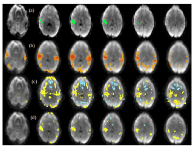 |
Detection of High-Frequency Resting-State Connectivity using Spectrally and Temporally Segmented Regression of High-Speed fMRI Data Video Permission Withheld
Bruno Sa de La Rocque Guimaraes1, Khaled Talaat1, and Stefan Posse2,3
1Nuclear Engineering, U New Mexico, Albuquerque, NM, United States, 2Neurology, U New Mexico, Albuquerque, NM, United States, 3Physics and Astronomy, U New Mexico, Albuquerque, NM, United States
This study investigates resting-state signal fluctuations at high-frequencies (>0.3Hz) using a novel regression method for high-speed fMRI data. Respiration and cardiac related signal changes and motion parameters were regressed using a spectral and temporal segmentation approach. This novel approach was shown to substantially remove physiological noise and motion effects. It reduces artificial high-frequency correlations compared with a recently developed sliding window regression approach. High frequency connectivity maps showed comparable localization to low frequency connectivity maps.
|
|||
3130.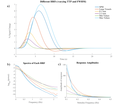 |
Predicting the temporal dynamics of the hemodynamic response using the spectrum of resting state fMRI signals
Sydney Bailes1 and Laura D. Lewis1
1Biomedical Engineering, Boston University, Boston, MA, United States
Given the substantial variations in the shape and timing of the hemodynamic response function (HRF) across the brain, it is critical to develop methods to characterize these variations for proper interpretation of the BOLD fMRI signal. Here, we identified significant differences in spectral properties of resting state fMRI signals between voxels with fast and slow hemodynamics. We found that these spectral properties can be used to classify fast and slow voxels, suggesting that information from the resting state can provide a way to understand and predict the temporal dynamics of the HRF across the brain.
|
|||
3131.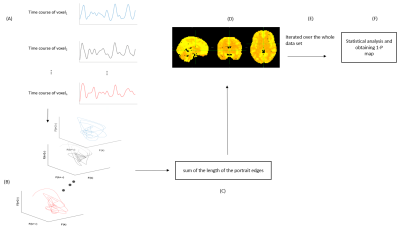 |
Mapping of phase-space dynamics for fMRI data
Zhenhai Zhang1, Kaiming Li2, and Xiaoping Hu2
1Department of Electrical and Computer Engineering, University of California,Riverside, Riverside, CA, United States, 2Department of Bioengineering, University of California,Riverside, Riverside, CA, United States
This work aims to map the dynamics of resting state fMRI data. With phase space embedding, we reconstructed the phase portrait of BOLD signal and calculated the sum of the length of the edges of the convex hull to capture the dynamics of fMRI BOLD time courses. Our statistical parametric maps on multiple datasets of mental disorder suggest that SE could be a viable disease biomarker.
|
|||
3132.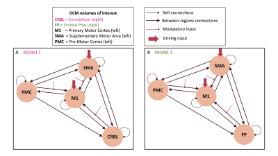 |
Cerebellum integration in motor network improves Dynamic Causal Modeling performance
Roberta Maria Lorenzi1, Letizia Casiraghi1,2, Adnan Alahmadi3,4, Anita Monteverdi1,5, Egidio D'Angelo1,5, Fulvia Palesi1,5, and Claudia A.M. Gandini Wheeler-Kingshott1,4,5
1Department of Brain and Behavioral Sciences, University of Pavia, Pavia, Italy, 2Azienda Socio Sanitaria Territoriale (ASST) di Pavia, Pavia, Italy, 3Department of Diagnostic Radiology, College of Applied medical sciences, King Abdulaziz University, Jeddah, Saudi Arabia, 4NMR Research Unit, Queen Square MS Centre, Department of Neuroinflammation, UCL Queen Square Institute of Neurology, Faculty of Brain Sciences, University College London (UCL), London, United Kingdom, 5Brain Connectivity Centre Research Department, IRCCS Mondino Foundation, Pavia, Italy
Dynamic Causal Modelling (DCM) is a framework enabling to quantify the causal relationship between functionally connected regions of the brain. Here we investigate cerebellum role in motion generation by adding a cerebellar node to an already validated motor network. We assessed how this operation improved DCM fMRI prediction. First- and second-level analyses were performed and the network including the cerebellum predicted the observed data with higher efficiency at single subject and group level. This network is important to study people with motor impairment where one could compare DCM predictions in heathy subjects and patients.
|
|||
3133.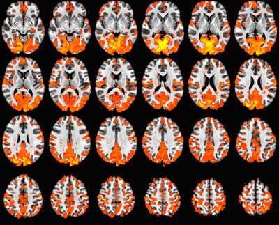 |
Reproducibility Test of Global Functional Connectivity
Jian Lin1, Wanyong Shin1, Stephen E Jones1, Katherine A Koenig1, and Mark J Lowe1
1Radiology, Cleveland Clinic, Cleveland, OH, United States
We evaluated the reproducibility of resting state global functional connectivity in scan to re-scan.
|
|||
3134.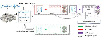 |
Deep Linear Modeling of Hierarchical Functional Connectivity in the Human Brain
Wei Zhang1, Eva M Palacios1, and Pratik Mukherjee1
1UCSF, San Francisco, CA, United States
To better map hierarchical brain connectivity networks, we introduce a novel class of deep (multilayer) linear models of fMRI that bridge the gap between conventional methods such as independent component analysis and more complex deep nonlinear models. These deep linear models do not require the manual hyperparameter tuning, extensive fMRI training data or high-performance computing infrastructure needed by deep learning, such as convolutional neural networks, and their results are more explainable from their mathematical structure. These benefits gain in importance as continual improvements in the spatial and temporal resolution of fMRI reveal more of the hierarchy of spatiotemporal brain architecture.
|
|||
3135.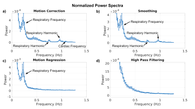 |
The effect of common resting-state fMRI preprocessing steps on the signal power spectrum
Jacob J.L. Matthews1, Jillian Krotz1, and J. Jean Chen1,2
1Rotman Research Institute, Baycrest, Toronto, ON, Canada, 2Medical Biophysics, University of Toronto, Toronto, ON, Canada
In resting-state functional MRI (rs-fMRI), the outcome measures, such as the amplitude of low-frequency fluctuation and functional connectivity, depend on the preprocessing steps applied. The mechanisms by which these steps modulate rs-fMRI outcomes are unclear, although it has long been established that the frequency composition of the rs-fMRI signal influences functional connectivity and related measures. In this work, we demonstrate the effect of commonly used preprocessing steps on rs-fMRI spectra, showing the potential interactions between these steps and cardiac/respiratory frequencies.
|
|||
3136.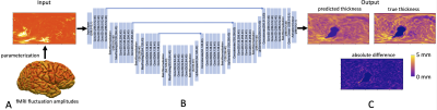 |
Testing for regional anatomical and physiological biases in fMRI signal fluctuations using surface-based deep learning
Olivia Viessmann1, Divya Varadarajan1, Adrian V Dalca1,2, Bruce Fischl1,2, Michael Bernier1, Lawrence L Wald1,2, and Jonathan R Polimeni1,2
1Massachusetts General Hospital, Harvard Medical School, Charlestown, MA, United States, 2Division of Health Sciences and Technology, Massachusetts Institute of Technology, Cambridge, MA, United States
We provide a proof of concept that surface-based CNNs can predict anatomical and physiological data from fMRI signals. Specifically, we trained CNNs to predict local cortical thickness, cortical orientation to the B0-field and MR angiography data to demonstrate that this information exists in the resting-state timeseries and can be extracted and possibly used for variance and bias reduction. Our results suggest that deep learning is able to identify non-linear relationships between the fMRI data and these anatomical and physiological biases.
|
|||
3137.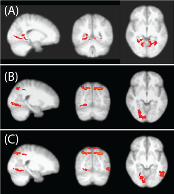 |
A human brain number system model based on fMRI connectivity and deep-learning network
Ray F. Lee1
1Zuckerman Mind Brain Behavior Institute, Columbia University, New York, NY, United States
Brain’s number system is essential for humans to gauge magnitudes, but has not been explicitly explained. An fMRI experiment was designed to quantitatively measure BOLD responses during numbering processes. The functional connectivity elucidates a deep-learning network model that decomposes the numbering to subitizing and counting, where subitizing is modeled as a CNN, and counting is modeled as an RNN where the subitizing is its recurrent cell. The initial success rate of this model is >95% for subitizing and >90% for counting in 500 tests. Further matching between the brain parcellates and the recurrent cell may fully reveal our number system.
|
|||
3138.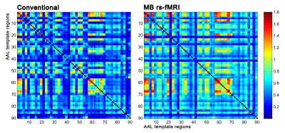 |
Investigation of altered brain activation based on MultiBand fMRI: A Graph Theory study
pengfei Zhang1, wanjun Hu1, jun Wang1, guangyao Liu1, jing Zhang1, and kai Ai2
1LanZhou University Second Hospital, lanzhou, China, 2Philips Healthcare, Xi'an, China
To test the advantages of multiband (MB) over single band echo planar imaging (EPI) acquisition schemes from the whole brain network level by using graph theory. Our results demonstrated that MB resting-state functional MRI (MB rs-fMRI) may provide much more insightful information than conventional rs-fMRI.
|
|||
3139.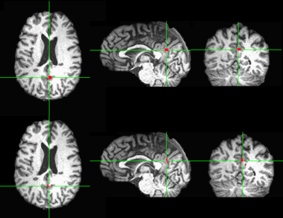 |
Locating seed automatically in posterior cingulate cortex for resting state fMRI data analysis by using unsupervised machine learning
Mingyi Li1, Katherine Koenig1, Jian Lin1, and Mark Lowe1
1Imaging Institute, Cleveland Clinic, Cleveland, OH, United States
We developed an automatic pipeline to generate seed clusters and corresponding connectivity maps for rs-fMRI data analysis by using unsupervised machine learning method. It only needed manual participation in the very end to review the candidate seed cluster locations and connectivity maps to make decision. Seeds in our pipeline were determined functionally within large pre-defined ROI which could be derived by using automatic brain segmentation tools like FreeSurfer or image registration. Successful application of the pipeline to locate seeds in PCC of control subjects and patients will be presented in this abstract.
|
|||
3140.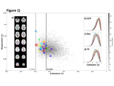 |
Group cohesive parcellation results in superior functional-based parcellation with greater parcel-level parsimony than current approaches
Ajay Nemani1 and Mark Lowe1
1Cleveland Clinic, Cleveland, OH, United States
Cohesive parcellation aims to produce parcels whose member voxels highly correlate to the parcel’s mean (exemplar) time series. The previously presented single subject version of cohesive parcellation is improved and extended to the group level using a novel hybrid parallel hierarchical framework. The resulting group parcellation compares favorably to traditional anatomical and connectivity-based parcellations over several measures of cluster validity at both the group level and when projected to individual subjects.
|
|||
3141.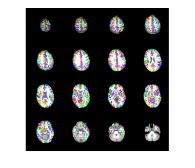 |
Validation of group cohesive parcellation of rsfMRI
Xuemei Huang1, Ajay Nemani1, and Mark J. Lowe1
1Cleveland Clinic, Cleveland, OH, United States
Single-subject cohesive parcellation were done for nine healthy subjects’ rsfMRI data. Based on motion quality, the best data for six subjects was used for group cohesive parcellation. The parcellation was in a leave one out cross validation. DICE scores were similar to intra-subject cross-validation and significantly better than random parcellation.
|
|||
3142.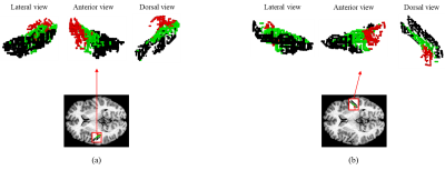 |
Individual-level parcellation of the human auditory cortex and comparison of their functional connectivity pattern
Hyebin Lee1,2, Kyoungseob Byeon1,2, Sean H. Lee3, and Hyunjin Park2,4
1Department of Electrical and Computer Engineering, Sungkyunkwan University, Suwon, Korea, Republic of, 2Center for Neuroscience Imaging Research, Institute for Basic Science (IBS), Suwon, Korea, Republic of, 3Department of Neuroscience, Max Planck Institute for Empirical Aesthetics, Frankfurt am Main, Germany, 4School of Electronic and Electrical Engineering, Sungkyunkwan University, Suwon, Korea, Republic of
Determination of the exact location and extent of a brain area at an individual level is crucial but challenging in in-vivo neuroimaging studies. We developed an analysis pipeline to parcellate the primary auditory cortex and its vicinity at an individual level with diffusion tensor imaging. Resulting three clusters, the central cluster and the two other flanking clusters, exhibited distinct functional connectivity patterns with the rest of the whole brain indicating that these clusters contribute to different functional networks.
|
|||
3143.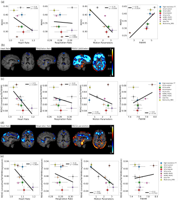 |
Inter-group Heterogeneity of Regional Homogeneity (REHO)
Yan Jiang1, Mohammed Ayoub Alaoui Mhamdi1, and Russell Butler1,2
1Bishop's University, sherbrooke, QC, Canada, 2Diagnostic Radiology, University of Sherbrooke, sherbrooke, QC, Canada
Differences in REHO across groups may not be indicative of differences in neuronal activity. In this study, we investigate physiological contributions to REHO across 412 subjects in 9 separate datasets downloaded from OpenNeuro. Overall, we find the following: Inverse correlation between heartrate and REHO across subjects, inverse correlation between respiration and REHO across time, and differences in REHO across groups is driven primarily by FWHM of data and motion. We conclude that, due to REHO’s highly significant correlation with motion, heartrate, and respiration, REHO should be used with caution to infer differences in neuronal activity across groups.
|
|||
3144.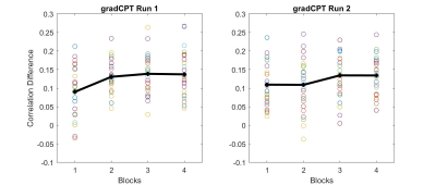 |
Optimizing connectome-based real-time neurofeedback for improved attentional control
Xiangrui Li1, Oyetunde Gbadeyan 1, and Ruchika Shaurya Prakash1
1Department of Psychology and Center for Cognitive and Behavioral Brain Imaging, The Ohio State University, Columbus, OH, United States
In this proof-of-concept study, we conducted an optimization assessment of predictive models for use with real-time functional magnetic resonance imaging. Here, we utilized two existing connectome-based models of sustained attention and mind-wandering derived in independent datasets, with each model comprising connections predictive of positive or negative associations with target behavior. Both models showed significant networks strengths across the blocks of the sustained attention task, with the combined model showing significant network strengths and capturing vigilance decrements. Our results suggest that our two models are representative of high and low attentional states, thus making them appropriate targets for neurofeedback.
|
The International Society for Magnetic Resonance in Medicine is accredited by the Accreditation Council for Continuing Medical Education to provide continuing medical education for physicians.