Digital Posters
Pediatrics: Neuro Topics
ISMRM & SMRT Annual Meeting • 15-20 May 2021

| Concurrent 3 | 17:00 - 18:00 |
 |
2239.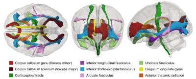 |
General factors of white matter microstructure in the newborn human brain
Kadi Vaher1, Paola Galdi1, Manuel Blesa Cabez1, Gemma Sullivan1, Gill Black1, David Q Stoye1, Alan J Quigley2, Michael J Thrippelton3, Simon R Cox4, Mark E Bastin3, Debby Bogaert5, and James P Boardman1
1MRC Centre for Reproductive Health, University of Edinburgh, Edinburgh, United Kingdom, 2Department of Paediatric Radiology, Royal Hospital for Sick Children, Edinburgh, United Kingdom, 3Centre for Clinical Brain Sciences, University of Edinburgh, Edinburgh, United Kingdom, 4Lothian Birth Cohort Studies group, Department of Psychology, University of Edinburgh, Edinburgh, United Kingdom, 5Centre for Inflammation Research, University of Edinburgh, Edinburgh, United Kingdom
Preterm birth associates with diffuse white matter dysmaturation, which is linked to neurocognitive impairment. We applied principal component analysis to tract-averaged diffusion tensor (DTI) and neurite orientation and density imaging (NODDI) measures to derive single- and multimodal general factors describing neonatal white matter microstructure. We report substantial shared variance of DTI and NODDI metrics across 16 white matter tracts and between the different metrics; the derived general factors associate with preterm birth. These results suggest that general factors are efficient markers of generalised white matter microstructure in early life, and are useful for investigating the determinants of white matter development.
|
||
2240.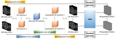 |
Learning 3D structures from 2D slices with scan-specific data for fast and high-resolution neonatal brain MRI
Yao Sui1,2, Onur Afacan1,2, Ali Gholipour1,2, and Simon K Warfield1,2
1Harvard Medical School, Boston, MA, United States, 2Boston Children's Hospital, Boston, MA, United States
Neonatal brain MRI is a resolution-critical task due to the small brain of neonates. Among post-acquisition resolution-enhancement techniques, deep learning has shown promising results. Most state-of-the-art deep learning-based super-resolution methods work on 2D slices, so ignore the 3D nature of the brain anatomy. Learning on 3D images requires large-scale training datasets of high-resolution volumes that are, unfortunately, difficult to acquire. We developed a methodology that enables learning 3D gradient structures from 2D slices for an individual subject without the need for large, auxiliary high-resolution datasets. Experiments on clinical data from ten neonates demonstrate our approach outperformed state-of-the-art MRI super-resolution methods.
|
|||
2241.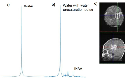 |
Relationship between brain temperature and prognosis during hypothermia in newborns with hypoxic-ischemic encephalopathy
Moyoko Tomiyasu1,2, Jun Shibasaki3, Yasuhiko Terada4, Katsuaki Toyoshima3, Tatsuya Higashi1, Takayuki Obata1, and Noriko Aida2
1Department of Molecular Imaging and Theranostics, National Institute for Quantum and Radiological Science and Technology, Chiba, Japan, 2Department of Radiology, Kanagawa Children’s Medical Center, Yokohama, Japan, 3Department of Neonatology, Kanagawa Children’s Medical Center, Yokohama, Japan, 4Institute of Applied Physics, University of Tsukuba, Tsukuba, Japan
We examined brain temperatures of 81 neonates with hypoxic-ischemic encephalopathy during and/or after hypothermia therapy. The brain temperature was obtained from magnetic resonance spectroscopy data (3T). The subjects also had a neurological developmental test at the age of 18–22 months, and were divided into favorable and adverse outcome groups. Brain temperatures for each outcome were significantly lower during hypothermia compared with those after hypothermia. Although the poor prognosis group tended to have a large dispersion of brain temperature after hypothermia, there was no significant difference in brain temperature between the favorable and adverse outcome groups.
|
|||
2242.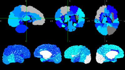 |
Association between multi-modal term MRI biomarkers and early cerebral palsy risk in very preterm infants
Julia E. Kline1, Weihong Yuan1, Karen Harpster1, and Nehal A. Parikh1
1Cincinnati Children's Hospital Medical Center, Cincinnati, OH, United States
Very preterm (VPT) infants are at risk for serious motor impairments, including cerebral palsy (CP). Motor interventions are most effective if delivered early, but early accurate biomarkers of CP are lacking. In a cohort of 345 VPT infants, we extracted cortical morphometrics and brain volumes from term structural MRI and applied graph theoretical methods to term diffusion MRI connectomes. We observed consistent negative associations between early CP risk and cortical surface area, subcortical volumes, and graph metrics, including global efficiency and local efficiency of motor regions. These early biomarkers enhance our understanding of VPT motor impairments and may predict CP.
|
|||
2243.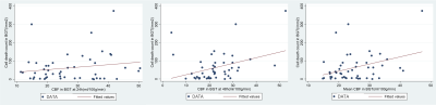 |
Optimizing Brain Injury Biomarkers in a piglet model of Neonatal Encephalopathy: combining perfusion with proton MRS
Hillary Idogwu1, Magdalena Sokolska2, Pang Raymand 3, Saiful Islam4, Christopher Meehan 3, Adnan Avdic-Belltheus 3, Kathryn Marinello 3, Ingran Lingam 3, Tatenda Mutshiya 3, Alan Bainbridge 2, Nikki Robertson 3, David Thomas1, and Xavier Golay1
1Brain Repair and Rehabilitation, Institute of Neurology, UCL Queen Square Institute of Neurology, London, United Kingdom, 2Medical Physics and Biomedical Engineering, UCLH NHS Foundation Trust, London, United Kingdom, 3Neonatology, UCL EGA Institute for Women's Health, London, United Kingdom, 4UCL Queen Square Institute of Neurology, London, United Kingdom
Despite therapeutic hypothermia, some survivors of neonatal encephalopathy (NE) still have adverse outcomes. Surrogate outcome measures or biomarkers are needed for early detection of babies with adverse outcome so that adjunct therapies can be considered. Basal ganglia and thalamus (BGT) lactate/ N-Acetyl-Aspartate (Lac/NAA) peak area ratio acquired with proton magnetic resonance spectroscopy (MRS) is a reliable predictor of developmental outcome at two years. Cerebral blood flow (CBF) changes in parallel with the metabolic changes after hypoxia ischaemia (HI). We hypothesised that the combination of CBF with BGT Lac/NAA would be more closely associated with quantitative cell death than either alone.
|
|||
 |
2244.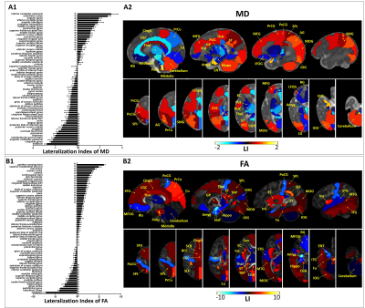 |
Brain asymmetry and development of healthy preterm infants within half-year-old: a diffusion MRI study based on ROI and fixel methods
Tingting Liu1, Weihao Zheng1, Yuqing You2, Ying Lv2, Weijun Chen2, Zhiyong Zhao1, Fusheng Gao2, Hongxi Zhang2, Chai Ji2, and Dan Wu1
1ZheJiang University, Hangzhou, China, 2Children's Hospital, ZheJiang University School of Medicine, Hangzhou, China
The present study aimed to investigate brain asymmetry of healthy preterm-born infants within half-year-old using high angular resolution diffusion MRI. ROI-based analysis revealed most brain regions showed significant asymmetry and the asymmetry changed with brain development. The MD-based lateralization index showed an center-versus-peripheral pattern with remarkable lateralization in the posterior cortex. Moreover, in contrast to the adult brain, we found a rightward asymmetry in language processing regions and leftward asymmetry in visuospatial processing regions. Besides, consistent leftward lateralization in white matter was observed at both ROI-based and fixel-based analysis. These findings advanced our understanding of brain asymmetry during early development.
|
||
2245.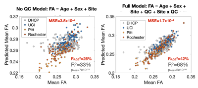 |
Accounting for Measurement Noise in a Multi-Site Newborn Diffusion Weighted Imaging Study
Jerod M Rasmussen1, Alice M Graham2, Pathik D Wadhwa1, Sonja Entringer3, Martin Styner4, Beatriz Luna5, Thomas G O'Connor6, Damien A Fair7, and Claudia Buss3
1UC Irvine, Irvine, CA, United States, 2Oregon Health Sciences University, Portland, OR, United States, 3Charité – Universitätsmedizin Berlin, Berlin, Germany, 4University of North Carolina, Chapel Hill, NC, United States, 5University of Pittsburgh, Pittsburgh, PA, United States, 6University of Rochester, Rochester, NY, United States, 7University of Minnesota, Minneapolis, MN, United States
We leverage an exemplar relationship (FAWM vs. postmenstrual age [PMA]) to quantify the benefits conferred by adjustment for QC measures in a multi-site infant DWI study. Data was preprocessed using a common pipeline (dHCP) and the FAWM-PMA association tested without and with controlling for SNR, CNR and mean FD. All three QC measures were broadly associated with FAWM within sites. Within and across sites, inclusion of QC measures greatly increased FAWM variance explained by PMA. Accounting for QC measures results in an improved capability for identifying reproducible small-effect size relationships in large multi-site infant imaging studies.
|
|||
2246.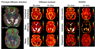 |
High resolution diffusion imaging in the unfixed post-mortem neonatal brain
Wenchuan Wu1, Luke Baxter1,2, Eleri Adams2,3, Foteini Andritsou2, Matteo Bastiani1,4,5, Ria Evans Fry2, Robert Frost6,7, Sean Foxley8, Chris Gallagher1, Fiona Moultrie2, Vaneesha Monk2, David Andrew Porter9, Anthony Price10,11,
Sebastian W Rieger1,12, Michael Sanders1, Anthony David Edwards11, Joseph V Hajnal10,11, Rebeccah Slater*1,2, and Karla L Miller*1
1Wellcome Centre for Integrative Neuroimaging, Nuffield Department of Clinical Neurosciences, University of Oxford, Oxford, United Kingdom, 2Department of Paediatrics, University of Oxford, Oxford, United Kingdom, 3Newborn Care Unit, John Radcliffe Hospital, Oxford University Hospitals NHS Foundation Trust, Oxford, United Kingdom, 4Sir Peter Mansfield Imaging Centre, School of Medicine, University of Nottingham, Nottingham, United Kingdom, 5NIHR Biomedical Research Centre, University of Nottingham, Nottingham, United Kingdom, 6Athinoula A. Martinos Center for Biomedical Imaging, Massachusetts General Hospital, Charlestown, MA, United States, 7Department of Radiology, Harvard Medical School, Boston, MA, United States, 8Department of Radiology, University of Chicago, Chicago, IL, United States, 9Imaging Centre of Excellence, College of Medical, Veterinary & Life Sciences, University of Glasgow, Glasgow, United Kingdom, 10Department of Biomedical Engineering, School of Biomedical Engineering and Imaging Sciences, King’s College London, King’s Health Partners, St. Thomas’ Hospital, London, United Kingdom, 11Centre for the Developing Brain, School of Biomedical Engineering and Imaging Sciences, King’s College London, King’s Health Partners, St. Thomas’ Hospital, London, United Kingdom, 12Department of Psychiatry, University of Oxford, Oxford, United Kingdom
In this work, we develop a 7T experimental platform to acquire high resolution diffusion MRI images in unfixed post-mortem infant brains. Here we present results from a first infant. The post-mortem structural images demonstrate superior SNR and improved depiction of small structures than age-matched low-resolution in vivo data. The post-mortem diffusion MRI images show sufficient SNR to support the considerably smaller voxel volume compared to in vivo. The high spatial resolution of the post-mortem data enables the depiction of brain features (e.g., cortical radiality, subplate organization) that are less prominent in low-resolution in vivo data.
|
|||
2247.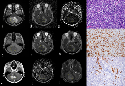 |
Grading of pediatric intracranial tumors: are intravoxel incoherent motion and diffusion kurtosis imaging superior to conventional DWI?
dejun she1, dairong cao1, shan lin1, zhongshuai zhang2, and Robert Grimm3
1The First Affiliated Hospital of Fujian Medical University, Fujian Fuzhou, China, 2SIEMENS healthcare diagnostic imaging, Shanghai, Pudong, Zhouzhu Highway 278, China, 3SIEMENS Healcare, Erlangen, Germany
To explore the correlations between parameters derived from conventional diffusion-weighted imaging (DWI), intravoxel incoherent motion (IVIM), and diffusion kurtosis imaging (DKI) with histopathologic features of pediatric intracranial tumors.
|
|||
2248.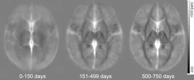 |
Quantitative susceptibility mapping of the brain in neonates and infants
Sayo Otani1, Yasutaka Fushimi1, Satoshi Nakajima1, Akihiko Sakata1, Takuya Hinoda1, Sonoko Oshima1, Krishna Pandu Wicaksono1, Hiroshi Tagawa1, Yang Wang1, and Yuji Nakamoto1
1Kyoto University Graduate School of Medicine, Kyoto, Japan
We evaluated magnetic susceptibility of the brain in 202 neonates and infants. We manually placed volumes of interest of the caudate nucleus, globus pallidus, putamen and ventral posterior lateral nucleus of the thalamus in the MNI space. Our study showed that elevation of magnetic susceptibility probably associated with iron deposition of the basal ganglia and thalamus increased with chronological age from birth to 2 years using volume-of-interest analysis.
|
|||
2249.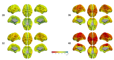 |
Mapping developmental trajectories of the cortex and its adjacent white matter for preterm neonates using DTI
Shiyu Yuan1, Jingda Yang1, Mengting Liu1, Duan Xu2, James Barkovich2, and Hosung Kim1
1USC Stevens Neuroimaging and Informatics Institute, Keck School of Medicine of USC, University of Southern California, Los Angeles, CA, United States, 2Department of Radiology and Biomedical Imaging, University of California, San Francisco, San Francisco, CA, United States
The brain development of cyto- and myeloarchitecture undergoes rapid and intensive changes during the third trimester. Diffusion tensor imaging (DTI) probes the microstructural organization of brain tissue by measuring the three-dimensional spatial diffusion of water molecules1. Using mean diffusivity (MD) and fractional anisotropy (FA), we show the spatiotemporal pattern of cortical and superficial white matter maturation in preterm neonates. This result might be useful in the prediction of neurodevelopmental outcome and functional impairments in preterm survivors.
|
|||
|
2250. |
Disentangling neurite and soma contributions to cortical microstructural development in vivo
Sila Genc1, Maxime Chamberland1, Gareth Ball2, Erika Raven1, Isobel Ward1, Chantal Tax1, Marco Palombo3, and Derek Jones1
1Cardiff University Brain Research Imaging Centre (CUBRIC), Cardiff University, Cardiff, United Kingdom, 2Developmental Imaging, Murdoch Children's Research Institute, Parkville, Australia, 3Centre for Medical Image Computing and Department of Computer Science, University College London, London, United Kingdom MRI studies of cortical development have revealed macroscopic changes in morphology, with thickness/volume decreasing over childhood and adolescence. Recent microstructural modelling advances hold promise for quantifying the cellular changes driving these observations. We characterised cortical neurite and soma microstructure in 88 participants aged 8-18 years. Key findings:
Overall, our study provides in vivo evidence of distinct developmental patterns of cortical neurite and soma microstructure. |
||
2251.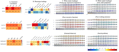 |
Multivariate Associations Among Symptoms, Cognition, Behavior, and White Matter across ADHD and typically developing children
Xuan Bu1,2, Yingxue Gao1, Kaili Liang1, Weijie Bao1, Lu Lu1, Ying Chen1, Lanting Guo3, Qiyong Gong1, Susumu Mori2, and Xiaoqi Huang1
1Radiology Department, Sichuan University, Chengdu, China, 2The Russell H. Morgan Department of Radiology and Radiological Science, Johns Hopkins University School of Medicine, Baltimore, MD, United States, 3Psychiatry Department, Sichuan University, Chengdu, China
In current study, we discovered several multivariate patterns of white matter properties linked to clinical symptoms, cognition and behavior across children with and without ADHD. Each dimension of phenotype was linked to a specific pattern of white matter microstructure alterations. Furthermore, these associations captured deviations between children with and without ADHD and displayed developmental effects. Together, these results suggest that complex ADHD phenotypes are associated with specific patterns of abnormal white matter microstructure during brain development.
|
|||
2252.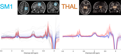 |
Increased glutamate + glutamine correlates with altered tactile perception and sensory responsivity in children with autism spectrum disorder
Georg Oeltzschner1,2, Jason He3, Mark Mikkelsen1,2, Alyssa DeRonda4, Deana Crocetti4, Stewart H. Mostofsky4,5,6, Richard A.E. Edden1,2, and Nicolaas A.J. Puts3
1Russell H. Morgan Department of Radiology and Radiological Science, The Johns Hopkins University School of Medicine, Baltimore, MD, United States, 2F. M. Kirby Research Center for Functional Brain Imaging, Kennedy Krieger Institute, Baltimore, MD, United States, 3Department of Forensic and Neurodevelopmental Sciences, Sackler Institute for Translational Neurodevelopment, Institute of Psychiatry, Psychology, and Neuroscience, King's College London, London, United Kingdom, 4Center for Neurodevelopmental and Imaging Research, Kennedy Krieger Institute, Baltimore, MD, United States, 5Department of Psychiatry, The Johns Hopkins University School of Medicine, Baltimore, MD, United States, 6Department of Neurology, The Johns Hopkins University School of Medicine, Baltimore, MD, United States
Abnormal perception of sensory input is among the core symptoms of autism spectrum disorders (ASD). Despite evidence for altered excitation-inhibition balance contributing to states of hyper- or hypo-excitability in ASD, findings have been mixed. In this study, we used magnetic resonance spectroscopy (MRS) in a large sample of children with ASD and typically developing children to measure levels of GABA and glutamate+glutamine (Glx). Elevated sensorimotor Glx levels in ASD correlated with vibrotactile frequency discrimination thresholds and caregiver-reported hyper- and hyporesponsivity to sensory stimuli, symptoms that are central to ASD. These results provide support for the excitation-inhibition imbalance theory of ASD.
|
|||
2253.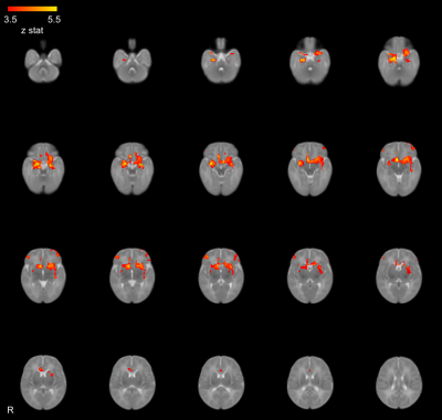 |
Prenatal maternal distress during the COVID-19 pandemic and its effects on infant brain connectivity
Kathryn Y. Manning1,2,3, Xiangyu Long1,2,3, Lianne Tomfohr-Madsen2,4,5, Gerald Giesbrecht2,4,5,6, and Catherine Lebel1,2,3
1Radiology, University of Calgary, Calgary, AB, Canada, 2Alberta Children's Hospital Research Institute, Calgary, AB, Canada, 3Hotchkiss Brain Institute, Calgary, AB, Canada, 4Psychology, University of Calgary, Calgary, AB, Canada, 5Pediatrics, University of Calgary, Calgary, AB, Canada, 6Community Health Sciences, Calgary, AB, Canada
We investigated the effects of prenatal maternal psychological distress in pregnant Canadian mothers during the COVID-19 pandemic on the early development of the infant brain. We scanned infants using the 3T MRI at the Alberta Children's hospital including resting state functional and diffusion MRI. We found that amygdala microstructural pathways were significantly related to functional connectivity between the right and left amygdala at 2-months of age. These infant MRI measures were correlated with maternal anxiety and psychological distress where we found relatively less mature tracts and lower amygdala-prefrontal functional connectivity were related to higher prenatal maternal psychological distress.
|
|||
2254.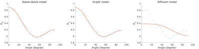 |
Orientation dependence of T2 in newborn white matter shows dipole-dipole interaction effects
Lara Bartels1,2, Jonathan Doucette1,2, Christoph Birkl2,3,4, Yuting Zhang5,6,7,8, Alexander Mark Weber2,9, and Alexander Rauscher1,2,9,10
1Department of Physics & Astronomy, University of British Columbia, Vancouver, BC, Canada, 2UBC MRI Research Centre, University of British Columbia, Vancouver, BC, Canada, 3Department of Neuroradiology, Medical University of Innsbruck, Innsbruck, Austria, 4Department of Neurology, Medical University of Graz, Graz, Austria, 5Department of Radiology, Children's Hospital of Chongqing Medical University, Chongqing, China, 6Ministry of Education Key Laboratory of Child Development and Disorders, Chongqing Medical University, Chongqing, China, 7Key Laboratory of Pediatrics in Chongqing, Chongqing Medical University, Chongqing, China, 8Chongqing International Science and Technology Cooperation Center for Child Development and Disorders, Chongqing, China, 9Division of Neurology, Department of Pediatrics, University of British Columbia, Vancouver, BC, Canada, 10Department of Radiology, University of British Columbia, Vancouver, BC, Canada
In adult White Matter (WM), tissue orientation effects on $$$R_2$$$ are best described by diffusion within field inhomogeneities created by the myelin sheath. Here we studied how $$$R_2$$$ relaxation depends on white matter fibre orientation in the human newborn brain in vivo. We found that the orientation dependency is very different from adults and best described by a model of dipole-dipole interaction.
|
|||
2255.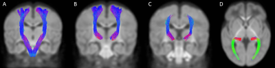 |
Early Sensorimotor Tract Integrity and Development of Cerebral Palsy in High Risk Infants
Rahul Chandwani1, Julia Kline1, Karen Harpster2,3,4, Jean Tkach5,6,7, and Nehal Parikh1,2
1Perinatal Institute, Cincinnati Children's Hospital Medical Center, Cincinnati, OH, United States, 2Department of Pediatrics, University of Cincinnati College of Medicine, Cincinnati, OH, United States, 3Division of Occupational Therapy and Physical Therapy, Cincinnati Children's Hospital Medical Center, Cincinnati, OH, United States, 4Department of Rehabilitation, Exercise and Nutrition Sciences, University of Cincinnati College of Allied Health Sciences, Cincinnati, OH, United States, 5Department of Radiology, Cincinnati Children's Hospital Medical Center, Cincinnati, OH, United States, 6Imaging Research Center, Department of Radiology, Cincinnati Children's Hospital Medical Center, Cincinnati, OH, United States, 7Department of Radiology, University of Cincinnati College of Medicine, Cincinnati, OH, United States
Very preterm (VPT) infants are at high risk of motor impairments such as cerebral palsy (CP). Currently, qualitative findings from structural MRI and outcomes from clinical assessments are insufficient for early, accurate diagnosis of CP. In a multicenter cohort of 263 VPT infants, we derived macro and microstructural biomarkers of sensorimotor white matter tracts from term-equivalent age diffusion MRI. We observed consistent negative associations between these metrics – fiber density, fiber cross-section, and combined fiber density and cross-section – and early diagnosis of CP. These findings enhance our understanding of the pathophysiology of CP in VPT infants.
|
|||
2256.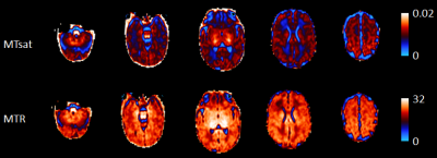 |
Breastmilk exposure is associated with improved MRI biomarkers of myelination in preterm infants
Gemma Sullivan1, Manuel Cabez1, Kadi Vaher1, Paola Galdi1, Gill Black1, David Q Stoye1, Alan J Quigley2, Elizabeth N York3, Michael J Thrippleton3, Mark E Bastin3, and James P Boardman1,3
1MRC Centre for Reproductive Health, University of Edinburgh, Edinburgh, United Kingdom, 2Department of Paediatric Radiology, Royal Hospital for Sick Children, Edinburgh, Edinburgh, United Kingdom, 3Centre for Clinical Brain Sciences, University of Edinburgh, Edinburgh, United Kingdom
Preterm birth is closely associated with altered white matter microstructure and dysconnectivity of developing neural networks with increased risk of long-term neurodevelopmental impairment. Nutritional exposures in the weeks after preterm birth affect head growth, brain macro- and micro- structure, and are associated with neurocognitive ability; the mechanisms underlying these associations are uncertain. By combining nutritional data with myelin-weighted imaging, we show that early breast milk exposure after preterm birth is associated with improved white matter myelination at term-equivalent age.
|
|||
2257.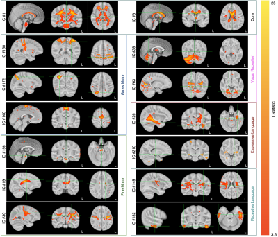 |
Patterns of De Novo Myelination Identify Functionally Relevant Brain Networks
Sean Deoni1, Lexie Volpe2, Jennifer Beauchemin2, and Viren D'Sa3
1Bill & Melinda Gates Foundation, Seattle, WA, United States, 2Advanced Baby Imaging Lab, Rhode Island Hospital, Providence, RI, United States, 3Pediatrics, Rhode Island Hospital, Providence, RI, United States
Infancy and childhood are important developmental periods punctuated by rapid brain development and cognitive growth, however, direct and longitudinal analysis of the relationships between maturing brain structure and evolving cognitive skill in human infants is sparse. Here we used longitudinal and myelin-sensitive MRI measures from a large cohort of neurotypical children to examine patterns of myelination throughout the brain and link these patterns to concurrently evolving motor, visual, and language skills. Results revealed a core system of central brain regions that contributed broadly across cognitive domains as well as domain-specific regions that align with known functional specialization.
|
|||
2258.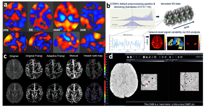 |
Alterations in vascular injury, rsfMRI signal variability and verbal memory in young patients treated with radiation therapy for a brain tumor.
Melanie Morrison1, Sivakami Avadappian1, Angela Jakary1, Erin Felton2, Schuyler Stoller3, Sabine Mueller3, and Janine Lupo1
1Radiology, UCSF, San Francisco, CA, United States, 2UCSF, San Francisco, CA, United States, 3Neurology, UCSF, San Francisco, CA, United States
This is a multimodal 7T MRI study evaluating imaging biomarkers of cognition in young brain tumor patients.
|
The International Society for Magnetic Resonance in Medicine is accredited by the Accreditation Council for Continuing Medical Education to provide continuing medical education for physicians.