Digital Posters
Pediatrics: Body Topics
ISMRM & SMRT Annual Meeting • 15-20 May 2021

| Concurrent 3 | 17:00 - 18:00 |
2259.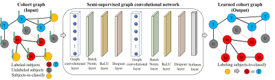 |
A semi-supervised graph convolutional network for early prediction of motor impairments in very preterm infants using brain connectome
Hailong Li1, Ming Chen1,2, Jinghua Wang3, Nehal A. Parikh4,5, and Lili He1,5
1Imaging Research Center, Department of Radiology, Cincinnati Children's Hospital Medical Center, Cincinnati, OH, United States, 2Department of Electrical Engineering and Computer Science, University of Cincinnati, Cincinnati, OH, United States, 3Deep MRI Imaging Inc., Lewes, DE, United States, 4The Perinatal Institute and Section of Neonatology, Perinatal and Pulmonary Biology, Cincinnati Children's Hospital Medical Center, Cincinnati, OH, United States, 5Department of Pediatrics, University of Cincinnati College of Medicine, Cincinnati, OH, United States
About 32−42% of very preterm infants develop minor motor impairments around the world. Unfortunately, large MRI datasets with clinical outcome annotations/labels are typically unavailable, especially in neonates. To address this challenge of limited training data, we developed a semi-supervised graph convolutional network model to utilize both labeled and unlabeled data during model training to predict motor impairments at 2 years corrected age using brain structural connectome derived from diffusion MRI obtained at term-equivalent age in very preterm infants. The proposed model was able to identify infants with motor impairments with an accuracy of 68.1% and an AUC of 0.67.
|
|||
2260.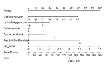 |
Predicting Early Neonatal Bilirubin Encephalopathy Based on Radiomics Nomogram of T1weighted Imaging
Jinhong Yu1, Yanwei Miao1, Yangyingqiu Liu1, Bingbing Gao1, and Yu Bing1
1The First Affiliated Hospital of Dalian Medical University, Dalian, China
The T1WI hyperintensity of bilateral globus pallidus(GP)is considered to be a typical imaging manifestation of bilirubin encephalopathy(BE), but the diagnosis ability is insufficient for the early stage of the disease (hyperbilirubinemia and T1WI negative). In this study, a better early prediction model of BE based on T1WI radiomics was obtained, which provided a new image marker for early diagnosis and monitoring of BE.
|
|||
2261.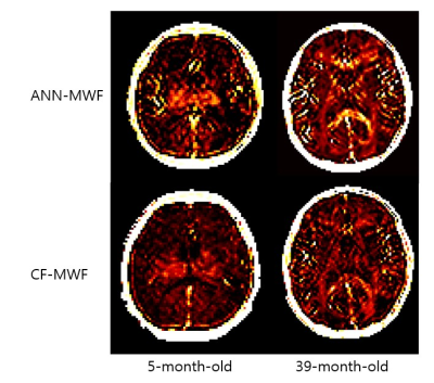 |
Artificial neural network derived myelin water fraction map with multi-echo gradient echo signal: brain development from infants to adults.
Hyun Gi Kim1, Jae Eun Song2, Dongyeob Han3, Jee Young Kim1, Se Won Oh1, and Dong-Hyun Kim2
1Radiology, The Catholic University of Korea, Seoul, Korea, Republic of, 2Yonsei University, Seoul, Korea, Republic of, 3Siemens Healthcare, Seoul, Korea, Republic of
Myelin water fraction (MWF) values were obtained by an artificial neural network (ANN-MWF) and complex model fitting (CF-MWF) with 3D multi-echo gradient-echo (mGRE) signal. Linear regression test showed high correlation between ANN-MWF and CF-MWF values (R2 = 0.802, p < .001). Bland-Altman plot showed higher ANN-MWF values compared to CF-MWF values in the areas with high MWF values. The ANN-MWF values in the white matter showed a high association with age (R2 = 0.821, p =.005).
|
|||
2262.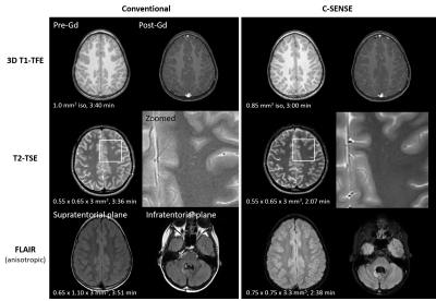 |
Evaluation of compressed sensing in pediatric neuro-oncological MR imaging: Impact on image quality and scan duration
Rieke Lisa Meister1, Shuo Zhang2, Michael Groth1, Julian Jürgens1, Christoph Katemann2, Jan-Hendrik Buhk3, and Jochen Herrmann1
1Department of Diagnostic and Interventional Radiology and Nuclear Medicine, Section of Pediatric Radiology, University Medical Center Hamburg-Eppendorf, Hamburg, Germany, 2Philips, Hamburg, Germany, 3Department of Diagnostic and Interventional Neuroradiology, University Medical Center Hamburg-Eppendorf, Hamburg, Germany
High-resolution MRI plays an important role in neuro-oncological exams in children. However, high-quality imaging remains challenging due to long scan times. In this study, we evaluate the compressed SENSE technique that employs compressed sensing with coil sensitivity information in a dedicated pediatric neuro-oncological scan protocol in children with brain tumor. The resulted image quality and scan duration are compared with the conventional techniques, showing promise for a wide adaption in the clinical routine practice.
|
|||
2263.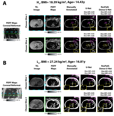 |
A Densely Connected Neural Network with Frequency Balancing Loss for Adipose Tissue Segmentation in Children using Free-Breathing Abdominal MRI
Sevgi Gokce Kafali1,2, Shu-Fu Shih1,2, Xinzhou Li1,2, Tess Armstrong1, Kelsey Kuwahara3, Sparsha Govardhan4, Karrie V Ly4, Shahnaz Ghahremani1, Kara L Calkins4, and Holden H Wu1,2
1Radiological Sciences, University of California, Los Angeles, Los Angeles, CA, United States, 2Bioengineering, University of California, Los Angeles, Los Angeles, CA, United States, 3Cognitive Science, University of California, Los Angeles, Los Angeles, CA, United States, 4Pediatrics, University of California, Los Angeles, Los Angeles, CA, United States
Obese children have larger amounts of subcutaneous and visceral adipose tissue (SAT, VAT) and are at high risk for cardiometabolic disease. The reference standard to analyze SAT/VAT uses breath-held (BH) abdominal MRI for manual annotation of SAT/VAT. In children, the BH requirement and spatially varying VAT distribution are major challenges for body composition analysis. This work proposed a densely connected neural network with a class frequency balancing, boundary emphasizing loss to segment SAT/VAT using free breathing abdominal MRI in children.
|
|||
2264.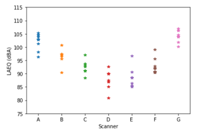 |
Survey of Acoustic Output in Neonatal Brain Protocols
Hannah Kurdila1, Tayeb Zaidi1, Ting Zhang1, Subha Maruvada1, and Sunder Rajan1
1Food and Drug Administration, Silver Spring, MD, United States
The purpose of this study was to determine the expected sound exposure to the neonate during neonatal brain protocols. To accomplish this, 7 neonatal brain protocols were recorded on 7 different MRI machines across 4 vendors. Neonatal protocol sound levels straddled existing notions of risk, exceeding sound levels known to cause non-auditory stress responses in neonates but not exceeding the IEC MRI hearing safety limit. These results indicate that these sound levels could be risky for the neonate, but that further work is required to clarify this.
|
|||
2265.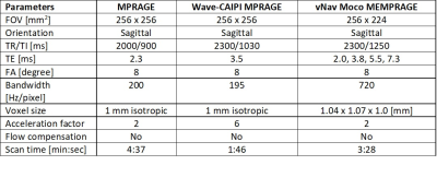 |
Prospectively motion-corrected multi-echo MPRAGE and wave-controlled aliasing in parallel imaging MPRAGE for pediatrics motion suppression
Emi Niisato1, Yung-Chieh Chen2, Bhat Himanshu3, Wei Liu4, Daniel Nicolas Splitthoff5, and Cheng-Yu Chen2
1Siemens Healthcare Limited, Taiwan, Taipei, Taiwan, 2Taipei Medical University Hospital, Taipei, Taiwan, 3Siemens Medical Solutions, Malvern, PA, USA, Malvern, PA, United States, 4Siemens Shenzhen Magnetic Resonance Ltd., Shenzhen, China, Shenzhen, China, 5Siemens Healthcare GmbH, Erlangen, Germany, Erlangen, Germany
Pediatric patients are preferred to be scanned with protocols which are resistant to motion artifact or which complete in a short scan time. We investigated the extent of motion artifact suppression in pediatric patients using modified multi-echo Magnetization Prepared Rapid Acquisition Gradient Echo (MEMPRAGE) with prospective motion correction using 3D EPI volumetric navigators (vNav Moco) by comparing with wave-controlled aliasing in parallel imaging (wave-CAIPI) MPRAGE. Visual assessment revealed vNav Moco MEMPRAGE significantly suppressed head motion artifacts compared with wave-CAIPI MPRAGE and conventional MRPAGE.
|
|||
2266.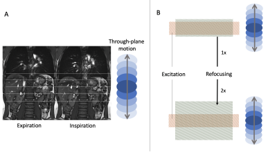 |
Free-breathing T2-weighted multi-shot TSE BLADE for pediatric abdominal imaging
Fedel Machado-Rivas1,2, Daniel J Park3, John Conklin1,2, John E Kirsch2,3, and Michael S Gee1,2
1Radiology, Massachusetts General Hospital, Boston, MA, United States, 2Radiology, Harvard Medical School, Boston, MA, United States, 3MGH Martinos Center for Biomedical Imaging, Boston, MA, United States
Abdominal MRI respiratory triggered acquisitions in pediatric populations often fail due to shallow or irregular breathing patterns, degrading image quality and increasing acquisition time. A free-breathing T2-weighted multi-shot TSE BLADE acquisition could overcome these limitations, but through plane motion of respiration causes substantial signal loss from long T2 (such as biliary ducts). We hypothesized that by modifying the refocusing pulse width of the sequence we could mitigate signal loss. Our results show that a 2x refocusing pulse modified free breathing sequence provides comparable image quality and contrast. Furthermore, it offers a fixed acquisition time independent of respiratory patterns.
|
|||
2267.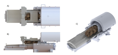 |
Design of a neonatal head and cardiac imaging system and parallel-transmit volume/receive phased array for 7T MRI during early infancy
Jérémie Clément1, Kathleen Colford2, Emer Hughes2, Tomoki Arichi2,3,4, David Edwards2,3,5, Joseph Hajnal1,2, and Ozlem Ipek1
1Biomedical Engineering, Kings College London, London, United Kingdom, 2Centre for the developing brain, Kings College London, London, United Kingdom, 3Bioengineering, Imperial College London, London, United Kingdom, 4Paediatric neurosciences, Evelina London children's hospital, Guy's and St Thomas' NHS Foundation Trust, London, United Kingdom, 5MRC Centre for Neurodevelopmental Disorders, Kings College London, London, United Kingdom Magnetic resonance imaging (MRI) of the developing brain and heart could greatly benefit from the higher signal levels and improved tissue contrast at 7T. However, there are currently no dedicated RF coils and associated patient handling for young infants (<3-months). We describe design criteria and solutions to build the first mechanical frame and coil formers for imaging the brain and heart in infants up to 3-months of age in an integrated manner at 7T. The different parts incorporate important design aspects including mechanical sturdiness/safety, toxicity, sanitization and ease of use. |
|||
2268. |
Damping of acoustic noise for neonatal MRI at 7 Tesla
Erik Huijing1, Evita Wiegers1, Dennis Klomp1, Fredy Visser1,2, Edwin Versteeg1, Koenraad Rhebergen3, Kim Annink4, Niek van der Aa4, Floris Groenendaal4, Jeroen Dudink4, Thomas Alderliesten4, Maarten Lequin1, Manon Benders4,
and Jannie Wijnen1
1Radiology, University Medical Center Utrecht, Utrecht, Netherlands, 2Philips HealthCare, Best, Netherlands, 3Otorhinolaryngology and Head & Neck Surgery, University Medical Center Utrecht, Utrecht, Netherlands, 4Neonatology, University Medical Center Utrecht, Utrecht, Netherlands
We designed a new acoustic hood for neonatal MRI exams at 7 Tesla. The new design is lightweight and allows easy access to the neonate during the MRI examination. We were able to optimize the new acoustic hood to a noise reduction of 10.6dB, exceeding the damping of a prototype with damping of 3.1dB. The new hood was designed to fit a head coil and allows the use of a ventilation tube.
|
|||
2269.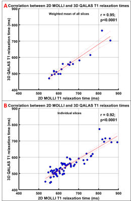 |
3D Liver T1 Quantification using Interleaved Look-Locker Acquisition with T2 Preparation Pulse Sequence (3D-QALAS): Comparison with 2D-MOLLI
Deep B. Gandhi1, Amol Pednekar1, Hui Wang2, Jean A. Tkach1, Andrew T. Trout1, and Jonathan R. Dillman1
1Imaging Research Center, Department of Radiology, Cincinnati Children's Hospital Medical Center, Cincinnati, OH, United States, 2MR Clinical Science, Philips, Cincinnati, OH, United States
Hepatic T1 relaxation values have been shown to correlate with hepatic fibrosis. The established 2D T1 mapping techniques require multiple breath-holds to quantify whole liver T1. 3D whole liver T1 quantification in a single breath-hold using an interleaved Look-Locker acquisition sequence with T2 preparation pulse (3D-QALAS) correlates very strongly (r=0.95) with T1 values obtained with a 2D Modified Look-Locker Acquisition (2D-MOLLI). However, 3D-QALAS underestimated T1 significantly (p<0.0001) compared to 2D-MOLLI with a mean bias of 92.5 ms (14.2%). 3D-QALAS has potential to measure T1 of the whole liver in a single breath hold, while simultaneously providing T2 relaxation values.
|
|||
2270.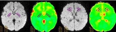 |
DWI and 3D SWAN improve the knowledge of pathophysiological mechanism of neonatal bilirubin encephalopathy
Jinhong Yu1, Yanwei Miao1, Yangyingqiu Liu1, Bingbing Gao1, and Yu Bing1
1The First Affiliated Hospital of Dalian Medical University, Dalian, China
Exposure to high levels of bilirubin can cause serious severe motor symptoms and cerebral palsy,but the pathophysiological mechanism of brain injury is still unclear.The aim of this study was to explore the possible pathophysiological mechanism of brain tissue injury using diffusion weighted imaging (DWI) and three dimensions T2* weighted angiography( 3D SWAN).
|
|||
2271.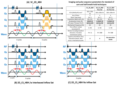 |
Liver Stiffness Measurement in Less Than Half the Conventional Breath-hold Time: Wave Polarity-Inversion Motion Encoding and Compressed SENSE
Amol Pednekar1, Deep B. Gandhi2, Hui Wang3, Jean A. Tkach1, Andrew T. Trout1, and Jonathan R. Dillman1
1Department of Radiology, Cincinnati Children's Hospital Medical Center, Cincinnati, OH, United States, 2Imaging Research Center, Department of Radiology, Cincinnati Children's Hospital Medical Center, Cincinnati, OH, United States, 3MR Clinical Science, Philips, Cincinnati, OH, United States
2D GRE MRE liver images acquired at 4 transverse levels in breath-hold times >13s per slice is currently standard of care (SC_2D_4BH). Inadequate breath-holds lead to inaccurate stiffness estimation and/or failed studies. The combination of wave polarity-inversion motion encoding and compressed-SENSE enables MRE images to be acquired in less than half the breath-hold time (2D_CS_HBH) e.g. <7s, with identical spatial resolution and field of view. In 19 participants, mean liver shear stiffness values estimated with SC_2D_4BH and 2D_CS_HBH correlated very strongly (ICC>0.96) with a bias of <0.15 kPa (<6%). 2D_CS_HBH MRE is beneficial in participants with compromised breath-holding capacity.
|
|||
2272.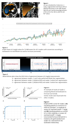 |
MRI T2* liver iron concentration measurement in children comparison using Wood or Garbowski (T2*-LIC) conversions and Ferriscan (R2-LIC)
Dianna ME Bardo1, Nicholas Rubert1, Mittun Patel1, Shiza Shahid1, and Robyn Augustyn1
1Radiology, Phoenix Children's Hospital, Phoenix, AZ, United States
LIC analysis may be performed with accurate and reproducible results in children using an ROI or a whole liver segmentation technique using locally available software and the FerriScan® technique.
|
|||
2273.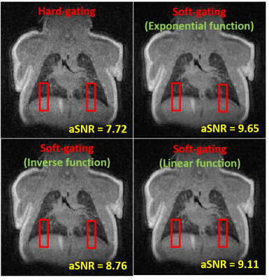 |
Comparison of Respiratory-Gating Weighting Algorithms in Neonatal Pulmonary UTE-MRI
Deep B. Gandhi1, Nara S. Higano2, Andrew D. Hahn3, Luis Torres3, Sean B. Fain3, Jason C. Woods2, and Alister J. Bates2
1Department of Radiology, Cincinnati Children's Hospital Medical Center, Cincinnati, OH, United States, 2Pulmonary Medicine, Cincinnati Children's Hospital Medical Center, Cincinnati, OH, United States, 3Department of Medical Physics, University of Wisconsin, Madison, WI, United States
Pulmonary 1H ultrashort echo-time (UTE)-MRI provides clinically relevant structural information in neonates with lung disease of prematurity (bronchopulmonary dysplasia, BPD) and has strong, unexplored potential to evaluate regional pulmonary function. Retrospective respiratory-gated UTE-MRI allows reconstructed images from different phases of the breathing cycle. Here, we compared four retrospective gating approaches in 3 BPD subjects with different weighted time intervals: 1 hard-gating and 3 soft-gating (exponential, inverse and linear weighting functions). Overall, linearly weighted soft-gating provided better compromise between apparent SNR (aSNR) and motion-blurring. This optimized respiratory-gating approach opens the door for improved understanding of regional pulmonary function deficits in neonates.
|
|||
2274.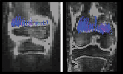 |
Dffusion tensor imaging of the physis and metaphisys as predictor of child growth
Diego M Jaramillo1, Phuong M Duong1, Jie C Nguyen2, Sogol Mostoufi-Moab2, Michael K Nguyen2, Andrew Moreau2, Christian A Barrera3, Shijie Hong2, and Jose M Raya4
1Columbia University Medical Center, New York, NY, United States, 2Children’s Hospital of Philadelphia, Philadelphia, PA, United States, 3Massachusetts General Hospital, Boston, MA, United States, 4New York University, New York, NY, United States
Prediction of growth potential in the pediatric population is critical to trace therapies targeting growth deficiencies and to inform surgical planning. Clinical models to predict growth potential are very inaccurate. We aim to validate DTI of the physis and metaphysis (DTI-P/M) as a prediction biomarker of growth potential in children. We compared in a cohort of 90 children the prediction accuracy of DTI-P/M and clinical models. Our data showed that DTI-P/M predicted growth potential more accurately than clinical models (over 40% reduction in error). Even more importantly, compared to clinical models DTI-MP predictions did not show any prediction bias.
|
|||
2275.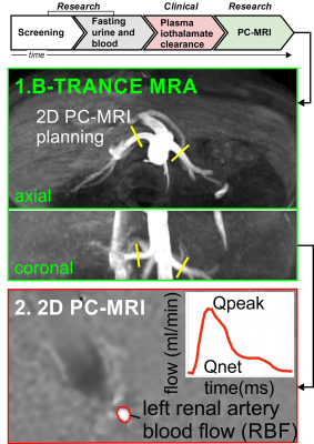 |
Estimation of Glomerular Filtration Rate in a Pediatric Population using Renal Phase Contrast MRI
Alex J Barker1, Lorna P Browne1, Michal Schafer2, Erin K Englund2, Takashi Fujiwara2, Kristen J Nadeau2, and Petter Bjornstad2
1Radiology, University of Colorado, Anschutz Medical Campus, Aurora, CO, United States, 2University of Colorado, Anschutz Medical Campus, Aurora, CO, United States
Pediatric patients may experience inaccurate estimates of glomerular filtration rate (GFR) by serum creatinine or cystatin C. Renal blood flow (RBF) is a key physiological determinant of GFR, thus, phase-contrast MRI (PC-MRI) is a promising non-invasive technique to investigate relationships to GFR. An interim prospective analysis (n=18) is presented, which investigates RBF as measured by PC-MRI in pediatric bone-marrow transplant candidates who also underwent gold standard measurement of GFR by 125I iothalamate clearance (GFR-iothalamate). The correlation between GFR-iothalamate and RBF by PC-MRI was markedly superior to that of the commonly used endogenous filtration markers serum creatinine or cystatin C.
|
|||
2276.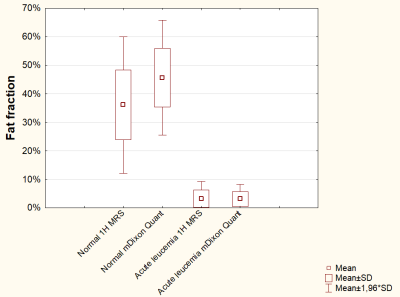 |
Quantitative bone marrow MRI in children with leukaemia
Nataliia Kriventsova1, Petr Menshchikov2, Dmitry Kupriyanov2, Dmitry Litvinov1, Galina Novichkova1, and Galina Tereshchenko1
1Dmitry Rogachev National Research Center of Pediatric Hematology, Oncology and Immunology, Moscow, Russian Federation, 2Philips Healthcare, Moscow, Russian Federation
In our study, we quantified the bone marrow fat fraction (FF) using mDixon-Quant MRI and MR Spectroscopy. The FF values in the bone marrow in children with acute leukemia compared with children of the same age without hematological diseases were significantly different. The value of the fat fraction may become an MR-biomarker of bone marrow in children with acute leukemia.
|
|||
2277.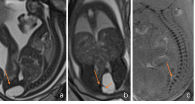 |
Diagnosis, Characterization of Fetal Sacrococcygeal Teratomas (Type IV) with Prenatal MRI and Its Outcome
Xianyun Cai1, Jinxia Zhu2, and Guangbin Wang1
1Shandong Medical Imaging Research Institute, Shandong University, Jinan, China, China, 2MR Collaboration, Siemens Healthcare Ltd., Beijing, China, China
This study investigated the prenatal diagnosis and prognosis of fetal sacrococcygeal teratomas (SCTs) (Type IV) with Prenatal MRI. Sixteen fetuses with suspected Type IV teratomas were included and followed up in this study. The results showed that prenatal MRI could effectly diagnosed fetal Type IV SCTs, which have a bright outcomes postnatally after surgery resection. This suggests correct diagnosis of teratoma on prenatal MRI and early operation after birth are important for prognosis.
|
|||
2278.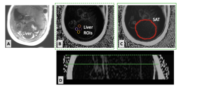 |
Quantifying Fetal and Maternal Body Composition Using 3-D Stack-Of-Radial Free-Breathing MRI
Katie M Strobel1, Sevgi Gokce Kafali2, Shu-Fu Shih2, Rinat Masamed2, Kara Calkins1, and Holden Wu2
1Pediatrics, University of California Los Angeles, Los Angeles, CA, United States, 2Radiological Sciences, University of California Los Angeles, Los Angeles, CA, United States
This pilot prospective cohort study investigated fetal and maternal body composition in the third trimester of pregnancy using 3D stack-of-radial free-breathing MRI. Nine women with healthy pregnancies, gestational diabetes, or fetal growth restriction were included. Fetal subcutaneous fat and liver fat, as well as maternal liver fat, subcutaneous fat, and visceral fat were successfully measured. Maternal body composition was associated with fetal body composition. Pregnancies complicated by gestational diabetes appear to alter fetal body composition.
|
The International Society for Magnetic Resonance in Medicine is accredited by the Accreditation Council for Continuing Medical Education to provide continuing medical education for physicians.