Digital Posters
Making MRI More Accessible
ISMRM & SMRT Annual Meeting • 15-20 May 2021

| Concurrent 3 | 15:00 - 16:00 |
3811.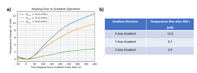 |
Investigation of Gradient-Induced Heating of a Cryogen-Free Magnet
Diego Felipe Martinez1, Arjama Halder1,2, Will Bradfield Handler1, and Blaine Alexander Chronik1,2
1The xMR Labs, Physics and Astronomy, Western University, London, ON, Canada, 2Medical Biophysics, Western University, London, ON, Canada
Cryogen-free low-field MR systems can address accessibility issues associated with MRI as they can relax siting requirements and lower operating costs. An important concern in these systems is heating of the magnet during imaging sequences, which could cause field stability issues or quench the magnet. The interaction between the gradient system and the main magnet was investigated using temperature probes. Trapezoidal based pulses were run on components of the gradient at various strengths, showing the x-axis coil as the most impactful on the main magnet. In all experiments, the heating was <0.015 K, with “worst case” heating guiding further study.
|
|||
3812.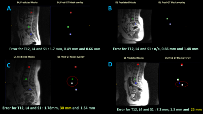 |
Deep Learning based spine labeling with three-plane 2D localizers without vertebrae segmentation
Dattesh Dayanand Shanbhag1, Arathi Sreekumari1, Soumya Ghose2, Chitresh Bhushan2, and Uday Patil3
1GE Healthcare, Bangalore, India, 2GE Global Research, Niskayuna, NY, United States, 3General Electric Company, Bangalore, India
In this work , we describe a deep learning-based methodology to generate vertebrae labels directly from the standard 2D tri-planar localizer images without the need any additional scanning or explicitly segmenting the vertebrae. This is accomplished by using deep-learning setup a to identify vertebrae labels directly on the localizer images. The method is demonstrated on lumbar spine localizer data to identify Thoracic-12 (T12), Lumbar-4 (L4) , and Sacral-1 (S1) vertebrae locations. In a test cohort of 50 lumbar MR spine exams, we report labeling accuracy of 92%, 98% and 96% for T12, L4 and S1 vertebrae respectively on localizer images.
|
|||
3813.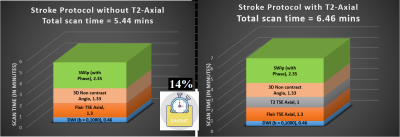 |
Evaluating the role of T2-weighted-imaging in acute ischemic stroke to accelerate routine Stroke-MRI protocol
Sanjay Dhawan1,2, Ishrat Afshan1,2, Rupsa Bhattacharjee3, and Indrajit Saha3
1Clearmedi Healthcare Pvt. Ltd., Gurugram, India, 2Department of Radiology, Paras Hospitals, Gurugram, India, 3Philips Health Systems, Philips India Limited, Gurugram, India
The focus of MRI stroke-research is concentrated in identifying relevant sequence for stroke-screening, accelerating the acquisitions by latest innovations like Compressed-SENSE and developing methodologies to extract meaningful clinical-relevance (i.e., penumbra or mismatch-ratio) using minimum number sequences. Our objective is to evaluate the impact of including T2-weighted-images in fast acute-ischemic-stroke MRI protocols, to comment on role of T2w vis-à-vis Flair in terms of clinical findings for making an informed decision on the inclusion-criteria of ideal MR sequences in stroke MR protocol. Flair proves to be better than T2, in depicting small/subtle signal changes in critical time-window, beneficial for acute-stroke patient-treatment-planning.
|
|||
3814.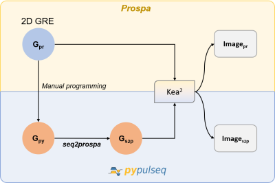 |
Seq2prospa: translating PyPulseq for low-field imaging
Keerthi Sravan Ravi1,2, Thomas O'Reilly3, John Thomas Vaughan Jr.2, Andrew Webb3, and Sairam Geethanath2
1Biomedical Engineering, Columbia University, New York, NY, United States, 2Columbia Magnetic Resonance Research Center, New York, NY, United States, 3Leiden University Medical Center, Leiden, Netherlands
Low-field MRI scanners are more viable in resource-strained regions such as Africa, sacrificing SNR for gains on the economics of purchase, siting and service. Pulse sequences need to be optimized for maximum efficiency on these scanners. This work introduces seq2prospa, a Python-based tool facilitating the conversion of pulse sequences designed using PyPulseq into a low-field spectrometer-friendly format. We perform an in-vitro and in-vivo acquisition experiment to image a structural phantom and a human hand respectively. Data acquired from a 2D GRE sequence designed natively in Prospa is compared with data acquired from a PyPulseq-converted 2D GRE sequence.
|
|||
3815. |
MRS metabolomics screening of human lung cancer with blood serum collected prior to disease diagnosis
L Cheng1, Tjada Schult1, Mara Lauer1, Yannick Berker2, Marcella Cardoso1, Lindsey Vandergrift1, Piet Habbel3, Johannes Nowak4, Martin Aryee1, Mari Mino-Kenudson1, and David Christiani5
1Mass General Hospital, Boston, MA, United States, 2German Cancer Research Center, Heidelberg, Germany, 3Charite Medical University, Berlin, Germany, 4Julius-Maximilians University, Wuerzburg, Germany, 5Harvard T.H. Chan School of Public Health, Boston, MA, United States
Lung cancer (LuCa), the leading cause of cancer deaths, are often diagnosed late due to the lack of screening. Low-dose spiral CT can detect small and early stage LuCa lesions, but cannot practically be used as an annual LuCa screening tool. Metabolomics detects global metabolite variations under physiology and pathology. Metabolomic profiles measured from blood may reveal LuCa at early stages as a screening tool to triage suspicious patients to CT tests. Blood sera obtained from LuCa patients prior to their diagnosis were studied with MRS to establish LuCa screening metabolomic profiles to discover LuCa earlier and reduce death rates.
|
|||
3816. |
MRI is superior to CT for liver staging in colon cancer and should be investigated as the definitive liver staging modality in colon cancer
Alexandra W. Acher1, Emily Winslow2, and Scott B. Reeder1
1University of Wisconsin School of Medicine and Public Health, Madison, WI, United States, 2Georgetown University, Washington DC, MD, United States
Colon cancer is the 3rd most common cancer in the United States. Unfortunately, most patients present with Stage III or IV disease and disease recurrence is high despite advanced in systemic therapies. Despite evidence that gadoxetic acid-enhanced MRI is superior for the detection and diagnosis of colon cancer liver metastases, computed tomography remains the recommended staging and surveillance modality. The purpose of this abstract is to synthesize and analyze the evidence regarding the accuracy of liver MRI to stage and surveil CCLM, and to identify knowledge gaps to inform future research on staging and surveillance imaging for CCLM.
|
|||
3817.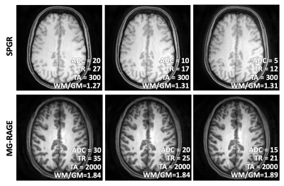 |
Towards Automated Scanning: A Framework for Maintaining Constant SNR and T1-contrast for SPGR and MP-RAGE
Dahan Kim1, Dinghui Wang1, and James G Pipe1
1Department of Radiology, Mayo Clinic, Rochester, MN, United States
We present a framework for maintaining constant SNR and T1 contrast in SPGR and MP-RAGE scans that employ non-identical scan parameters. Central to this work are the concept of time constant TA and simplified SNR expression. By adjusting the flip angle to keep TA constant and adjusting the number of spiral arms to match SNR, we demonstrated constant SNR and T1 contrast under varied ADC time, TR, and FOV. This work ultimately aims for reducing image variability, automating scanning, reducing technologist intervention, and increasing patient safety.
|
|||
3818.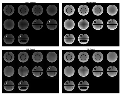 |
Validating Open-Source MR Sequences for Reproducible Research
Gehua Tong1, Sairam Geethanath2, Enlin Qian1, and John Thomas Vaughan, Jr. 2
1Biomedical Engineering, Columbia University, New York, NY, United States, 2Columbia Magnetic Resonance Research Center, Columbia University, New York, NY, United States
A framework for testing, validating, and sharing open-source MR sequences was developed to improve their accessibility, repeatability, and safety. Accessibility is improved by requiring documentation of sequence usage and data processing steps, repeatability by requiring test experiments and examples to replicate, and safety by requiring simulation or records of SAR and PNS levels. Forms and guidelines are provided to help developers and users package, share, and apply novel sequences efficiently. The framework was demonstrated for two common sequences, Inversion Recovery Spin Echo (IRSE) and Turbo Spin Echo (SE), and they were packaged and shared in an open-source repository.
|
|||
| 3819. | A National Clinical Research MRI Quality Assurance and Quality Control Survey by NCITA
Penny L Hubbard Cristinacce1,2, Damien J McHugh1,2, Matthew R Orton2,3, Arnold JV Benjamin2,4, Ross A Little1,2, James P. B. O'Connor1,2,3, and Maite Jauregui-Osoro2,5
1The University of Manchester, Manchester, United Kingdom, 2National Cancer Imaging Translational Accelerator (NCITA), CRUK, United Kingdom, 3Institute of Cancer Research, Sutton, United Kingdom, 4University of Cambridge, Cambridge, United Kingdom, 5Imperial College London, London, United Kingdom National Cancer Imaging Translational Accelerator (NCITA) ran a survey on quality assurance (QA) and quality control (QC) procedures for human MRI scanners and associated equipment at research institutions across the UK. The survey was conducted by the NCITA QA/QC Unit, which is developing an MRI core lab to facilitate the validation and standardisation of quality-assured imaging biomarkers from first-in-human studies to the assessment of multicentre reproducibility. The results showed institutions emphasise the earlier part of the QA/QC pipeline (i.e. scanner maintenance and data acquisition QA), with fewer institutions performing comprehensive QC on acquired/analysed data or any software quality management. |
|||
3820.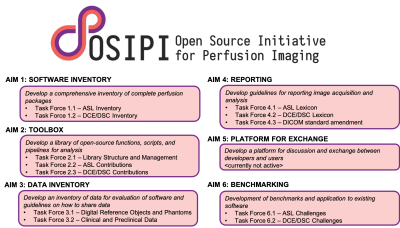 |
The Open Source Initiative for Perfusion Imaging (OSIPI)
Laura C. Bell1, Henk Mutsaerts2, Andrey Fedorov3, Zaki Ahmed4, Patricia Clement5, Simon Levy6, Frank G Zollner7, Jan Petr8, Sudipto Dolui9, Kathleen Schmainda10, Melissa Prah10, Matthias Schabel11, Ananth Madhuranthakam12, Li
Zhao13, Michael Thrippleton14, Petra van Houdt15, James Holmes16, C. Chad Quarles1, Greg Cron17, David L Thomas18, Yuriko Suzuki19, Ina Kompan20, David L Buckley21, Paula Croal22, Udunna Anazodo23, Anahita Fathi Kazerooni9,
Hamidreza Saligheh Rad24, Charlotte Debus25, and Steven P Sourbron26
1Barrow Neurological Institute, Phoenix, AZ, United States, 2Amsterdam University Medical Cente, Amsterdam, Netherlands, 3Brigham and Women’s Hospita, Boston, MA, United States, 4Mayo Clinic, Rochester, MN, United States, 5Ghent University, Ghent, Belgium, 6Aix-Marseille Univ, Marseille, France, 7Heidelberg University, Mannheim, Germany, 8Helmholtz-Zentrum Dresden-Rossendorf, Dresden, Germany, 9University of Pennsylvania, Philadelphia, PA, United States, 10Medical College of Wisconsin, Milwaukee, WI, United States, 11Oregon Health and Science University, Portland, OR, United States, 12UT Southwestern Medical Center, Dallas, TX, United States, 13Children’s National Medical Center, Washington DC, DC, United States, 14University of Edinburgh, Edinburgh, United Kingdom, 15the Netherlands Cancer Institute, Amsterdam, Netherlands, 16University of Wisconsin - Madison, Madison, WI, United States, 17Ottawa Hospital Research Institute, Ottawa, ON, Canada, 18University College London, London, United Kingdom, 19University of Oxford, Oxford, United Kingdom, 20German Cancer Research Center, Heidelberg, Germany, 21University of Leeds, Leeds, United Kingdom, 22University of Nottingham, Nottingham, United Kingdom, 23Western University, London, ON, Canada, 24Tehran University of Medical Sciences, Tehran, Iran (Islamic Republic of), 25Karlsruhe Institute of Technology, Karlsruhe, Germany, 26University of Sheffield, Sheffield, United Kingdom
Open Source Initiative for Perfusion Imaging (OSIPI) was founded by the ISMRM Perfusion Study Group as a community-driven initiative. Supported by six distinct aims, its mission is “to promote the sharing of perfusion imaging open-source software in order to eliminate the practice of duplicate development, improve the reproducibility of perfusion imaging research, and speed up the translation into tools for discovery science, drug development, and clinical practice”. OSIPI seeks to provide centralized resources to deliver reproducible perfusion research. In addition, it provides a platform for exchange where new and more advanced methods may be validated for perfusion accuracy.
|
|||
3821.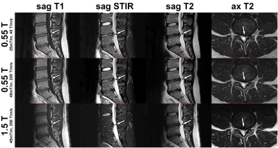 |
High-Performance Low-Field MRI of the Lumbar Spine: Comparison of 0.55T MRI With Two Gradient Systems To 1.5T MRI in Humans
Mohammad Samim1, Mahesh Bharath Keerthivasan2, Iman Khodarahmi1, Marisa Ilag1, Mary Bruno1, Hersh Chandrana1, Inge Brinkmann2, and Jan Fritz1
1Department of Radiology, NYU Langone Medical Center, New York, NY, United States, 2Siemens Medical Solutions USA Inc, Malvern, PA, United States
Next-generation low-field magnetic resonance imaging (MRI) systems hold great potential to reduce the cost of ownership and improve access to MRI worldwide. We compared MRI of the lumbar spine at 0.55T MRI with two different gradient performance modes to 1.5T MRI in 10 volunteer participants. Regardless of field strength and gradient performances, all MRI studies had sufficient signal-to-noise-ratios, contrast-to-noise-ratios, image quality, and visibility of anatomical structures with only small differences. Our initial results support the great potential of next-generation low-field MR systems to achieve similar image quality than state-of-the-art clinical 1.5-T MRI systems to detect the lumbar spine abnormalities.
|
|||
3822.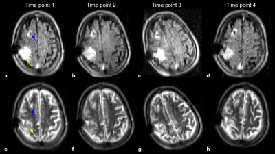 |
Point-of-Care Brain MRI: Clinical Application in a Stroke Rehabilitation Center
Samantha By1, E. Brian Welch1, Houchun Harry Hu1, Rafael O'Halloran1, Laura Sacolick1, Carole Lazarus1, Hadrien Dyvorne1, Jonathan Rothberg1, Khan Siddiqui1, and Lorraine Cullen2
1Hyperfine, Guilford, CT, United States, 2Gaylord Specialty Healthcare, Wallingford, CT, United States
Portable, point-of-care MRI offers many new opportunities, such as scanning at the bedside or serial monitoring of a patient (i.e. daily or weekly). We demonstrate feasibility and initial results of a portable 64 mT MRI in a stroke rehabilitation setting. Six recovering stroke patients received such exams; four of them were serially monitored, each receiving four exams. Each exam consisted of T2-W, T1-W, FLAIR and DWI imaging. Different cases are highlighted to demonstrate the utility of point-of-care MRI in a rehabilitation setting.
|
|||
3823.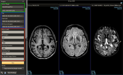 |
Advanced image processing outside the academia: The integration of ExploreASL into the workflow of an outpatient imaging center
Nandor K Pinter1,2, Sandeep Ganji3, Jan Petr4, Bela Ajtai 5, Harry Friel3, Joseph Fritz5, Laszlo Mechtler5, Alexander Fischer6, Frederik Barkhof7,8, and Henri Mutsaerts7,9
1Dent Neurologic Institute, Buffalo, NY, United States, 2Neurosurgery, University at Buffalo, Buffalo, NY, United States, 3Philips Healthcare, Gainesville, FL, United States, 4Helmholtz-Zentrum Dresden-Rossendorf, Dresden, Germany, 5Dent Neurologic Institute, Amherst, NY, United States, 6Philips Research Europe, Aachen, Germany, 7Dept of Radiology and Nuclear Medicine, Amsterdam University Medical Center, Amsterdam, Netherlands, 8Institute of Neurology, University College London, London, United Kingdom, 9Ghent Institute for Functional and Metabolic Imaging, Ghent, Belgium
While neurological imaging is mostly done in non-academic centers, the lack of academic resources, special image processing skillset and the fast paced workflow prevent radiologists in these data-rich environments from engaging in high quality clinical research that utilizes quantitative imaging. Clinical adoption of Arterial Spin Labeling could benefit from providing easy-to-use, PACS connected image processing solutions that do not require neuroscience background and provide truly quantified Cerebral Blood Flow values in any outpatient center or public hospital. Our goal is to create such a solution and bridge the gap between academic research and real world practice.
|
|||
3824.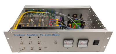 |
A low-cost battery-powered tri-axial current-controlled gradient power supply for sustainable and portable MRI
Thomas O'Reilly1, Danny de Gans2, Lars Leenheer2, and Andrew Webb1
1C.J. Gorter Center for High Field MRI, Leiden University Medical Center, Leiden, Netherlands, 2DEMO, Delft University of Technology, Delft, Netherlands
Low field MRI has gained interest in recent years as a way of providing MRI to under-served and remote settings. MR hardware designed for such applications should be robust, portable and easy to maintain. In this work we present the design of a three-axis gradient amplifier capable of an output current of up to 15 amperes. The gradient amplifier is designed to run off two car batteries and consumes less than 100 watts during imaging. The components in the gradient amplifier are designed to be easily repairable and commonly available.
|
|||
3825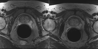 |
Repeatability of DWI and DCE-MRI of Prostate in an Active Surveillance Population Video Permission Withheld
Jack C. Williams1, Matthew Gibbons1, Janet E. Cowan2, Peter R. Carroll2, and Susan M. Noworolski1
1Radiology and Biomedical Imaging, University of California San Francisco, San Francisco, CA, United States, 2Urology, University of California San Francisco, San Francisco, CA, United States
This study attempted to fill the dearth of literature establishing the repeatability of DWI and DCE-MRI using a population of non-progressing men on active surveillance for prostate cancer. MRIs from men (n=26) with two DCE-MRI scans in one year were examined and mean ROI values on different imaging maps were analyzed for percent repeatability coefficient (%RC). %RC was lowest for ADC, DCE MRI peak enhancement, T2-weighted images and pharmacokinetically modeled ve and Ktrans.
|
|||
3826.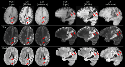 |
Low-to-High Field MR Image Quality Transfer
Hongxiang Lin1, Matteo Figini1, Felice D'Arco2, Godwin Ogbole3, David W Carmichael4,5, Ikeoluwa Lagunju6, Helen Cross4,7, Delmiro Fernandez-Reyes6,8, and Daniel C Alexander1
1Centre for Medical Image Computing, Department of Computer Science, University College London, London, United Kingdom, 2Department of Radiology, Great Ormond Street Hospital, London, United Kingdom, 3Department of Radiology, College of Medicine, University of Ibadan, Ibadan, Nigeria, 4Great Ormond Street Institute of Child Health, University College London, London, United Kingdom, 5Department of Biomedical Engineering, King’s College London, London, United Kingdom, 6Department of Paediatrics, College of Medicine, University of Ibadan, Ibadan, Nigeria, 7Great Ormond Street Hospital for Children, London, United Kingdom, 8Department of Computer Science, University College London, London, United Kingdom
We devise and demonstrate an image quality transfer (IQT) system to estimate, from low-field (<0.5T) magnetic resonance (MR) structural images, the corresponding high-field (1.5T or 3T) images. The intended application scenario is to improve clinical lesion-detection, classification, and decision-making in childhood epilepsy in lower and middle income countries where T1w, T2w and FLAIR sequences on 0.36T scanners remain the clinical standard. Results on synthetic and real data verify substantial resolution and contrast enhancements, aiding conspicuity of pathological lesions.
|
|||
3827.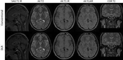 |
A Paradigm shift in MR physics with Deep Learning Reconstruction: higher image quality and spatial resolution in shorter scan time
Mo Kadbi1, Dawn Berkeley1, Brian Tymkiw1, Hung Do1, and Erin Kelly1
1Canon Medical System USA, Tustin, CA, United States
In MR physics, there is a fundamental tradeoff between image spatial resolution, signal to noise ratio, and scan time. To acquire images with high resolution and SNR, signal averaging is the most common solution, but results in longer scan time. In this study, a Deep Learning Reconstruction method was employed to remove the noise from clinical images and improve SNR. This SNR improvement was devoted to increase the spatial resolution without the need of signal averaging and increased scan time. Hence, higher resolution images with high image quality can be obtained in shorter time in clinical practice.
|
|||
3828.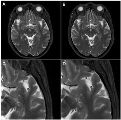 |
Accelerating Neuroradiology Protocols with Deep Learning MR Image Reconstruction. Which Methods Result in the Highest Perceived Image Quality?
Gregory Avey1, Laura Eisenmenger1, Nathan Kim1, Alexey Samsonov1, James Holmes1, Lloyd Estkowski2, and Tabassum Kennedy1
1University of Wisconsin School of Medicine and Public Health, Madison, WI, United States, 2GE Healthcare, Madison, WI, United States
Deep Learning (DL) MRI image reconstruction holds great promise in improving overall MRI image quality and decreasing examination time. Given the nonlinear properties of DL reconstruction it is unknown which methods of decreasing MRI examination time are most suitable when adapting current protocols for DL based examination acceleration. 10 volunteers were scanned with clinical baseline T1 and T2 weighted FSE exams, along with 5 accelerated exam types. The resulting exams were scored to determine differences in image quality. DL image reconstruction allowed for an up to 78% reduction in scan time while matching baseline subjective image quality.
|
|||
3829.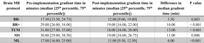 |
Value of Adopting Fast Brain MRI Techniques for Outpatient Brain MRI: Proof of Principle and Operational Impact During the COVID-19 Pandemic
Min Lang1, Samuel CD Cartmell1, Azadeh Tabari1, Daniel Briggs1, Oleg Pianykh1, John E Kirsch1, Stephen F Cauley1, Wei-Ching Lo2, Seretha J Risacher1, Augusto Lio Goncalves Filho1, Marc D Succi1,3, Otto Rapalino1, Pamela Schaefer1,
John Conklin1, and Susie Y Huang1
1Department of Radiology, Massachusetts General Hospital, Boston, MA, United States, 2Siemens Medical Solutions, Boston, MA, United States, 3Medically Engineered Solutions in Healthcare Incubator, Massachusetts General Hospital, Boston, MA, United States
We report our clinical experience of implementing fast MRI sequences into the most performed brain MRI protocols at a large academic center, which resulted in significant decrease of gradient times on both 1.5T and 3T scanners. The overall scan times were reduced by up to 40% for the longest examinations, with all included brain MRI protocols now being under 20 minutes. The potential benefits of reduced scan time include increased imaging volume throughput and improved patient access. During the ongoing COVID-19 pandemic, the decreased scan times also accommodated heightened infection control protocols to help protect both patients and healthcare workers.
|
|||
3830. |
Abdominal MR imaging on a prototype low-field 0.55T scanner in comparison to a conventional 1.5T scanner
Hersh Chandarana1, Barun Bagga1, Chenchan Huang1, Bari Dane1, Robert Petrocelli1, Mary Bruno1, Mahesh Keerthivasan2, Himanshu Bhat2, Kai Tobias Block1,3, David Stoffel1, and Daniel K Sodickson1
1Radiology, Center for Advanced Imaging Innovation and Research, NYU Grossman School of Medicine, New York, NY, United States, 2Siemens Medical Solutions USA Inc, Malvern, PA, United States, 3Siemens Healthcare GmbH, Erlangen, Germany
MRI is a powerful imaging modality for abdominal examination. However, high costs and accessibility limit its utilization. Recent advances in acquisition and reconstruction techniques coupled with considerations of value have reignited interest in low-field (≤ 1T) MRI systems. In this study, we developed an abdominal imaging protocol on a prototype 0.55T scanner operating with higher or regular (45 mT/m; 200 T/m/sec) and lower (25 mT/m; 40 T/m/sec) gradients in order to investigate the level of tolerable cost reduction. Our study shows that diagnostic non-contrast abdominal imaging with T2W, DW, and T1W contrast can be performed within 10 minutes or less.
|
The International Society for Magnetic Resonance in Medicine is accredited by the Accreditation Council for Continuing Medical Education to provide continuing medical education for physicians.